
Abstract
The use of autologous fat grafting in breast reconstruction still requires optimization. Fat survival and calcification are the main issues that affect the outcomes of the procedure. In this study, a cell-based therapy utilizing laminin-alginate beads (LABs) as carriers was proposed to promote cell survival and adipogenesis by providing short-term physical support and facilitate nutrient diffusion of the implants. Laminin-modified alginate beads were fabricated by immobilizing laminin onto ring-opened alginate, used to encapsulate 3T3-L1 preadipocytes, and evaluated in vitro and in vivo. LABs as preadipocyte carriers showed better biocompatibility and stability than unmodified alginate beads. Preadipocytes in LABs had higher survival rate and enhanced adipogenesis than those in unmodified alginate beads. In vivo studies showed that LABs gradually degraded and the sites were replaced by newly formed fat tissues, and new blood vessels were also observed. 7T-MRI study mimicking clinical fat grafting showed that LABs carrying adipose stem cells improved the results of conventional fat grafts. Therefore, we believe that LABs represent promising cell carriers and can be potentially used for the reconstruction of breasts or other soft tissues in the future.
Introduction
B
Unfortunately, autologous fat grafting is reported to have highly unpredictable resorption rates ranging from 25% to 80%, and may result in deformities or asymmetry. 3 Necrosis of grafted fat is a common cause for the difference in resorption rates; Khouri et al. reported fat necrosis in 16% of patients by magnetic resonance imaging (MRI) at the 1-year follow-up. 4 Other associated complications include calcification and cyst formation. These complications are likely due to inefficient blood supply. 3 Eto et al. found that the cell survival in fat grafts show a distance effect as the ones closer to the edge survived, whereas the ones in the middle tend not to survive due to lack of blood supply. 5 Therefore, enhanced graft tolerance to hypoxic environment and increased neoangiogenesis may improve the outcome of fat grafting.
One clinically tested strategy to improve graft survival is cell-assisted lipotransfer (CAL), a technique introduced by Matsumoto et al. 6 and Moseley et al. 7 in 2006. It involves separating adipose-derived stem cells (ASCs) from the stromal vascular fraction to enrich the fat grafts. 8 ASCs are mesenchymal stem cells that are not only multipotent but also able to secrete growth factors, angiogenic signals, and immunomodulatory cytokines. 9 Although some studies showed favorable results with CAL, a recent meta-analysis found that CAL and conventional fat grafting yield similar results for breast augmentation. 10 The major complicating factor is likely related to the inconsistent performance of ASCs especially in vivo. 3T3-L1 cells are fibroblast-like cells that can be induced to differentiate into mature adipocytes. 3T3-L1 cells can secrete vascular endothelial growth factor (VEGF) to promote angiogenesis, and the culture supernatant of 3T3-L1 cells increases vein formation of endothelial cells. 11 Moreover, expression of the angiogenic factor Ang-1 is upregulated when 3T3-L1 cells are differentiated to adipocytes, suggesting that 3T3-L1 cells promote angiogenesis during the adipogenic process. 12 In this study, we tested whether 3T3-L1 preadipocytes would facilitate graft survival.
As for choosing materials as scaffolds for physical support in the desirable place of lost surrounding tissue, biodegradable polymers have been shown to provide both physical support and chemical cues to facilitate stem cell survival and differentiation.
13
Many naturally derived materials such as Matrigel™, collagen, hyaluronan, alginate, and gelatin have been tested in adipose tissue engineering.
14
Alginate is a natural polysaccharide comprising 1,4-linked β-
In the study, alginate was oxidized by NaIO4 to create dialdehyde groups for laminin immobilization. The material was fabricated into laminin-alginate beads (LABs) and used as cell carriers for 3T3-L1 preadipocytes to form cell-laden LABs. Evaluations were performed in vitro and in vivo to determine the biocompatibility and adipogenic potential of cell-laden LABs. Finally, cell-laden LABs were tested in combination with conventional fat grafts in vivo and monitored by 7T-MRI to provide preclinical evidence for the application of LABs in the future.
Materials and Methods
Materials
Alginic acid sodium salt (Cat. No. A2158), insulin, NaIO4, and natural mouse laminin (Cat. No. 23017-015) were purchased from Sigma-Aldrich (St. Louis, MO). Dulbecco's modified Eagle's medium (DMEM), calf serum, fetal bovine serum (FBS), mouse fibroblast growth factor basic, and growth supplements were purchased from Invitrogen (Carlsbad, CA).
Preparation of laminin alginate
To prepare oxidized alginate, 500 μL of 100 mM NaIO4 solution was added to 100 mL of 1.5% (w/v) alginate (Sigma-Aldrich) with stirring at room temperature for 10 min, and the oxidation reaction was then blocked by ethylene glycol. Dialysis (molecular weight cut off [MWCO]: 6000–8000 Da) was performed after oxidation followed by freeze-drying to yield oxidized alginate (referred to as “oxi-alginate”). Oxi-alginate was dissolved in 20 mM 4-(2-hydroxyethyl)-1-piperazineethanesulfonic acid (HEPES) buffer at 1.5% (w/v), and then mouse laminin was added to a final concentration of 60 mM, allowing the amine group of laminin to crosslink to the aldehyde group of oxi-alginate. The product was designated as “laminin-alginate.” Finally, the polymerization process of alginate gelation was formed by dropping laminin-alginate into 0.1 M CaCl2. Bicinchoninic acid (BCA) assay was used to quantify the amount of laminin immobilized on oxi-alginate.
Fourier transform infrared spectrometer spectrophotometric analysis
Alginate, oxi-alginate, laminin, and laminin-alginate were each mixed with potassium bromide and pressed into discs. Each disc was mounted in the detection chamber of Fourier transform infrared spectrometer (FTIR) spectrophotometer (JASCO FTIR-4200 with ATR PRO450-S) for functional group analysis. The infrared spectra were recorded from 4000 to 400 cm−1, and pure potassium bromide was used as the reference material.
Encapsulation of preadipocytes in LABs (cell-laden LABs)
Murine 3T3-L1 preadipocytes (ATCC Cat. No. CL-173) were seeded at 3 × 10⁵ cells per 10-cm Petri dish in DMEM supplemented with 10% calf serum and cultured at 37°C in a humidified chamber with 5% CO2 for 4 days or until confluence. The preadipocytes were then collected and induced by 10 μg/mL insulin, 1 mM 3-isobutyl-1-methyl-xanthine (IBMX), and 1 μM dexamethasone, and this was defined as day 0 (D0). Two days later (D2), induced cells were suspended in 10 mL of 1.5% laminin-alginate solution, loaded into a syringe capped with a 27-gauge needle, and released dropwise by a peristaltic pump into 0.1 M CaCl2 in a beaker with gentle stirring. LABs instantly polymerized when fallen into the polymerization solution, entrapping the induced preadipocytes. Polymerization was allowed to complete for an additional 10 min with gentle agitation. The beads were carefully filtered, washed thrice with 0.9% NaCl solution, and collected with a spatula. An average of 60 LABs were formed from 1 mL of laminin-alginate solution.
Cell-laden LABs were then cultured in lipid accumulation medium composed of 90% DMEM with 10% FBS with 10 μg/mL insulin added every 2 days. The numbers of encapsulated cells were estimated by dissolving 60 beads in 55 mM EDTA solution, and the released cells were harvested by centrifugation and quantified. The average cell density was 833 cells/bead.
The percentage of LAB degradation in phosphate-buffered saline (PBS) was monitored every week with a UV-visible spectrometer. The sizes of the beads were measured, and the in vitro degradation rate of LABs was determined by weight loss of the beads over time. Sixty beads were incubated in 1 mL HEPES buffer at 37°C. Every 7 days, the LABs were recovered from the solution, freeze-dried, and weighed. The percentage of degradation over time was determined using the following equation:
where Wt is the weight of the dried beads at time t, and W0 is the initial dry weight of the beads. All procedures were conducted under sterile conditions.
Biocompatibility of LABs
Cytotoxicity of LABs was evaluated by a LDH-Cytotoxicity Colorimetric Assay Kit (BioVision, Inc.) according to the manufacturer's instruction. LABs were soaked in PBS for 24 h, and the solution was then collected and added to the culture medium for 3T3-L1 cells. The amounts of lactate dehydrogenase (LDH) released were quantified after 1 or 3 days. Cells in culture medium only were used as a control group (n = 7).
The proliferation of cells enclosed in LABs was determined by a WST-1-based Cell Proliferation Colorimetric Assay (BioVision, Inc.) according to the manufacturer's instruction. 3T3-L1 cells encapsulated in LABs were evaluated (n = 7) after 3, 7, or 14 days in culture. The assays were performed in 96-well plates, and optical densities were measured at 450 nm.
The live or dead 3T3-L1 cells were evaluated by staining with Calcein-AM for live cells and propidium iodide for dead cells. Cell-laden LABs were cultured for 7 or 14 days. For staining, beads were gently rinsed with PBS and dissolved in 55 mM EDTA. Cells that were released were then harvested by centrifugation at 1000 rpm for 5 min. Differentiating cells were stained by carefully adding 120 μL of well-mixed dyes and incubated in the dark for 20 min. Unbound dye was washed off by PBS after the incubation. Cells were transferred to a glass slide and observed at 490 and 535 nm under a fluorescent microscope.
The morphology of cells in LABs was observed using a scanning electron microscope on day 7. In brief, cell-laden LABs were fixed with 2% paraformaldehyde and underwent a series of ethanol dehydration. The samples were then subjected to supercritical drying followed by gold sputtering before being observed under a scanning electronic microscope (Hitachi Table Top Microscope TM3000).
AdipoRed assay
Cell-laden LABs were cultured for 2, 4, 8, or 14 days, and the intracellular triglyceride contents were evaluated according to the instructions for AdipoRed™ Assay Reagent, Lonza (Walkersville, MD). 3T3-L1 cells without chemical induction served as a control group (n = 6). First, culture medium was discarded, and the cell-laden LABs were rinsed twice with PBS. LABs were then dissolved with 55 mM EDTA, and the released cells were collected by centrifugation. The cells were resuspended in 200 μL PBS, and 5 μL of assay reagent was added into each well of a 96-well plate. After 20 min, absorbance was measured with excitation at 485 nm and emission at 535 nm and the results were expressed in relative fluorescence units (RFU).
In vivo study of cell-laden LABs in NOD SCID mice
Eighteen female NOD/SCID mice at 5 weeks of age were used to evaluate the effects of LAB implantation. Animals were housed in the Laboratory Animal Center at the National Taiwan University College of Medicine and handled according to a protocol approved by the Institutional Animal Care and Use Committee (IACUC, No. 20100300). General anesthesia was induced by intraperitoneal injection of chloral hydrate. A 2-cm longitudinal incision was made on each side of the back, and each mouse was implanted with 1 mL of LABs containing 5 × 10⁴ induced 3T3-L1 cells subcutaneously. Four mice were euthanized at 1, 2, and 4 weeks for subsequent analysis. Weights of the implanted materials were measured for each animal. Hematology and biochemistry tests were performed, including red blood cell (RBC) count, white blood cell (WBC) count, platelet count (PLT), and the levels of hemoglobin (Hb), glutamate oxaloacetate transaminase (GOT), glutamate pyruvate transaminase (GPT), total bilirubin (Bil-T), blood urea nitrogen (BUN), creatinine (CRE), and triglyceride (TG). In addition, general histology was assessed by hematoxylin and eosin (H&E) stain to evaluate the morphology of the implants and the surrounded tissues.
In vivo fat grafts analyzed by 7T-MRI
Female SD rats at 5 weeks of age were divided into three groups and received mature adipose tissue only, cell-laden LABs only, or mature adipose tissue mixed with cell-laden LABs. To prepare mature adipose tissue for grafting, fat pads from the left groins of SD rats were collected, shredded by scissors, and then aspirated into a syringe for autologous grafting (Huang et al. 23 ). The rats were anesthetized while the materials were grafted onto the backs through subcutaneous injections.
Animals were monitored by MRI at weeks 1 and 12 using a 7T scanner (Bruker BioSpec 70/30 USR), with a 116-mm shielded gradient insert that is capable of producing a maximum gradient amplitude of 400 mT/m and an 80-μs rise time. After acquisition of an orthogonal scout image for animal positioning, all anatomic images were obtained using a T2-weighted spin-echo sequence with TE/TR = 2500/33 ms, a 14 × 14 cm2 field of view, and at 1-mm thick.
Statistics
One-way ANOVA was used for analysis of variance by comparing the means between groups, and statistical significance was accepted at the 95% confidence interval (p < 0.05).
Results
Analysis of material characteristics
In this study, alginate was oxidized using sodium periodate to form oxi-alginate, and then cross-linked with laminin to give rise to laminin-alginate chains. The presence of functional groups was analyzed by Fourier transform infrared spectroscopy (FTIR), and the patterns are shown in Figure 1 for laminin (a), alginate (b), oxi-alginate (c), and laminin-alginate (d). The absorption bands at the wavelength of 1639 cm−1 were detected in alginate (b), oxi-alginate (c), and laminin-alginate (d), corresponding to the carbonyl stretching (C = O) of aldehyde groups that are known to appear between 1750 and 1625 cm−1. 24 Comparing the absorption bands for alginate (b) and oxi-alginate (c), the increase of aldehyde groups (1639 cm−1) in oxi-alginate (c) supports the successful oxidation of alginate. The presence of amide bonds that are only found in laminin but not in alginate was used to verify the formation of laminin-alginate. The appearance of a C–N bond (1047 cm−1) was only observed in laminin (a) and laminin-alginate (d), but not in alginate (b) or oxi-alginate (c). Two NH2 bands were also observed at wave numbers of 1290 and 800 cm−1 in laminin-alginate (d) rather than in oxi-alginate (c). Laminin-alginate cross-linking was further confirmed by BCA assay, and the average amount of immobilized laminin was 10.6% ± 2.14%.
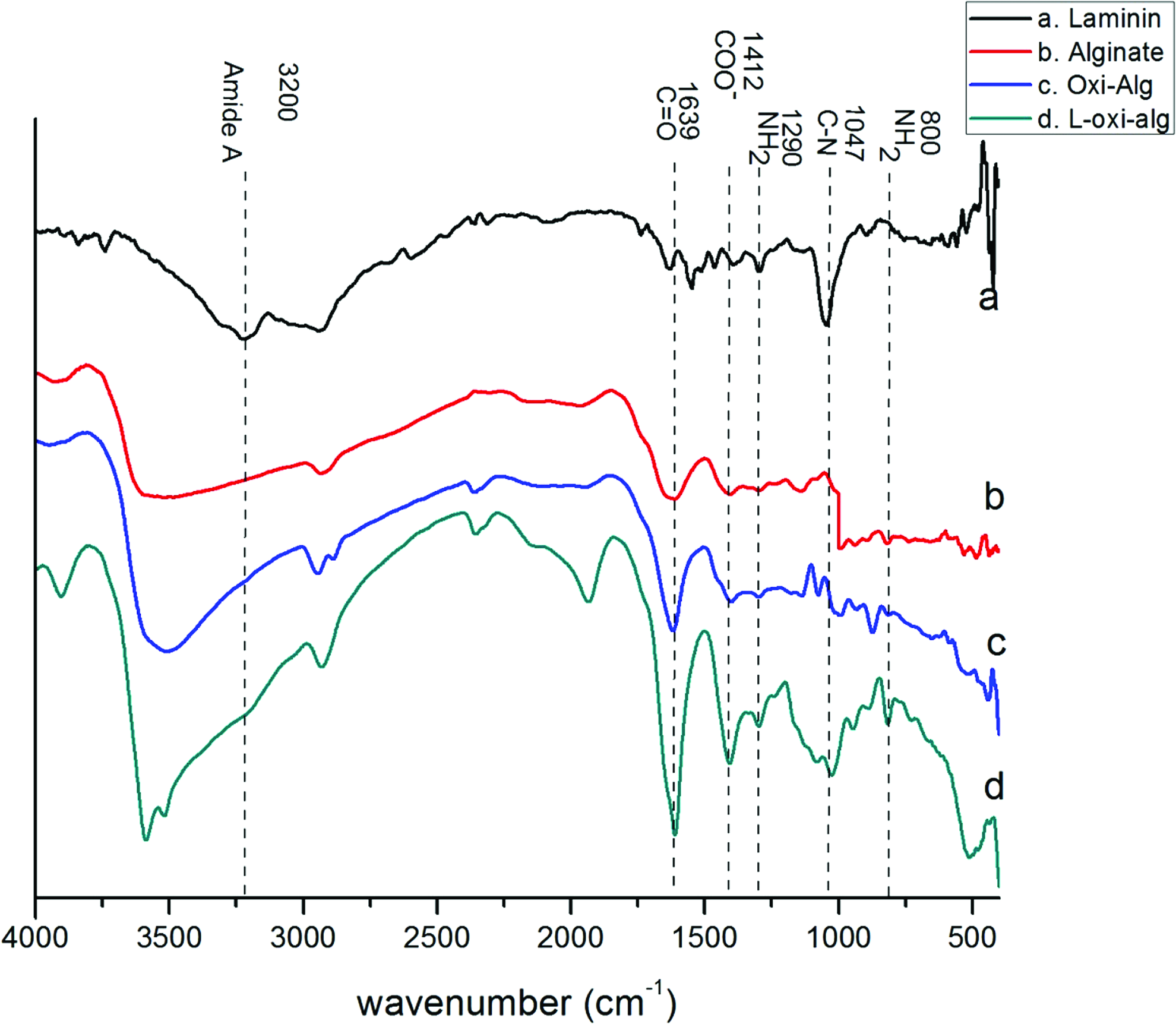
The FTIR of spectra of laminin (a), alginate (b), oxi-alg (c), and laminin-oxi-alginate (d). The absorption band of 1639 cm−1 corresponds to the carbonyl stretching C = O vibration of aldehyde groups that were detected in (b), (c), and (d). The absorption band at 1047 cm−1 corresponds to the stretching mode of C–N bond in laminin. Immobilizing laminin onto alginate created a new absorption band at 1047 cm−1 in (d), and this linkage consumed the amino group of laminin at the wavelength of 1290 cm−1. The ratio –NH2 (1047 cm−1)/COO– (1412 cm−1) showed that the amino group presence decreased in (d), supporting the cross-linking of laminin onto oxi-alginate (d). The characteristic NH2 group of laminin could also be observed at 800 and 1290 cm−1 (d). FTIR, Fourier transform infrared spectrometer. Color images available online at www.liebertpub.com/tea
Laminin-alginate was added dropwise into CaCl2 to form spherical beads that had an average diameter of 1.593 ± 0.07 mm (n = 10), and 1 mL of LABs contained 5 × 10⁴ 3T3-L1 cells. The weight of the LABs reduced gradually in vitro and retained 90% ± 1% and 72% ± 4% of the original weight at day 30 and day 60, respectively (Fig. 2).
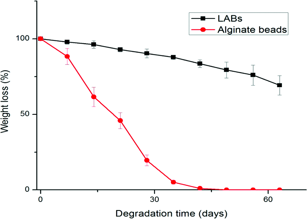
In vitro degradation of LABs compared to unmodified alginate beads. The alginate beads were completely degraded by day 42, while the LABs retained more than 70% of the original weight after 60 days. LAB, laminin-alginate bead. Color images available online at www.liebertpub.com/tea
Biocompatibility of LABs
Cytotoxicity of LABs was determined by LDH activity assays (Fig. 3). There was no significant difference in LDH activities between the control group and the LAB group on day 1 or day 3. The LDH activity of both the experimental group and control group was less than 10% of the total lysis group whether on day 1 or day 3. These results support that the LABs had little toxicity toward 3T3-L1 cells in vitro.
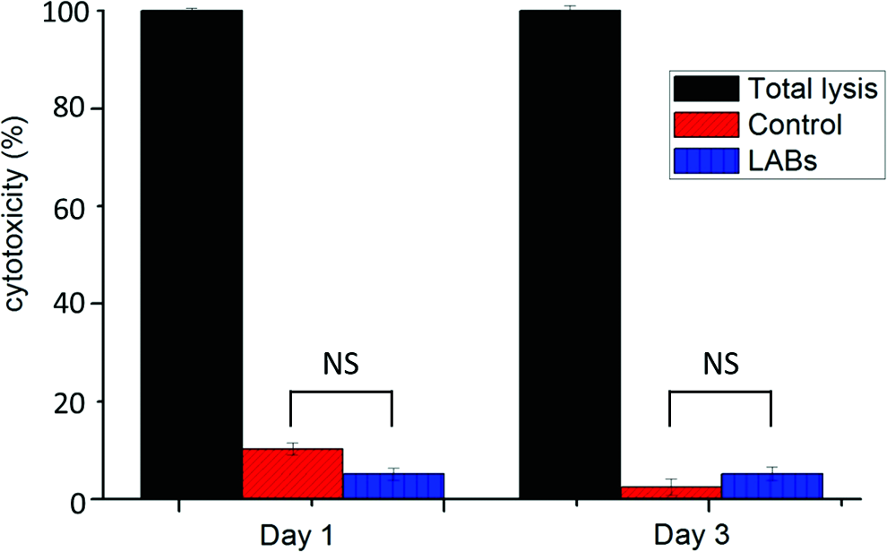
Cytotoxicity assayed by the activities of released LDH. 3T3-L1 cells in PBS (control) or in LABs were tested after 1 and 3 days in culture, respectively. The O.D. values of the total lysis group were used as references and set to be 100%. Both the control and LAB groups had O.D. measurements of lower than 10% either on day 1 or day 3.There was no significant difference between the cells in the control group and cells enclosed in LABs. (NS, not significant, p > 0.05 for all, n = 7). LDH, lactate dehydrogenase; PBS, phosphate-buffered saline. Color images available online at www.liebertpub.com/tea
The proliferation of preadipocytes encapsulated in LABs was evaluated by the WST-1 assay as shown in Figure 4. Induced 3T3-L1 cells encapsulated in LABs were compared to those enclosed in alginate beads. Cells in LABs showed greater increases in absorbance than those in alginate beads, with the O.D. values being 1.35 ± 0.14 and 1.05 ± 0.32 on day 7 (p < 0.05), respectively. The difference between the two groups grew larger, with the O.D. values being 1.62 ± 0.11 for the LAB group and 1.21 ± 0.16 for the alginate group on day 14 (p < 0.01). These results suggested that LABs were slightly better in supporting the growth of 3T3-L1 cells than beads formed by unmodified alginate.
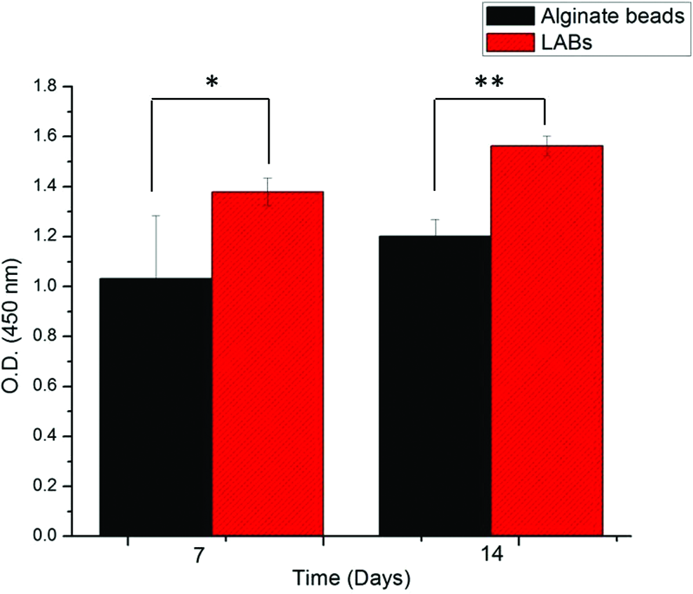
Proliferation of 3T3-L1 cells as determined by the WST-1 assay. The O.D. values of preadipocytes encapsulated in alginate beads were compared to those enclosed in LABs on day 7 and day 14, respectively. (*p < 0.05, **p < 0.01, n = 7). Color images available online at www.liebertpub.com/tea
The live and dead 3T3-L1 cells were further assayed by stain with Calcein-AM to label living cells and propidium iodide for detecting dead cells (Fig. 5). Comparing to cells enclosed in alginate beads, those encapsulated in LABs had broad and more even signals for both Calcein-AM and propidium iodide (PI). This difference made it difficult for a proper quantification. However, it is clear that LABs were similar, if not better, in supporting the survival of 3T3-L1 cells. Taken together, these results supported that LABs represent a suitable environment for 3T3-L1 survival comparing to unmodified alginate.

Cell survival monitored by the live/dead stain on day 7 and day 14. Preadipocytes were encapsulated in unmodified alginate beads or in LABs and analyzed on day 7
The morphology of induced 3T3-L1 cells in LABs was observed on day 7 by scanning electron microscopy (Fig. 6). The surface of LABs consisted of numerous micropores interconnected by worm-like nanopores (Fig. 6a). These are structures known to allow diffusion of nutrients for the survival of the encapsulated cells. 25 Entrapped cells were easily observed from the surface of LABs (Fig. 6b).

Characterization of the microstructure of LAB encapsulating preadipocytes by SEM on day 7.
Adipogenesis in vitro
The in vitro adipogenesis of 3T3-L1 cells in LABs was evaluated by the fluorescence-based AdipoRed assay as shown in Figure 7. In the first 4 days after adipogenic induction, there was no significant difference in RFUs between uninduced preadipocyte controls, induced preadipocytes in alginate beads, and induced preadipocytes in LABs (p > 0.05). On day 8, the intracellular lipid contents of the induced 3T3-L1 cells in alginate beads and that of cells in LABs were higher than those of uninduced preadipocytes. On day 14, the signals detected for induced 3T3-L1 cells in LABs were 16,500 ± 1050 RFUs, almost twice of the signals of cells in alginate beads (8500 ± 950 RFUs) (p < 0.05). Thus, LABs better supported adipogenesis of induced 3T3-L1 cells in vitro comparing to beads composed of unmodified alginate.
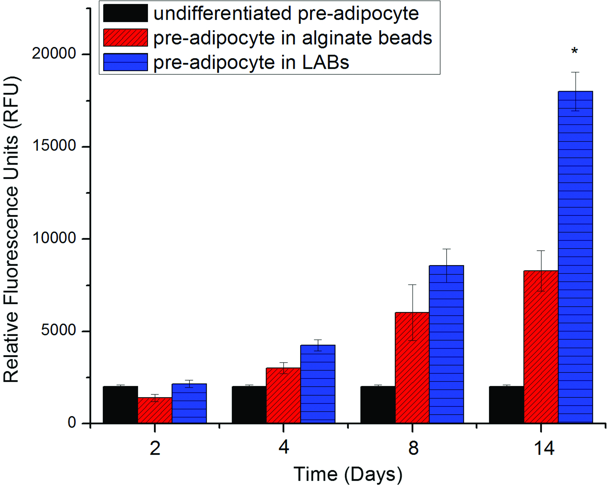
In vitro adipogenesis of preadipocytes in unmodified alginate beads and those in LABs. Intracellular lipid accumulation was determined by AdipoRed™ assay, and preadipocytes that were not induced for adipogenesis were used as controls. The RFU values for cells in LABs and cells in alginate beads on day 8 were 8800 ± 1000 and 6100 ± 1300, respectively. The values on day 14 were 16,500 ± 1050 and 8500 ± 950, respectively, with cells in LABs having significantly higher lipid contents than those in alginate beads (*p < 0.05, n = 6). RFU, relative fluorescence unit. Color images available online at www.liebertpub.com/tea
In vivo study
Each NOD/SCID mice received 1 mL of cell-laden LABs (5 × 10⁴ induced 3T3-L1 cells) subcutaneously, and neoadipogenesis and possible subchronic toxicity were determined. Histological analysis showed that, 1 week after implantation, the site of introduction was still mostly occupied by LABs that remained structurally evident (Fig. 8a). After 2 weeks, noticeable adipogenesis occurred, with newly formed adipose tissue and oil droplets readily detectable (Fig. 8b). After 4 weeks, LABs were mostly degraded, and the space was now replaced by fat tissues, and new blood vessels were observed sporadically (Fig. 8c).

Histological analysis of the grafts by H&E staining. As shown by arrow in
Hematological and blood biochemical assessments showed mostly normal results at 1, 2, and 4 weeks after implantation (Table 1). However, we did notice slightly fewer RBCs and reduced levels of Bil-T and TG. BUN was slightly elevated at 1 week, but quickly lowered and became normal by week 2. These results suggest that, for mice, 1 mL of cell-laden LABs may be a large enough quantity to become a temporary burden for the kidney and liver. Because these effects were minor and transient, our data indicated that LABs showed little subchronic toxicity in NOD/SCID mice.
Values are presented as mean ± standard deviation, and the normal ranges are provided as reference values.
Bil-T, total bilirubin; BUN, blood urea nitrogen; CRE, creatinine; GOT, glutamate oxaloacetate transaminase; GPT, glutamate pyruvate transaminase; Hb, hemoglobin; RBC, red blood cell; TG, triglyceride; WBC, white blood cell.
7T-MRI analysis
Incorporation of the grafted materials on the animal's back was compared by 7T-MRI at 1 and 12 weeks after implantation (Fig. 9). We compared the results of grafting mature adipose tissue composed of shredded fat pads (Fig. 9a, a′), cell-laden LABs only (Fig. 9b, b′), and mature adipose tissue mixed with cell-laden LABs (Fig. 9c, c′). Comparing to the MRI image at 1 week after grafting (Fig. 9a), grafted mature adipose tissues became more homogenous, but showed signs of inflammation and fat necrosis (Fig. 9a′). Better results were obtained from the animals that received cell-laden LABs or a mixture of mature adipose tissue and cell-laden LABs. At 1 week after implantation, the signals of LABs were obvious (Fig. 9b), which is gone by week 12, suggesting the degradation of LABs (Fig. 9b′). Furthermore, the implanted materials were replaced by a homogenous layer of tissue with no signs of calcification or inflammation (Fig. 9b′, c′). Therefore, cell-laden LABs were well tolerated and allowed the formation of new adipose tissues in vivo.
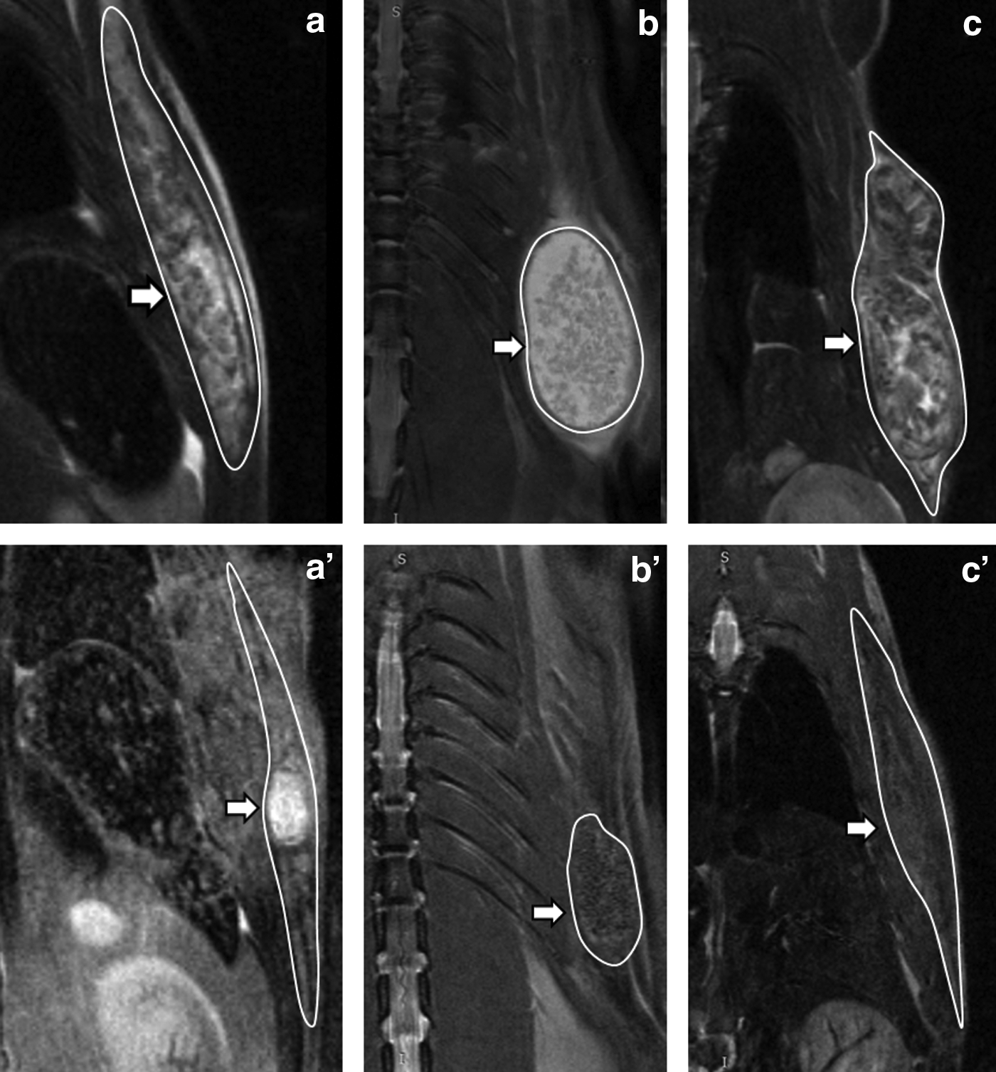
Representative 7T magnetic resonance images for each group at weeks 1 and 12. The arrow indicates the sites of implants in each panel.
Discussion
In the study, alginate was partially oxidized by sodium periodate to create aldehyde groups for laminin immobilization. The linkage of laminin was confirmed by FTIR patterns based on the presence of amide bonds that are specific for laminin. Changes in the ratio of –NH2 (1047 cm−1) to COO– (1412 cm−1) supported the successful cross-linking between alginate and laminin. Decrease in the ratio of C = O to C–N is consistent with the consumption of C = O during cross-linking in the FTIR spectrum of laminin-oxi-alginate. The LABs showed better stability than alginate beads in aqueous environment. It is likely that the addition of laminin increased the overall molecular weight and structurally stabilized the beads. Alternatively, laminin immobilization further cross-linked alginate molecules together, thus slowed down the degradation process. Since LABs were freeze-dried and underwent extensive washing and dialysis to remove unreacted laminin, our data indicated that laminin was immobilized onto oxidized alginate, and this modification resulted in improved stability and improved bioactivity in vitro.
We showed that the addition of laminin improved the biocompatibility of alginate. While both materials showed minimal cytotoxicity, 3T3-L1 cells encapsulated in LABs had enhanced survival and proliferation over those in unmodified alginate beads. Consistently, in vivo LAB implantation caused negligible effects on hematology and plasma biochemistry. Most importantly, the results of the AdipoRed assay demonstrated that LABs were superior in promoting adipogenesis compared to unmodified alginate. Laminin can interact with a series of cell surface receptors, including integrins, and these interactions indicate important biological functions such as cell attachment, spread, growth, and motility. 18 Laminin can self-assemble into lattices in vitro; studies show that the formation of laminin polymers is required for basement membrane assembly and plays a crucial role for cell-anchoring during early embryogenesis. 18 Moreover, interactions between laminin and its cellular receptors are required to establish cell polarity and to trigger differentiation in embryoid body. 18 For mesenchymal stem cells, adipogenic induction upregulates laminin expression, 21 and stem cell adhesion to the ECM strongly correlates with adipogenic differentiation. 26 These evidence supports that laminin is a critical component of the ECM for stem cell differentiation. Indeed, we found that cell-laden LABs successfully induced new adipose tissue formation and neovascularization in vivo. By cross-linking laminin onto alginate, the LABs showed better biocompatibility and induced higher level of adipogenesis than alginate alone, strongly supporting that the addition of laminin is beneficial for graft survival.
The histological results showed that the implanted materials were partially replaced by newly formed adipose tissue by week 2 and new blood vessels appeared near the implanted LABs by week 4. MRI images also identified the formation of homogeneous adipose tissue at 12 weeks after cell-laden LABs were implanted together with conventional fat grafts. Our results suggested that the degradation of LAB scaffold was more efficient when 3T3-L1 cells were present. 3T3-L1 cells are not only capable of adipogenic differentiation but also well known for their biologic modulatory ability such as secreting VEGF to promote angiogenesis and vein formation of endothelial cells. 11 Therefore, LABs can be as supportive biodegradable scaffolds for preadipocytes forming newly normal adipose tissue in graft.
Various modifications have been tested to enhance cellular interaction with alginate-based biomaterials. For example, cross-linking of RGD (Arg-Gly-Asp) peptides to alginate has been found to promote cell adhesion and cell survival.27,28 RGD is an integrin binding motif first identified from fibronectin and, subsequently, found in many ECM proteins. 29 Incorporation of RGD peptides to biomaterials yields promising results in vitro; however, in vivo studies showed more variable results. 30 The processes for RGD immobilization may involve cytotoxic conditions, and the conjugated RGD moieties are sensitive to hydrolysis. 31 RGD have been shown to retain only 10%–30% of the bioactivity compared with whole integrin-binding proteins 32 and do not elicit the same cellular response. These findings suggest that integrin-binding proteins contain additional information that dictates downstream integrin signaling. Therefore, it is possible that incorporating an integrin-binding protein, such as laminin, may better serve the purpose. In this study, we showed data supporting the use of LABs as a stem cell carrier for adipose tissue engineering. It will be important to compare the efficacies of LABs and RGD-modified alginate for adipogenesis in vitro and in vivo.
Conclusion
In the study, LABs as preadipocyte carriers showed better biocompatibility and stability than unmodified alginate beads. Preadipocytes encapsulated in LABs had higher survival rates than those in alginate beads. Cell-laden LABs were well tolerated in vivo and were gradually replaced by newly formed fat tissues with blood vessels sporadically observed. The addition of cell-laden LABs improved the outcomes of mature fat grafts as observed by 7T-MRI. Our findings demonstrated that LABs are efficient cell carriers that are capable to promote adipose tissue formation in a subclinical setting.
Footnotes
Acknowledgments
The project is supported by National Taiwan University Hospital (Grant No.: NTU 101-M1960). The authors sincerely thank the 8th core laboratory of National Taiwan University Hospital (NTUH) for their instruments. The authors also acknowledge the Central European Research Abroad Program for PhD students granted by National Science Council and International Visegrad Fund for clinical research.
Disclosure Statement
No competing financial interests exist.