
Abstract
Three-dimensional (3D) bioprinting is a rapidly emerging technique in the field of tissue engineering to fabricate extremely intricate and complex biomimetic scaffolds in the range of micrometers. Such customized 3D printed constructs can be used for the regeneration of complex tissues such as cartilage, vessels, and nerves. However, the 3D printing techniques often offer limited control over the resolution and compromised mechanical properties due to short selection of printable inks. To address these limitations, we combined stereolithography and electrospinning techniques to fabricate a novel 3D biomimetic neural scaffold with a tunable porous structure and embedded aligned fibers. By employing two different types of biofabrication methods, we successfully utilized both synthetic and natural materials with varying chemical composition as bioink to enhance biocompatibilities and mechanical properties of the scaffold. The resulting microfibers composed of polycaprolactone (PCL) polymer and PCL mixed with gelatin were embedded in 3D printed hydrogel scaffold. Our results showed that 3D printed scaffolds with electrospun fibers significantly improve neural stem cell adhesion when compared to those without the fibers. Furthermore, 3D scaffolds embedded with aligned fibers showed an enhancement in cell proliferation relative to bare control scaffolds. More importantly, confocal microscopy images illustrated that the scaffold with PCL/gelatin fibers greatly increased the average neurite length and directed neurite extension of primary cortical neurons along the fiber. The results of this study demonstrate the potential to create unique 3D neural tissue constructs by combining 3D bioprinting and electrospinning techniques.
Introduction
D
In our previous studies, we have separately employed stereolithography (SL) and electrospinning to create microporous three-dimensional (3D) printed constructs or highly aligned, fibrous neural scaffolds respectively.8,9 SL is a laser-based 3D printing system capable of fabricating aligned micro-macro size 3D constructs via a layer by layer assembly method. We have shown that polyethylene (glycol) diacrylate (PEG-DA) 3D scaffolds printed by our customized table top SL printer are mechanically stable and have ideal hydrophilic properties. Furthermore, we observed enhanced PC-12 cell and primary cortical neuron growth and differentiation on the scaffold. 8 In addition, electrospinning is an extremely versatile technique for fabrication of nano to micro fibers for tissue engineering applications. Particularly, electrospun polymeric fibers were previously employed for the production of scaffolds for neural tissue engineering.9–14 Highly aligned, fibrous, electrospun scaffolds serve as excellent substrates, directing neural cells to differentiate along the fibers. For example, our previous research has shown that highly aligned electrospun fibers can enhance neural cell proliferation and differentiation along the fiber. 9
Despite the aforementioned advantageous features of both fabrication techniques, each method has its limitation. Many current 3D bioprinting including SL printing techniques exhibit difficulty in achieving a nano resolution. Electrospun fibers often have lack of pore interconnectivity resulting in poor cell infiltration and migration within the scaffold. Currently, one interesting trend in scaffold design is to combine the electrospinning technique and 3D printing system to fabricate a novel tissue scaffold with both nano and well-designed micro architecture. For example, Xu et al. have successfully combined electrospinning and inkjet bioprinting to fabricate a cartilage construct. 15 Such scaffold showed enhanced mechanical properties and biocompatibilities compared to conventional hydrogel constructs generated using inkjet printing alone. This implies that micron to nano size features of electrospun fiber can enhance the specificity and accuracy of 3D printed scaffolds for diverse tissue engineering applications including fabrication of neural scaffold. 16
In this study, we introduced electrospun fibers into 3D printed microporous tissue constructs, and investigated neural stem cell (NSC) and primary neuronal cell growth in the neural tissue construct for the first time. The primary objectives of this study are to (1) evaluate the feasibility of integrating SL 3D printing and electrospinning to create a highly aligned and microporous scaffold, and to (2) examine the proliferative capability and differentiation potential of neural cells seeded in the resultant composite scaffold in vitro. NSC cloned from early embryonic (E9) mouse neuroectoderm (NE-4C cell) was used in this study as they can display nestin and tubulin beta III immunoreactivity, self renewal, and differentiation into distinct neural cell types upon appropriate induction. In the presence of nerve growth factor (NGF), NSCs differentiate into neurons and astrocytes in a progressive process, through well-defined stages displaying specific morphological and cell physiological characteristics. 17 Additionally, primary culture of rat cortical neurons on our various scaffolds will be performed as they mimic events occurring in vivo to some extent and permit detailed molecular analysis.
In regards to the printing materials, electrospun fibers can be made of many types of polymers and composite materials that can be utilized to mimic innate properties of native extracellular matrix. Previous studies have demonstrated enhanced neural cell attachment and proliferation on substrates such as polycaprolactone (PCL), PCL with laminin, and PCL with gelatin, created with electrospun nanofibers.9,18–20 PCL is a bioresorbable and biocompatible polyester, approved by the FDA for several medical devices and widely used as a biomaterial scaffold with slow degradation behavior. Additionally, PCL has been widely used as a nerve scaffold material.18,19,21,22 Furthermore, several studies have successfully demonstrated the potential of gelatin in neural tissue engineering.14,20,23,24 Gelatin is a natural extracellular matrix component and has shown excellent biocompatibility for various cell types, including neural cells. Alvarez-Perez et al. has shown the influence of gelatin cues in electrospun fibers on nerve outgrowth. 18 Altogether, we used PCL and PCL/gelatin composite materials in our electrospinning setup and PEG-DA for the SL 3D printing. We examined how the incorporation of gelatin into PCL fibers can affect neurite growth and differentiation compared to PCL fibers alone.
Materials and Methods
Fabrication of electrospun fibers
Aligned PCL and PCL/gelatin microfibrous scaffolds were fabricated via our lab's electrospinning setup. Briefly, PCL (Mw = 70,000–90,000; Sigma-Aldrich) was dissolved in chloroform under sonication to form a 15% (w/v) clear solution. For PCL/gelatin composite microfibrous scaffolds, both PCL and gelatin (Mw = 1000; Sigma-Aldrich) were separately prepared in 2,2,2-trifluoroethanol (TFE) to form 15% (w/v) and 20% (w/v) solutions, respectively. Both solutions were then mixed to obtain a solution of PCL/gelatin 50:50 (w/w). Then, each solution was dispensed from a 10 mL standard syringe attached to a 21G blunt needle. The solution was extruded at a flow rate of 2.5 mL/h and electrospun under a 10–15 kV voltage. Electrospun microfibers were collected on a rotating mandrel at a distance of 12 cm from the needle tip creating the aligned scaffolds.
Fabrication of electrospun fiber-3D printed composite scaffolds
Our customized SL-based 3D printing platform was based on the existing Printrbot® rapid prototyping platform for addictive manufacturing of photosensitive hydrogel scaffolds. The 3D printer with movable Z-stage consists of a 110 μm fiber optic-coupled solid-state UV (355 nm) laser (MarketTech, Scotts Valley, CA) mounted on an X-Y toolhead for a three-axis motion. The laser beam of energy output of ∼20 μJ at 15 kHz was focused to a spot diameter of 190 ± 50 μm. Specifically, Printrun software package controlled the printing configuration. The laser frequency was optimized in the 8–11 kHz range and the printing speed was set to 25 mm/s. Prepared electrospun fibers were placed on the bottom of the petri dish. Then, printable hydrogel inks, composed of 40 wt% PEG (Mn 300), 60 wt% PEG-DA (Mn 700), and photoinitiator (0.5 wt% of PEG-DA concentration), covered the electrospun fiber. Scaffold geometries were designed with 66% large porosity and a square pore geometry using computer-aided design (CAD) software. Figure 1 shows the overall fabrication process of the study.
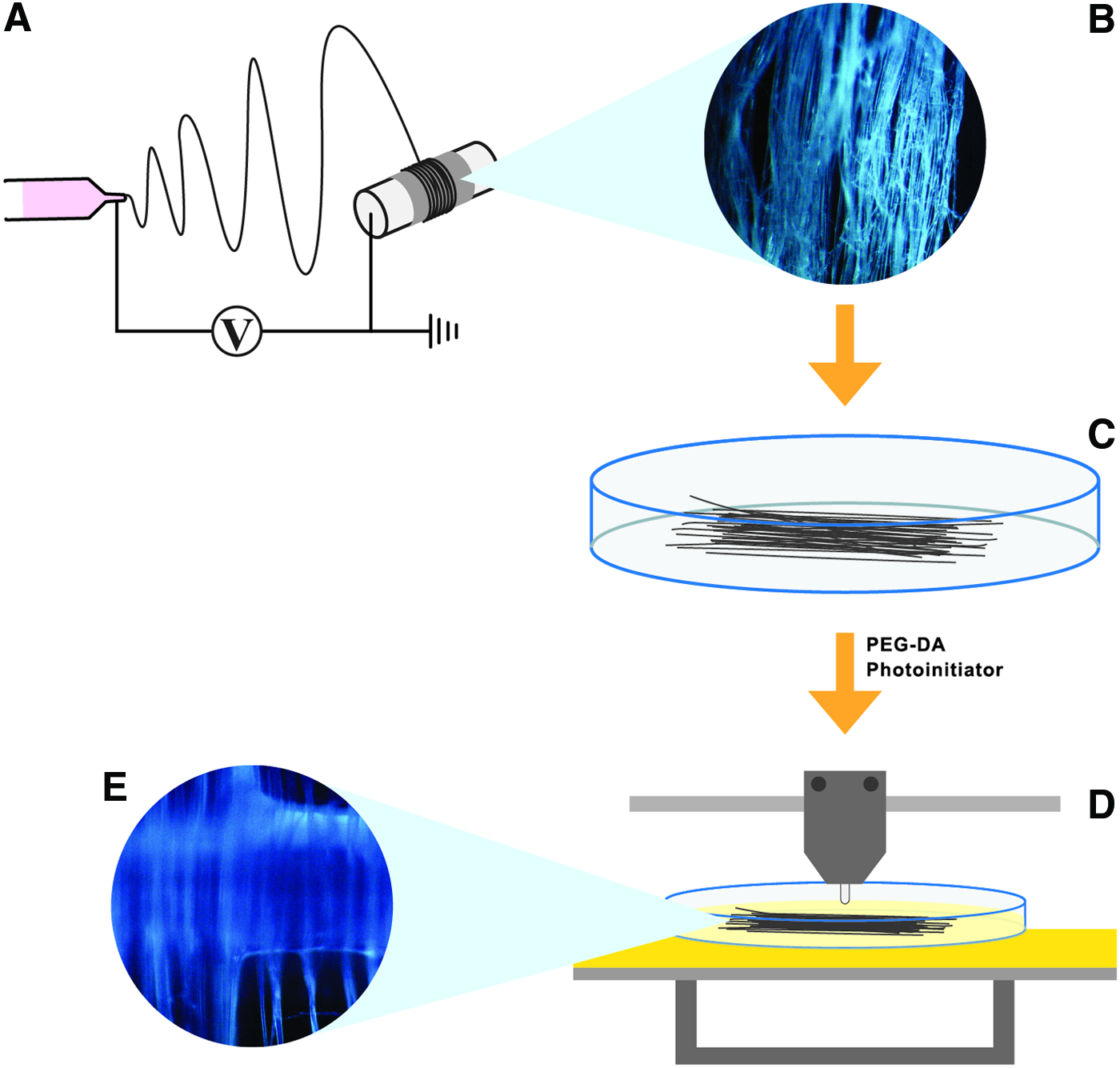
Schematic diagram of our 3D printed neural scaffold fabrication.
3D printed scaffold characterization
Scanning electron microscopy (SEM) was employed to assess morphology and channel size of SL printed scaffolds with or without electrospun fibers. All samples were sputter coated with gold for 10 s (sputter set point 80 mTorr Vacuum pressure) and visualized with a Zeiss SigmaVP SEM. MTS Criterion Model 42 conducted tensile mecanical tesing of various scaffolds under a constant crosshead rate of 2 mm/min. Rectangular sheets (10 mm by 20 mm) were cut from all scaffolds including electrospun fibers and 3D printed scaffolds with or without electrospun fibers. The ends of the rectangular sheets were clasped on each end by mechanical grips. Young's modulus was calculated by the linear portion of the resulting tensile stress–strain curve.
A drop shape analyzer (DSA4; Krüss) equipped with a camera measured the contact angles of the resultant scaffolds to determine surface hydrophilicity of the scaffolds. Briefly, the scaffolds were cut into circular disks with diameter of 10 mm and placed on glass slides. Ultra pure water (2.0 μL) was pumped automatically onto the samples' surface using a syringe. Temporal images immediately after the droplet fell off were selected from the captured videos for contact angle measurement. All experiments were conducted in room temperature and were performed at least three times per group. A Raman spectrometer (Horiba Scientific) was used to analyze the chemical composition of 3D printed scaffold with electrospun fibers using a 532.06 nm Ar+ laser with excitation ranging from 0 to 4000 cm−1. Experiments were conducted in ambient conditions.
Neural cell culture and growth studies
NSCs (NE-4C; ATCC) were utilized to evaluate cell response on various printed nerve scaffolds. NSCs were culture in Eagle's minimum essential medium (ATCC), 5% fetal bovine serum (ATCC) and 1%
Primary embryonic rat cortical cultures were performed. All protocols involving animals were approved by the Georgetown University Institutional Animal Use and Care Committee and were in compliance with the standards stated in the Committee on Care and the Use of Laboratory Animals of the Institution of Laboratory Resources DHEW Publication. Specifically, cortices from embryonic day 18 (E18) Sprague-Dawley rat embryos (Charles River, Wilmington, MA) were dissected from the meninges and blood vessel in calcium-free Hank's buffered saline solution (HBSS). Then, using sterile fine-tipped forceps and microdissecting scissors, all cortices were minced and dissociate. The minced tissues were incubated in Trypzean for 3 min. Immediately after the removal of Trypzean, PBS containing 40 μL/mg DNAse was added to the tissue and tissues were triturated through a 5 mL pipet. Then, the supernatant was gently centrifuged at 300 g for 10 min at room temperature. The cell pellet was resuspended in Neurobasal media (Gibco) with 1% B 27 supplement (Gibco), 0.5 mM glutamine, and 50 ng/mL NGF and cultures maintained in this growth medium with various materials at 37°C, 5% CO2 until immunocytochemistry was conducted.
Immunocytochemistry of NSCs and primary cortical neurons
For differentiation studies, NSCs and primary cortical neurons were cultured in respective standard medium with 50 ng/mL NGF on bare scaffold, scaffold with PCL fibers, and scaffold with PCL/gelatin fibers for 3 and 11 days for NSCs and 7 days for primary neurons. All scaffolds were coated with laminin (Sigma-Aldrich) for 4 h to enhance cellular attachment. Samples were rinsed with PBS and fixed with 10% formalin for 12 min at room temperature at specified time. The cells were further permeabilized with 0.3% Triton X-100 in PBS for 6 min. Diluted primary antibodies, mouse anti-TuJ1 (1:1000; Covance), and rabbit anti-MAP2 antibody (1:500; Abcam), were gently added in scaffolds and incubated at 4°C in a moist environment overnight. This was followed by secondary antibodies incubation with Alexa Fluor 488 (Abcam) goat anti-rabbit and Alexa Fluor 594 goat anti-mouse (Life technologies) at room temperature. The cell nuclei were stained by 10 μg/mL 4′-6-diamidino-2-phenylindole dihydrochloride (DAPI) (Life technologies). Laser scanning confocal microscopy (LSCM 710; Zeiss) was employed to visualize and monitor the fluorescent images of 3D neural cell growth and neurite extension of cells.
Neurite angle distribution and length quantification
Image J was used to calculate the angle between the fibers or channels of the scaffold and the neurite extension of primary cortical neurons. All angle measurements were constrained between 0° and 90°.
Image analysis software (ImageJ; National Institutes of Health, Bethesda, MD) and the NeuriteTracer plugin 26 were used to quantify neuronal cells and neurite length. At least four areas were randomly selected for analysis on each sample (four independent samples/group) using a 10× or 20× objective. Mean of each independent sample was used. A total of 89 to 641 cells/group were observed with nuclear staining (DAPI) and the percentage of TUJ-1-positive cells were calculated. The NeuriteTracer quantified the neuronal differentiation by tracing the neuronal marker TUJ-1. The total neurite length was determined as the total measured length of all the neurites in a field of view. The average neurite length is defined by dividing the total neurite length by the total neurite count. Comparably, the average total neurite length per cell was calculated by dividing the total measured lengths of all neurites in the field of view by the total cell count. Lastly, the average length of the longest neurite per cell was calculated.
Statistical analysis
All quantitative data are expressed as average ± standard error of the mean. Numerical data were analyzed via student's t-test to determine differences among the groups. All the data obtained for tensile strength and surface wettability of scaffolds were statistically evaluated using one-way ANOVA with Turkey's multiple comparison. Statistical significance was considered at p < 0.05.
Results
3D printed scaffold characterization
A highly aligned electrospun fiber is clearly embedded within the 3D printed construct as shown in a microscopic view in Figure 2. Figure 3 displays the SEM morphology of printed scaffolds with or without PCL or PCL/gelatin electrospun fibers at low magnification (Fig. 3A, C, E) and at high magnification (Figs. 3B, D, F). It shows that SL printed scaffolds exhibit uniformly oriented channels and pores having micrometer resolution. Also, micropores in the scaffolds were highly interconnected throughout the entire structure. Our previous study has shown that SL printed PEG-DA scaffold with 66% porosity (large porosity) enhanced neural cell attachment compared to other scaffolds with smaller porosity. 8 Hence, this study utilized the identical PEG-DA 3D printed scaffold with large porosity to evaluate the effect of incorporation of different electrospun fibers. The incorporation of either PCL or PCL/gelatin electrospun fibers on 3D printed scaffolds is shown in Figure 3C–F. Electrospun fibers are highly aligned and embedded in the 3D printed scaffolds.
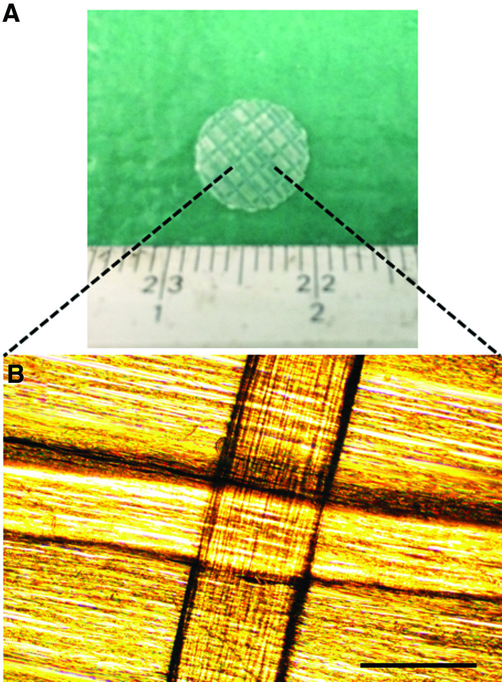
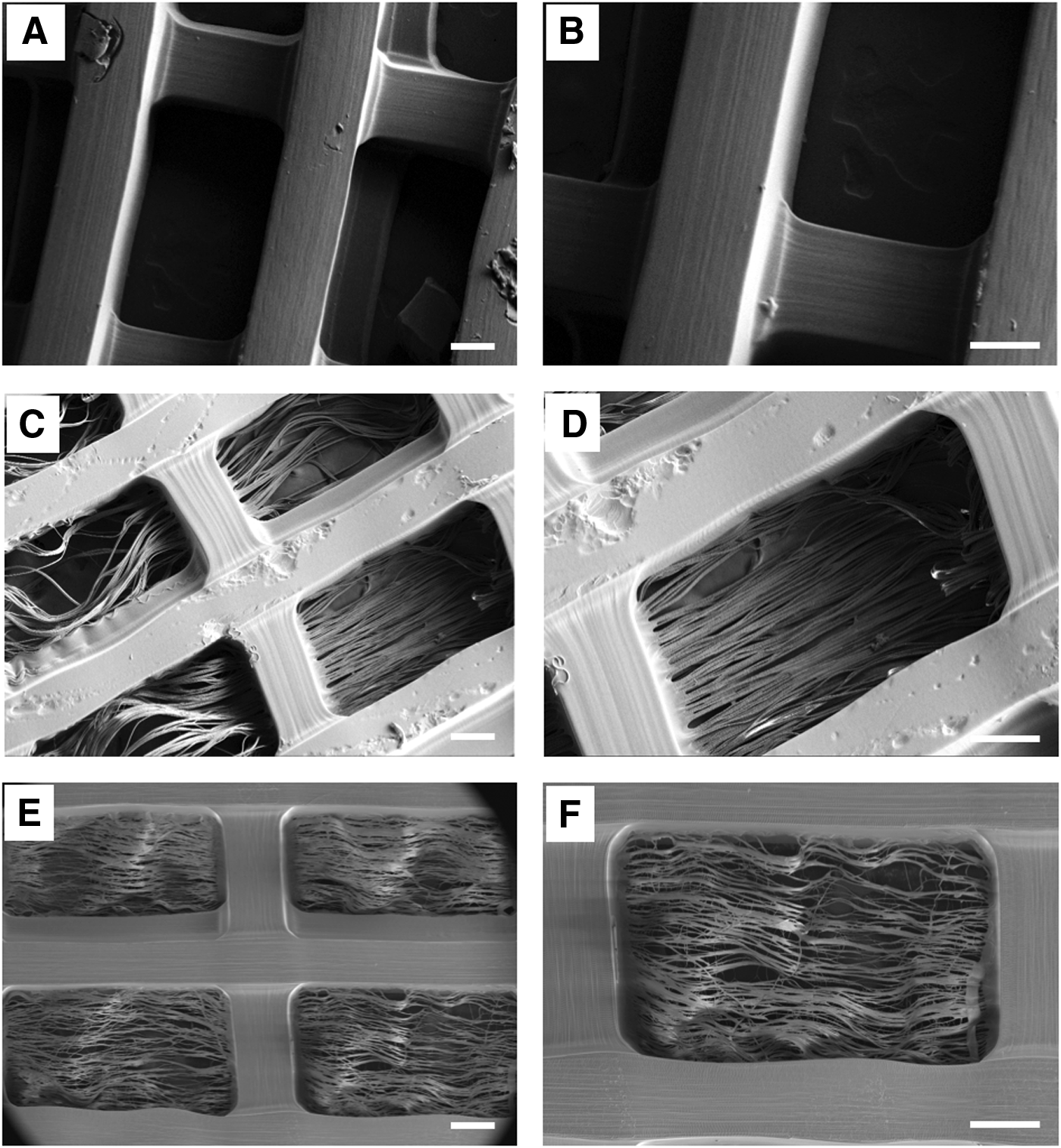
SEM images of 3D printed scaffolds with large porosity percentage of 66%
Table 1 shows the results of tensile strength of various scaffolds. As shown in the table, blending of PCL with with gelatin caused a significant increase in Young's modulus but a reduction in ultimate tensile strength. Similar phenomenon was also reported from a previous study 20 where the addition of gelatin (PCL/gelatin, 1:1 wt%) resulted in enhanced tensile properties. On contrast, The Young's modulus of the SL printed scaffolds was 0.45 ± 0.13 MPa (Table 1), which is 60% lower than PCL fiber. The addition of PCL and PCL/gelatin electrospun fibers onto SL printed scaffolds increased the Young's modulus by 151% and 217%, respectively over the plain large porosity scaffold without the fibers. Similarly, the use of PCL or PCL/gelatin electrospun fibers increased the ultimate tensile strength by 143% and 89%, respectively, over the plain scaffold. The apparent increases in Young's modulus and ultimate tensile strength are mainly because of the incorporation of the PCL and gelatin electrospun fibers. Overall, the mechanical properties of SL printed scaffolds with embedded PCL and PCL/gelatin fibers were almost nearing to bare PCL fibers, which implied that incorporation of PCL/gelatin fibers onto 3D printed hydrogel scaffold provided enhanced mechanical strength and biological properties.
Data are mean ± standard error of the mean; n = 4.
p < 0.05 when compared to control scaffold.
p < 0.05 when compared to all other scaffolds.
PCL, polycaprolactone.
Table 2 shows the contact angle of bare electrospun fibers and large porosity scaffolds with or without PCL and PCL/gelatin fibers. The contact angle obtained for PCL fibers was about 93° which implies that these scaffolds are highly hydrophobic. On the other hand, the PCL/gelatin fibers showed very hydrophilic property with their contact angle equals to 24°. The presence of electrospun fibers did not change the contact angle significantly when compared to the control SL printed scaffolds. Nonetheless, it should be noted that the contact angle decreased from 36.8 ± 5.1° to 24.2 ± 9.7° after incorporation of PCL/gelatin electrospun fibers on the scaffold with large porosity. On the contrary, the incorporation of PCL fibers on the scaffold increased the contact angle by 30%. In general, all scaffolds with and without electrospun fibers maintained a relatively high surface wettability. Surface Raman spectroscopy was conducted to characterize the functional groups present on the SL printed scaffolds with PCL fibers and PCL/gelatin fibers (Fig. 4). Representative Raman spectra of PEG-DA were observed on both scaffolds. These include typical double-bond carbon and oxygen stretching vibration υ(C O) that appeared at 1680 cm−1 and double-bond carbon and carbon υ(CC) was observed at around 1500 cm−1. In addition to the characteristic peaks of PEG-DA, PCL fiber-embedded scaffolds showed a peak at around 2900 cm−1, which corresponds to the typical υ(C—H) chains in PCL (Fig. 4A). Figure 4B shows a PCL/gelatin fiber-embedded scaffold with a new peak at around 3500 cm−1, which corresponds to the absorption of amine group υ(N—H) from NH of gelatin.
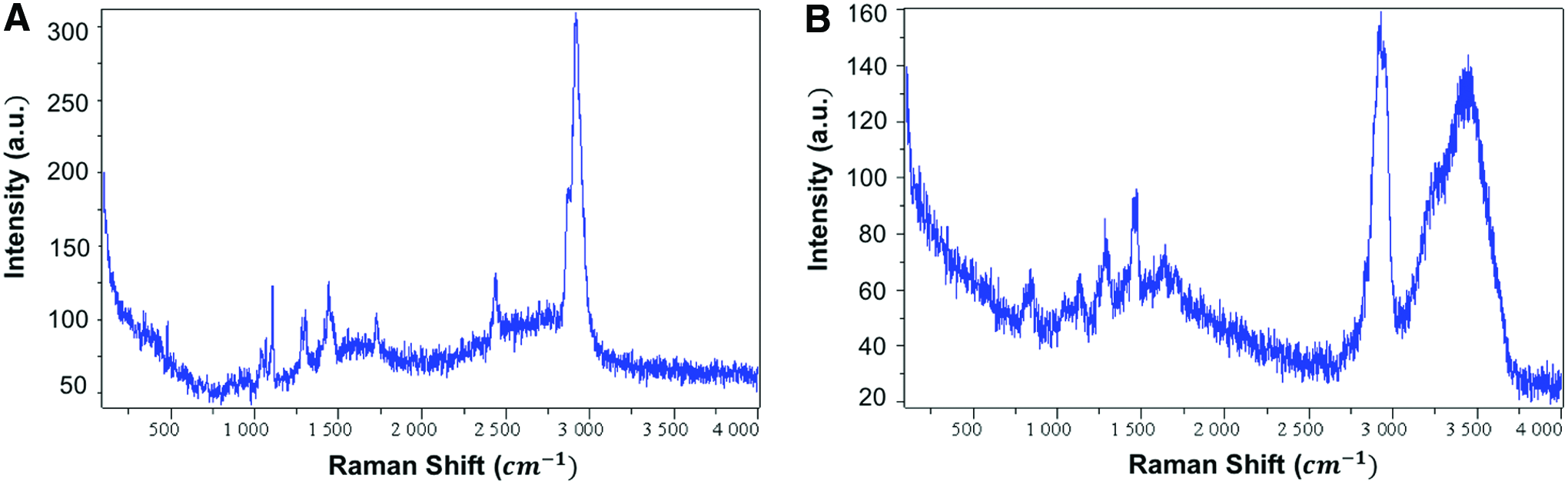
Raman spectroscopy of
Data are mean ± standard error of the mean; n = 4.
p < 0.05 when compared to all other scaffolds.
NSC growth and differentiation studies
NSCs were used to conduct the in vitro cell-scaffold interaction studies. Specifically, PEG-DA 3D printed scaffolds with or without electrospun fibers were evaluated for NSC growth and differentiation studies. The NSCs adhered well on all scaffolds, evidenced in Figure 5. The cell attachment study showed that scaffolds with PCL and scaffolds with PCL/gelatin fibers enhanced the NSC attachment significantly compared to the plain scaffold without fibers. However, there was no significant difference between the scaffolds with electrospun fibers. The proliferation studies (Fig. 6) examined the cell growth on the same constructs over 1, 3, and 5 days periods. Similarly, scaffolds with electrospun fibers significantly increased NSCs growth on 1, 3, and 5 days time points. However, there was again no noticeable difference in cell growth between PCL and PCL/gelatin fibers.
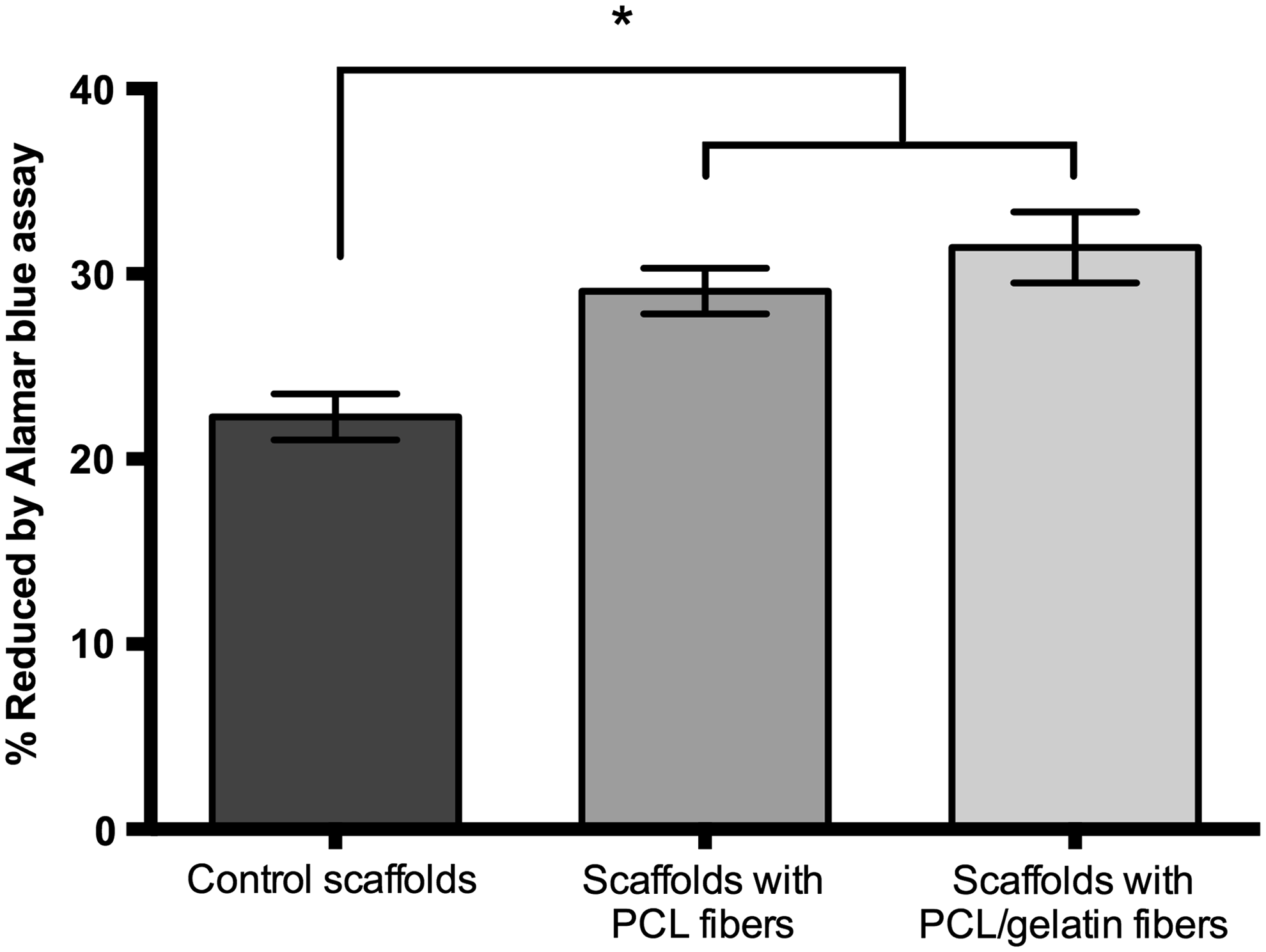
NSC adhesion on 3D printed scaffolds with or without electrospun fibers after 4 h of culture. Data are mean ± standard error of the mean; n = 9. *p < 0.05 when compared to all other scaffolds. NSC, neural stem cell.

Enhanced NSC proliferation in 3D printed scaffolds with or without electrospun fibers after 5 days culture. Data are mean ± standard error of the mean, n = 9; *p < 0.05 when compared to the corresponding scaffolds at day 1, 3, and 5, respectively.
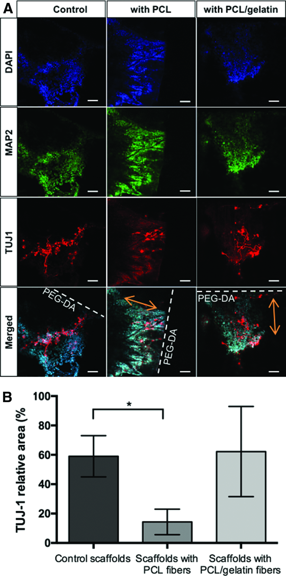
Figures 7 and 8 shows the confocal micrographs of NSCs cultured on control scaffolds with or without electrospun fibers for 3 and 11 days, respectively. Dashed lines shows the boundary of SL printed scaffold whereas orange arrows show the direction of embedded fibers. Various scaffolds showed positive evidence of NSC attachment and growth. Two neuronal markers, TuJ1 and MAP2 that reveal the early and late stages of neuronal differentiation were used to image the neural differentiation of NSCs. All scaffolds did not trigger significant neurite growth and extension by day 3 (Fig. 7) even in the presence of NGF in media. Both TUJ1 and MAP2 markers stained spheres of undifferentiated NSCs. On contrary, at day 11, differentiation of NSCs was greatly enhanced on control scaffolds and scaffolds with PCL/gelatin fibers (Fig. 8A). Differentiated neurons were clearly labeled with TUJ1 within the undifferentiated population of NSC stained by MAP2. As shown in Figure 8B, compared with scaffolds with PCL fibers, NSCs cultured on control scaffolds exhibited significantly higher TUJ-1 expression by ∼4.0-folds. Additionally, there was no significant difference in TUJ-1 expression between the scaffolds with electrospun fibers (PCL and PCL/gelatin).
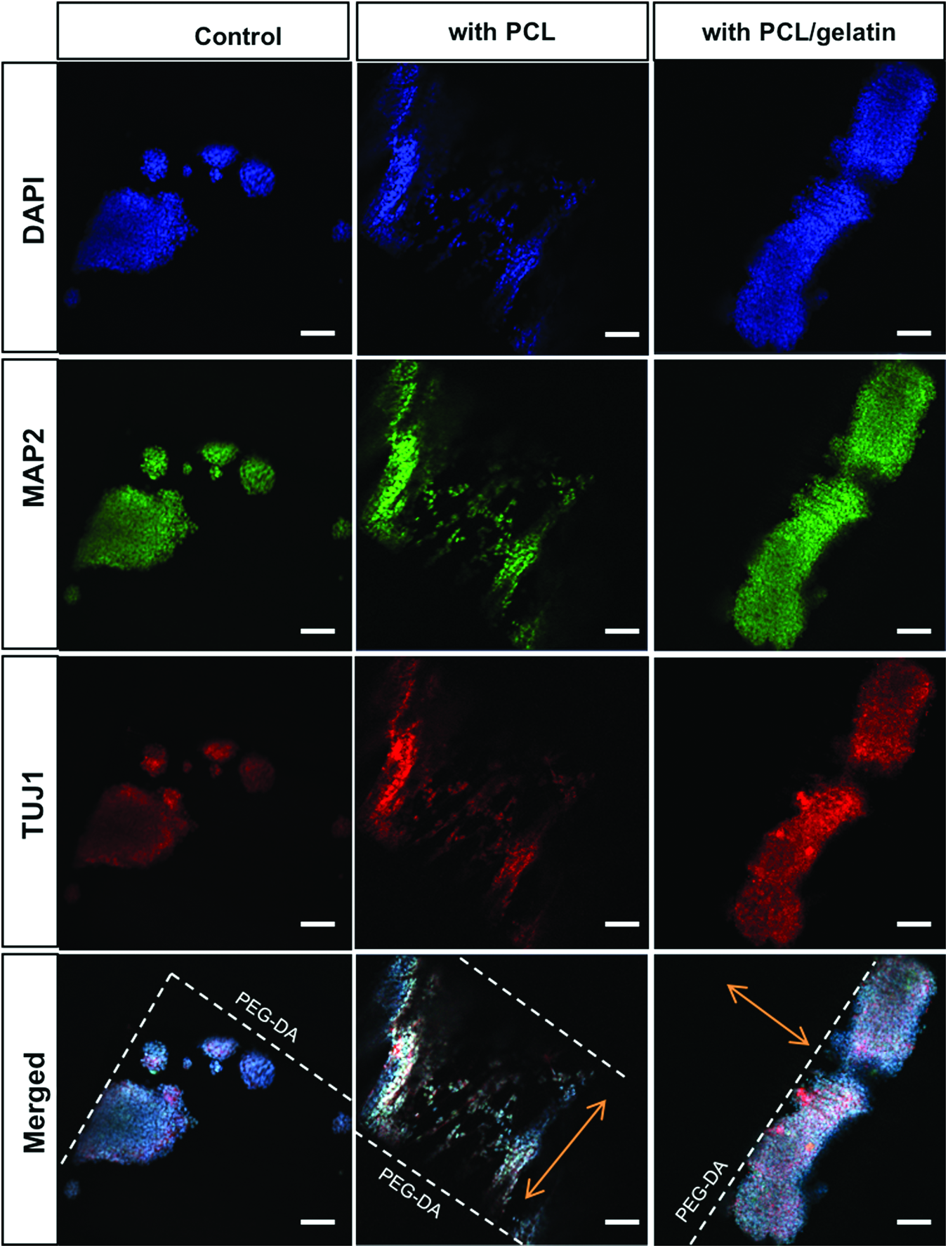
Confocal microscopy images of undifferentiated NSC growth and alignment morphology on scaffolds with or without electrospun fibers after 3 days of culture. Last row shows boundaries of SL printed scaffold (dashed line) and direction of embedded aligned fibers (orange arrow). Cell nuclei were stained by DAPI. Double staining of MAP2 and TUJ1 were used to detect neurite outgrowth of NSCs on various scaffolds. Scale bar = 100 μm. Color images available online at www.liebertpub.com/tea
Figures 9 and 10 shows the confocal micrographs of primary cortical neurons cultured on plain scaffolds with or without electrospun fibers for 7 days. All scaffolds supported the growth and differentiation of primary neurons. Additionally, highly aligned neurite extension along the fibers was observed on scaffolds with fibers. Figure 11 shows that scaffolds with PCL/gelatin fibers provide the greatest degree of neurite alignment on fibers, with 20% of neurite segment angles directly matching the direction of fibers and 59% within five degrees of the fiber direction (Fig. 11C). Neurite angles tended 0°, along the fiber, indicate that the neurites have a high tendency to orient in the direction of fiber alignment. Control scaffolds without fibers also supported some level of neurite alignment. Approximately 40% of all neurites were distributed within 5° of aligned fibers. However, about 42% of all neurites are differentiated at greater than 20° of aligned fibers. Scaffolds with aligned PCL fibers also showed an enhanced neurite alignment compared to the control plain scaffold. Forty-one percent of neurites match the direction of fibers within 5° of the fiber direction. About 85% of neurites on the scaffold with PCL fibers were extending within 20° of the fiber direction.

Confocal microscopy images of neurite growth of primary cortical neurons on
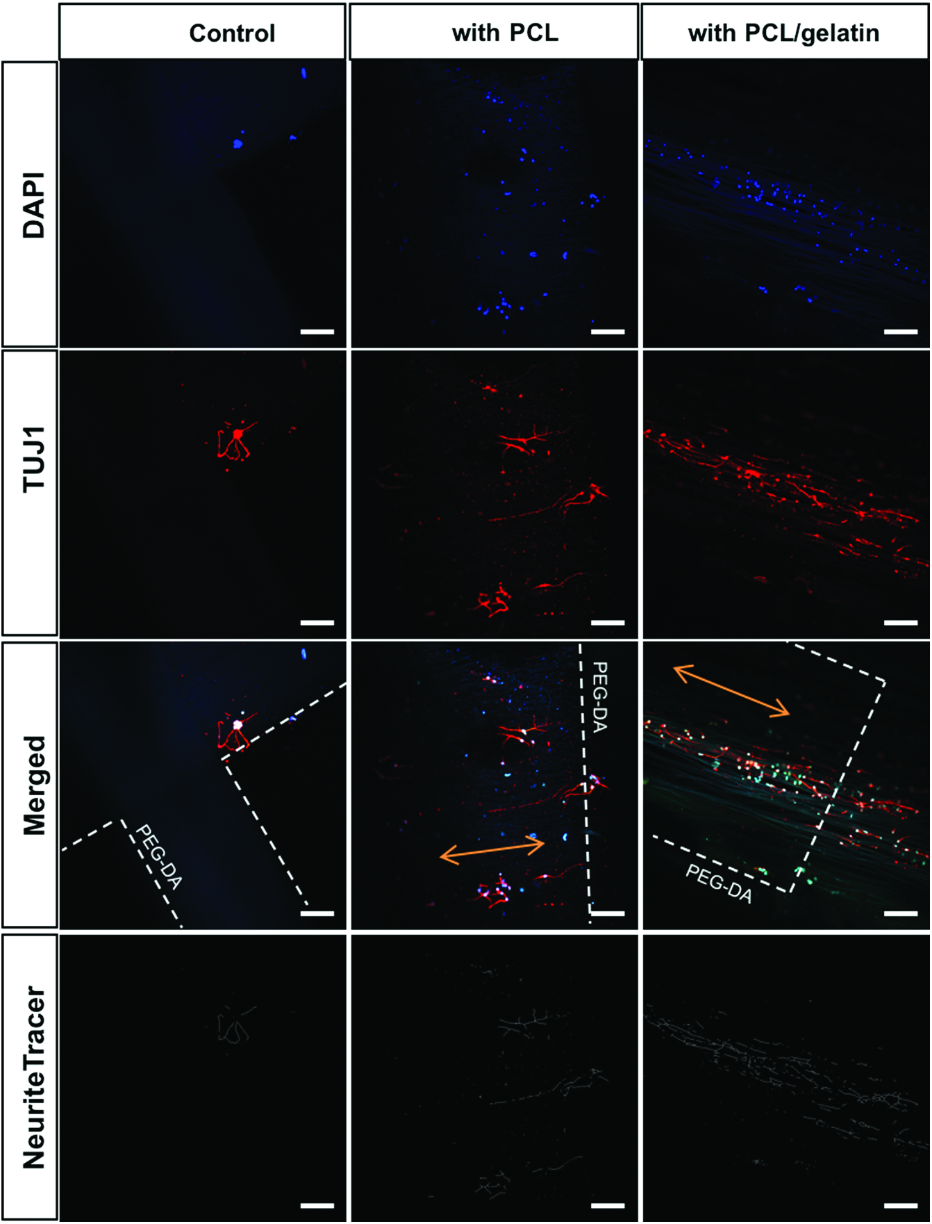
Confocal microscopy images of neurite growth of primary cortical neurons on various 3D printed scaffolds with or without electrospun fibers at day 7. Third row shows boundaries of SL printed scaffold (dashed line) and direction of embedded aligned fibers (orange arrow). DAPI was stained to detect nuclei. TuJ1 was stained to detect neurite outgrowth of primary cells on various scaffolds after 7 days of culture. The associated neurite outgrowth was traced automatically by NeuriteTracer. Scale bar = 100 μm. Color images available online at www.liebertpub.com/tea
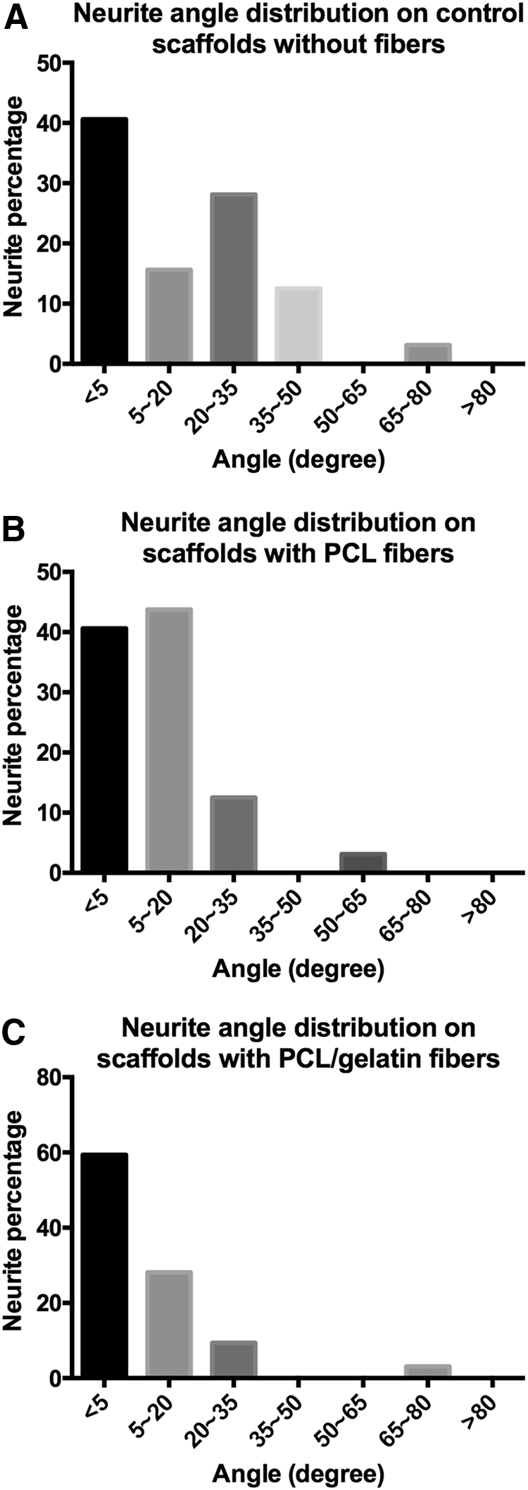
Neurite angle distributions on
Primary neuron outgrowth on all scaffolds was quantified by Image J and NeuriteTracer (Fig. 12). The incorporation of electrospun fibers into 3D printed scaffolds greatly enhanced neurite outgrowth. Particularly, scaffolds with PCL/gelatin fibers significantly improved total neurite length and average length of the longest neurites when compared to any other group at day 7 (Fig. 12A–D). Similar trends were noted when looking at average neurite lengths (Fig. 12C). The 3D printed scaffolds with PCL/gelatin fibers significantly increased the total neurite length by 66.5% over the control scaffold and by 111.4% compared to the scaffolds with PCL electrospun fibers. Furthermore, the 3D scaffold with PCL/gelatin fibers increased the average length of the longest neurites by 58.3% over the bare control scaffold. The average total length of neurites per neuron was not statistically significant among the experimental scaffold groups (Fig. 12B).
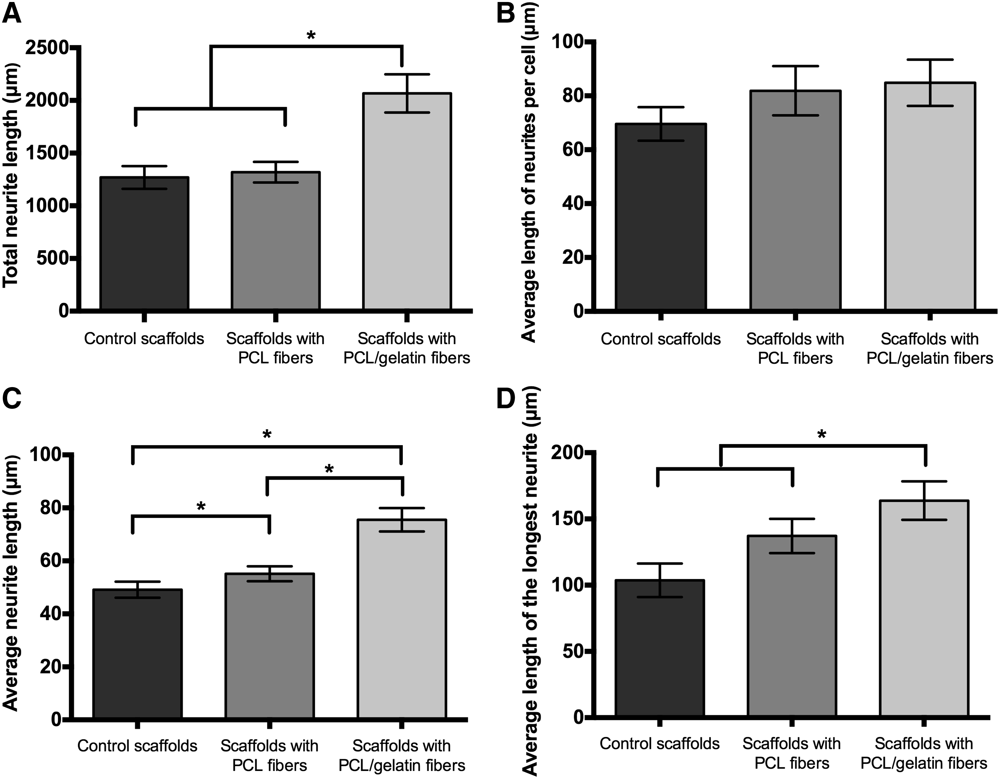
Quantification of neurite length of primary cortical neurons on 3D printed scaffolds with or without electrospun fibers after 7 days of culture. Neurite length was analyzed using ImageJ software and NeuriteTracer.
Discussion
Incorporation of electrospun fibers onto 3D scaffold for neural regeneration
The development of 3D addictive manufacturing processes for use in the engineering of nerve grafts is a promising alternative, which obviates the need for immunosuppression or surgery involving autografts or allografts. 27 Many traditional approaches explored various biofabrication techniques to fabricate neural scaffold. However, lack of adequate bioinks and limited printing resolution often result in less than ideal scaffold for nerve regeneration. In an effort to address these concerns, we demonstrated the feasibility of creating a highly aligned and porous 3D scaffold combining the SL 3D bioprinting and electrospinning. A PCL/gelatin composite fibers were embedded in 3D printed hydrogel scaffold to improve neural cell behavior and mechanical properties of the scaffold.
All scaffolds maintained hydrophilic surface properties except bare PCL fibers. The addition of PCL/gelatin fibers greatly decreased the contact angle compared to the scaffolds without gelatin. This might be attributed to characterized functional group such as amine and carboxylic groups found in the gelatin structure. According to Ghasemi-Mobrakeh et al., the occurrence of the amide group in the Raman spectrum of PCL/gelatin scaffolds indicate that the PCL chains were chemically bonded to gelatin sidewalls leading to the introduction of functional groups such as NH2 on the surface of the scaffolds embedded with PCL-gelatin fibers. 28 The tensile stress obtained for hydrogel scaffolds with aligned electrospun fibers of PCL/gelatin was 1.43 MPa and for PCL was 1.23 MPa. Tensile strength for scaffolds without aligned electrospun fibers was 0.45 MPa. Peak tensile strength of gelatin alone is weak and blending with PCL produced a microfibrous scaffold with intermediary mechanical strength more suitable for nerve regeneration.
Low neurite outgrowth was expected in the SL printed hydrogel scaffolds without fibers since PEG-DA hydrogels do not possess functional groups to which cells can easily attach. The addition of electrospun fibers throughout the hydrogel, however, significantly enhanced neurite outgrowth along the fiber. As shown in Figures 5 and 6, 3D scaffolds with aligned electrospun fibers have shown to greatly increase cell attachment and proliferation compared to those without electrospun fibers. In addition, it was found that aligned microfibers enabled significant directional control of neurite outgrowth in the 3D constructs and the use of laminin-coated microfibers in the 3D printed scaffold significantly enhanced neurite length and directional outgrowth of primary cortical neurons (Figs. 11 and 12).
Confocal images of NSCs on various scaffolds showed some important behaviors. NSC are immortalized neuroectodermal progenitor cell line, which have been established to differentiate into neurons and astrocytes in presence of all trans retinoic acid (RA) or NGF17,29 However, these cells differentiate when RA or NGF administration is in concert with formation of adequate intercellular contacts via aggregation. 17 In this work, NGF was administrated after 4 h post cell seeding. We found out that it takes about 4 days for NSC to exhibit distinctive aggregation properties. To have mass appearance of neurons, NE-4C cells must have grown as aggregates and treated with appropriate growth factor. As shown in Figure 7, only aggregates of undifferentiated NSCs were visible by day 3. Immature postmitotic neurons stained by TUJ1 were visualized within the undifferentiated NSC aggregates by day 11 (Fig. 8A).
We were also interested in how NE-4C cell will behave on aligned construct since no previous studies have shown the effect of physical topography of scaffold. Although we hypothesized that the incorporation of aligned fibers would enhance NSC differentiation and alignment, we actually observed the opposite result. The addition of aligned electrospun fibers inhibited NSC communication and their ability to form clusters, thereby decreasing the levels of differentiation. It can be inferred from these experiments with NE-4C line that they do not differentiate in response to the topography of the aligned fibers.
On the other hand, the improved behavior of primary cortical neurons on various scaffolds was observed. Figures 9 and 10 show that neurites are present on both pores and on 3D printed scaffold. Particularly, primary neurons on scaffolds with PCL/gelatin appear to grow along the fibers and extending neurite between the isolated pores. This implies that neurons are not limited to a single pore of the scaffold but capable to grow and extend along the entire scaffold. In addition, the PCL/gelatin incorporated scaffolds greatly enhanced total neurite length compared to any other scaffolds (Fig. 12). The hydrophilic nature of the PCL/gelatin scaffolds could be another reason for the improved cell adhesion and differentiation of the primary cells. 20 The incorporation of gelatin into the SL printed hydrogel scaffold promoted more axon elongation compared to the gelatin-free hydrogel scaffolds. These findings suggest that gelatin provides better adhesion for primary neuron axons differentiating into the scaffold.
Conclusion
This study demonstrated a novel approach for creating neural scaffold using a combination of SL 3D printing and electrospinning techniques. The resultant scaffold exhibited 3D printed microporous channels embedded with PCL or PCL/gelatin electrospun fibers. The incorporation of electrospun fibers into the 3D constructs result in enhanced mechanical properties. In addition, it was found that 3D printed scaffolds with PCL/gelatin fibers enhanced the NSC differentiation compared to scaffolds with PCL alone fibers. Furthermore, a highly aligned PCL/gelatin fibers within the 3D printed scaffold improved the neurite outgrowth and directional control of primary cortical neurons.
Footnotes
Acknowledgment
The authors would like to thank March of Dimes Foundation's Gene Discovery and Translational Research Grant for financial support.
Authors' Contributions
The article was written through contributions of all authors. All authors have given approval to the final version of the article.
Disclosure Statement
No competing financial interests exist.