
Abstract
Tissue engineering, the application of stem and progenitor cells in combination with an engineered extracellular matrix, is a promising strategy for bone regeneration. However, its success is limited by the lack of vascularization after implantation. The concept of in situ tissue engineering envisages the recruitment of cells necessary for tissue regeneration from the host environment foregoing ex vivo cell seeding of the scaffold. In this study, we developed a novel scaffold system for enhanced cell attraction, which is based on biomimetic mineralized collagen scaffolds equipped with a central biopolymer depot loaded with chemotactic agents. In humid milieu, as after implantation, the signaling factors are expected to slowly diffuse out of the central depot forming a gradient that stimulates directed cell migration toward the scaffold center. Heparin, hyaluronic acid, and alginate have been shown to be capable of depot formation. By using vascular endothelial growth factor (VEGF) as model factor, it was demonstrated that the release kinetics can be adjusted by varying the depot composition. While alginate and hyaluronic acid are able to reduce the initial burst and prolong the release of VEGF, the addition of heparin led to a much stronger retention that resulted in an almost linear release over 28 days. The biological activity of released VEGF was proven for all variants using an endothelial cell proliferation assay. Furthermore, migration experiments with endothelial cells revealed a relationship between the degree of VEGF retention and migration distance: cells invaded deepest in scaffolds containing a heparin-based depot indicating that the formation of a steep gradient is crucial for cell attraction. In conclusion, this novel in situ tissue engineering approach, specifically designed to recruit and accommodate endogenous cells upon implantation, appeared highly promising to stimulate cell invasion, which in turn would promote vascularization and finally new bone formation.
Introduction
W
Following this strategy, the aim of our work was to develop a biomimetic scaffold capable of attracting cells from the surrounding tissue to migrate toward the scaffold center (Fig. 1). As recently outlined by Rouwkema and Khademhosseini, the control of vascularization is predominantly determined by the presence of growth factor gradients guiding vascular migration. 3 Directed cell migration could be achieved by integrating a central depot into the scaffold loaded with chemoattractive substances. The depot would dissolve slowly in humid milieu after implantation, leading to growth factor gradient formation. Through the interconnected pores within the scaffold, cells would migrate toward this gradient as far as they are sufficiently supplied. Forced by progressive hypoxic conditions in deeper scaffold regions, they will secrete angiogenic signaling molecules, which in turn would contribute to accelerate vascularization. The signaling molecule predominantly secreted under hypoxic stress is vascular endothelial growth factor A (VEGF-A), 5 which serves as chemoattractant for endothelial cells, 6 and also bone marrow-derived mesenchymal stem cells (hBMSC).7,8 The recruitment of these cells within the scaffold would promote the generation of new bone tissue and accelerate bone remodeling and fracture healing.
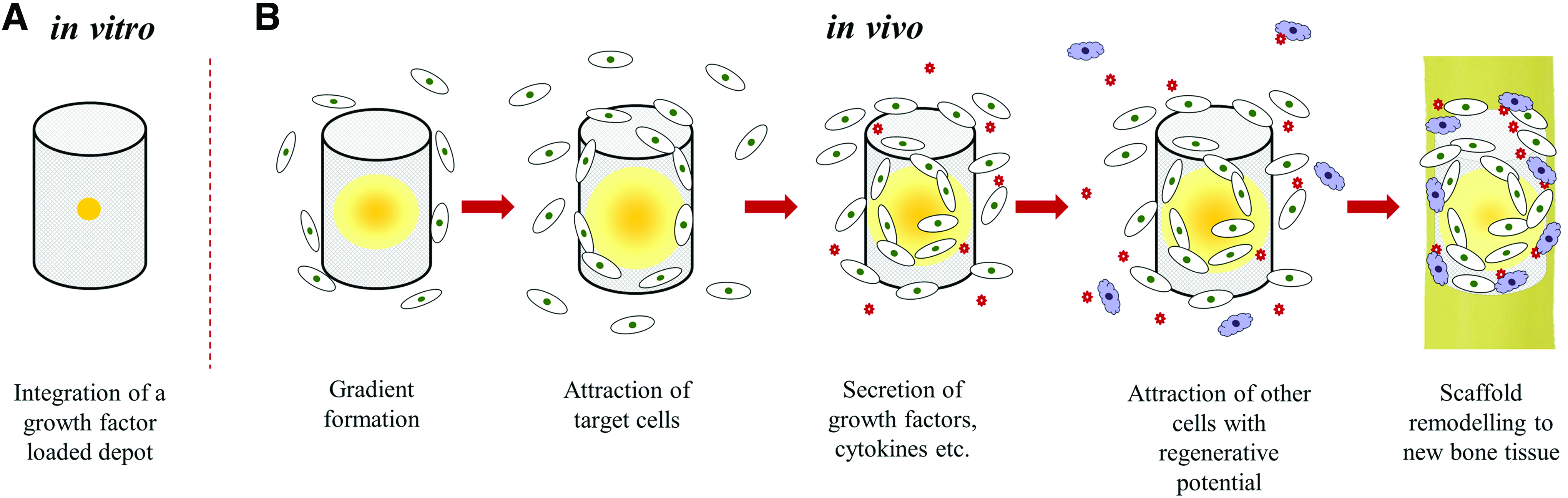
Concept of an in situ bone tissue engineering approach based on a biomimetic scaffold modified with a central signaling factor loaded depot for in situ cell attraction and accelerated vascularization and bone defect healing.
For this study, mineralized collagen, a nanocomposite of collagen type I and nanocrystalline hydroxyapatite, 9 was used as scaffold material. An open-porous microarchitecture suitable for cell ingrowth is achieved by freeze-drying of the mineralized collagen suspension, followed by chemical crosslinking. 10 The suitability of this synthetic bone graft material for bone tissue engineering and regeneration has been demonstrated in vitro and in vivo.11–13 To attract cells to the scaffold center and stimulate vascularization, a biopolymer-based depot enriched with chemoattractive signaling factors should be integrated in the scaffold (Fig. 1A). Several biopolymers have been investigated as depot components:
The anionic polysaccharide alginate is characterized by excellent biocompatibility and the ability of hydrogel formation by gentle crosslinking with multivalent cations, leading to a highly porous nanostructure 14 ; it has been widely used in several medical applications. VEGF encapsulated in alginate was shown to be released gradually and biologically active over long time periods.14,15 The anionic, nonsulfated glycosaminoglycan hyaluronic acid is one of the major components of the extracellular matrix (ECM) in vertebrates. 16 Due to its remarkable physicochemical properties as well as distinctive biological functions, for example, specific binding of proteins and regulating their activity, hyaluronic acid is a promising biopolymer for many medical applications.17,18 Synergistic effects of hyaluronic acid with VEGF have been observed regarding the stimulation of in vivo angiogenesis. 19 The highly anionic, sulfated glycosaminoglycan heparin, known for its affinity toward proteins with heparin binding domains, including growth factors and chemokines,20,21 was already used to modify scaffolds of mineralized collagen. In former studies, we could demonstrate a controlled release of biologically active VEGF 22 and an enhanced proliferation and osteogenic differentiation of hBMSC cultured on heparin-modified scaffolds. 23 In contrast to the previous work, where the scaffolds were completely modified, in this work, heparin was used to form a central depot within otherwise unmodified scaffolds.
Main focus of this study was on the establishment of a scaffold system containing a central growth factor loaded depot. Several depot variants were compared to realize a sustained release of VEGF, which was used as model factor. Finally, in vitro migration studies with endothelial cells were conducted with the modified scaffolds.
Materials and Methods
Preparation of porous scaffolds of mineralized collagen
Collagen type I from bovine tendon (Syntacoll, Germany) was used to produce porous scaffolds (diameter: 6 mm, height: 8 mm) consisting of fibrillated mineralized collagen as described. 22 In brief, collagen dissolved in 10 mM hydrochloric acid was mixed with a 0.1 M calcium chloride solution; the pH value was adjusted to 7.0 by the addition of 0.5 M Tris and 0.5 M Sørensen phosphate buffer. The neutralized mixture was incubated at 37°C for 12 h to run the process of synchronous collagen fibril reassembly and nanocrystalline hydroxyapatite precipitation. Thereafter, mineralized collagen fibrils were collected by centrifugation. The pellet was resuspended in distilled water (1.5 g/mL), the suspension was filled in cavities of a 96-well tissue culture polystyrene (TCPS) plate, frozen at −20°C, and freeze-dried. Crosslinking of the porous scaffolds was carried out by incubation in 1 wt% EDC (1-ethyl-3-(3-dimethyl aminopropyl) carbodiimide; Sigma-Aldrich) dissolved in 80% ethanol for 1 h. After thorough rinsing of the scaffolds in water, 1 wt% glycine solution, and again in water, final freeze-drying was conducted. Before injection of the VEGF depot, the scaffolds were sterilized by γ-radiation at 25 kGy.
Injection of a central VEGF-loaded depot into the scaffolds
Recombinant human VEGF-A165 (VEGF; Morphoplant GmbH, Germany) was used for loading of the central depots; the whole process was conducted under sterile conditions. A stock solution of 100 μg/mL VEGF in phosphate-buffered saline (PBS) was mixed into different biopolymer solutions listed in Table 1. The mixtures were injected into the center of dry mineralized collagen scaffolds (15 μL containing 400 ng VEGF for release studies and proliferation assay; 100 ng for migration assay) using a needle with an inner diameter of 250 μm (Globaco, Germany) connected to a multipipette (Brand, Germany). After injection of Alg-based depots, the scaffolds were transferred into a 0.1 M CaCl2 solution and incubated for 10 min for crosslinking of Alg. Finally, the loaded scaffolds were soaked in the cell culture medium to start the respective experiments.
VEGF, vascular endothelial growth factor.
Visualization of the central depot
The biopolymer components of the injected depots were visualized at different time points of 28 days of incubation under cell culture conditions. The modified scaffolds were cut in the longitudinal direction. Hep was stained by incubating the scaffolds in DMMB (1,9-dimethylmehtylene blue) solution 24 for 10 min while shaking, followed by rinsing with water. Toluidine blue (TB) was used to visualize Alg and Hya 25 : the scaffolds were incubated in TB solution for 10 min, while shaking, and afterward rinsed with water. The samples were analyzed with a Leica Microscope M205C (Leica, Germany).
Furthermore, FITC-labeled VEGF (Morphoplant GmbH) was applied to visualize the loaded depots as well as their stability during incubation under cell culture conditions for 7 days. Immediately after injection of the depot mixtures containing 400 ng FITC-VEGF, the scaffolds were incubated in 1.5 mL cell culture medium for different time periods, cut in the longitudinal direction, and analyzed by confocal laser scanning microscopy (cLSM) using a Zeiss cLSM 510 (Carl Zeiss, Germany), located in the Core Facility Cellular Imaging of Technische Universität Dresden.
VEGF release experiments
Scaffolds with an injected VEGF depot were transferred into a 24-well TCPS plate coated with bovine serum albumin (BSA) and 1 mL of release medium (Endothelial Cell Basal Medium MV; Promocell, Germany), containing 15% fetal calf serum (FCS; heat inactivated, 30 min at 56°C), 100 U/mL penicillin, and 100 μg/mL streptomycin, was added. Heat-inactivated FCS showed no measureable VEGF concentration. The release experiment was carried out under cell culture conditions for 28 days. At different time points, the release medium was collected from the samples and replaced by the fresh medium. Aliquots of the collected medium were used for quantification by VEGF ELISA and for assessment of the biological activity of released VEGF.
ELISA for VEGF quantification
For quantification of released VEGF, a sandwich ELISA procedure using goat anti-human VEGF (Sigma-Aldrich) as capture and biotinylated goat anti-human VEGF (R&D Systems) as detection antibody was applied as described previously. 22 For detection, streptavidin-horseradish peroxidase (R&D Systems) and tetramethylbenzidine (Sigma-Aldrich) as substrate were used. A calibration curve with a linear range between 2000 and 78 pg/mL was used to calculate the VEGF concentrations released from the scaffolds. Absorbance was measured spectrophotometrically (infinite M200 PRO, Tecan, Switzerland) at a wavelength of 450 nm and a reference wavelength of 570 nm. Dilutions of a VEGF standard solution was used as calibration line.
Endothelial cell culture
Human dermal microvascular endothelial cells (HDMEC), purchased from Promocell, were cultivated in the Endothelial Cell Growth Medium MV (Promocell) until 80% confluence. Cells in passage 4–6 were used for the proliferation assay to analyze the biological activity of released VEGF as well as for the migration assay to assess chemoattraction by the central depot.
Endothelial cell proliferation assay
The biological activity of VEGF released from the scaffolds was studied with an endothelial cell proliferation assay as described. 22 In brief, HDMEC were seeded in 96-well TCPS plates (2000 cells/well) and incubated in the Endothelial Cell Growth Medium MV. After cell attachment, the medium was removed and the cells were washed with the Endothelial Cell Basal Medium MV. The release solutions collected from the samples (scaffolds loaded with 400 ng VEGF-containing biopolymer depots and scaffolds loaded with VEGF-free biopolymer depots and nonloaded scaffolds; the latter served as negative control) were diluted in a 1:1 ratio with the fresh release medium and added to the cells. After cultivation for 72 h, the cells were washed with PBS and cell quantification was carried out by measurement of the DNA content with the Quant-iT™ PicoGreen® dsDNA reagent (Life Technologies, Germany). The cell numbers that were measured after incubation with the release solutions from the VEGF-loaded scaffolds were related to the cell numbers determined for the respective negative control.
Cell migration assay
Before seeding, the scaffolds loaded with a depot containing 100 ng VEGF were incubated in the Endothelial Cell Basal Medium MV for 30 min. A suspension of HDMEC (1 × 10⁵ cells in a small volume of 10 μL) was seeded on top of the fully soaked scaffolds. After incubation of the scaffolds for 45 min under cell culture conditions, the Endothelial Cell Basal Medium MV was carefully added to cover the scaffolds completely. After 72 h of culture, the scaffolds were washed with PBS and fixed with 4% formaldehyde; cell nuclei were stained with Hoechst dye (Invitrogen). After cutting of the scaffolds in longitudinal direction with a razor blade, both scaffold halves were transferred into Tissue-Tek® Cryomold®'s (Sakura Finetek, VWR), which were then filled up with the frozen section medium (Richard-Allan Scientific™ Neg-50™, Thermo Scientific, Waltham), put on dry ice for 30 min, and stored at −80°C. Fifty micrometers thin scaffold slices were prepared by cryosectioning with a Leica CM 1900 cryotom (Leica) and fixed on glass slides (Superfrost Ultra Plus, Thermo Scientific) for 1 h at 37°C. Finally, the sections were mounted with Roti®-Mount FluorCare (Roth, Germany). Fluorescence microscopic analysis of the fixed sections was performed with a Keyence BZ9000E (Keyence, Japan) to obtain a merged picture of the whole scaffold slice. Based on this, cell migration was analyzed by Arivis Vision 4D (Arivis AG, Germany): the scaffold slice was divided in frames that cover the scaffold section, starting from the surface, in 250 μm steps until a depth of 2500 μm. In each frame, the cell number was determined.
Statistical analysis
The cell culture experiments were performed using triplicates, the results are given as mean value ± standard deviation. Two-way analysis of variance (ANOVA) was used to evaluate statistical significance at a level of p < 0.05. Post hoc analysis using the Tukey method was used to determine multiple comparisons (Origin 8.5.0G, OriginLab).
Results
Injection of a biopolymer-based VEGF depot
Hep and Hya were either used alone or in combination with Alg to create a central depot within the scaffolds. Injectability of the depot components was found to be convenient for the tested compositions listed in Table 1. DMMB staining of injected Hep revealed the formation of a central core in the scaffolds with a well-defined interface between Hep-modified and Hep-free areas, which was stable over the incubation period of 28 days (Fig. 2). The combination Alg+Hep with the additional crosslinking step led to a similar result. Injection and crosslinking of Alg alone resulted in a central area that was visible over 28 days, but without a clearly pronounced interface area. In contrast, staining of Hya indicated the presence of a central area shortly after injection, which, however, was spread during further incubation (Fig. 2).
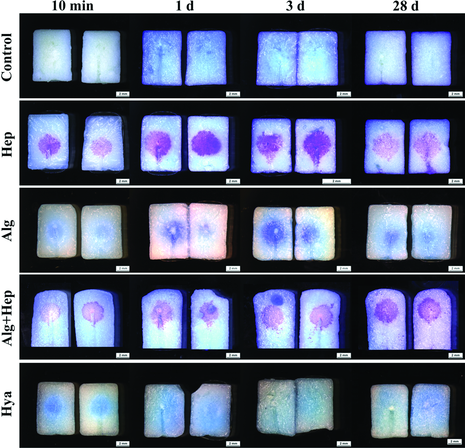
Central depots visualized by staining of the biopolymer components. After injection, and in case of Alg crosslinking, the modified scaffolds were incubated under cell culture conditions. At various time points, they were cut longitudinally into two halves and stained with DMMB (Hep and Alg+Hep) or TB (Hya and Alg). PBS was injected for the control. PBS, phosphate-buffered saline; TB, Toluidine blue. Color images available online at www.liebertpub.com/tea
To visualize that indeed a growth factor loaded depot can be integrated in the scaffolds, the biopolymers were injected together with FITC-labeled VEGF. Figure 3 clearly shows the distribution of VEGF and the retardation of its release over 7 days by the addition of biopolymers. While 10 min after injection the biopolymer-free depot shows a strong spreading of VEGF throughout the scaffold, the VEGF concentrations in case of the biopolymer variants appear locally more pronounced. All later time points clearly show VEGF retention in the biopolymer depots, whereby this effect appears to be more persistent in Alg and Alg+Hep variants.
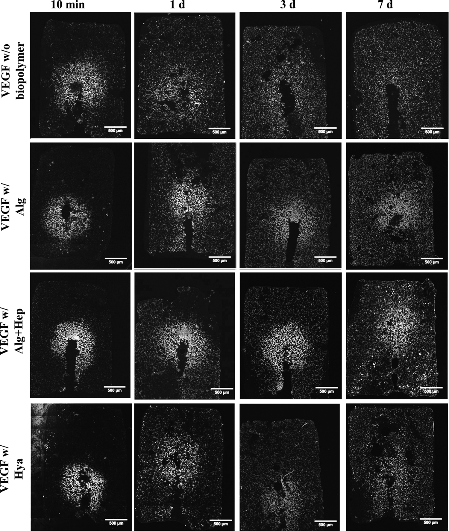
Mineralized collagen scaffolds with central depot loaded with FITC-labeled VEGF without additional biopolymer and with biopolymers for VEGF retention (Alg, Alg+Hep, and Hya). Fluorescence microscopic analysis of FITC-VEGF distribution 10 min, 1 day, 3 days, and 7 days after injection. Visualized the retarded VEGF release from the Alg+Hep depot. VEGF, vascular endothelial growth factor.
Release of VEGF from scaffolds containing a biopolymer-based depot
Release of VEGF from scaffolds with a central depot was studied in the cell culture medium over a period of 28 days and determined by ELISA. The release profiles shown in Figure 4A revealed that all biopolymers injected together with VEGF reduced the burst release, which resulted in a more sustained VEGF delivery. In addition, the data demonstrate that the release kinetics can be controlled by varying the depot composition. Injection of VEGF dissolved in PBS (biopolymer-free control) resulted in a high initial burst within the first days, which was clearly reduced later on (Fig. 4B provides a more detailed depiction showing the absolute amount of released VEGF for the early [day 0–4] and the later [day 5–28] phase). A near constant release of VEGF over the whole period was found for the scaffolds containing Hep and Alg+Hep depots, which was significantly different from those of the control. Alg alone also had a significant effect on VEGF delivery—the respective release profile can be considered an intermediate condition between those of the control and the Hep-containing depots. VEGF released from depots consisting of Hya, and Alg+Hya also showed a reduced release in the first days and higher release rates at the later time points compared to the control. These release kinetics clearly show the retarded VEGF release with different extents for the various tested depot variants, which also inevitably implies the generation of gradients with varying steepness, that is, different VEGF concentrations in the scaffold center (c1) and the surrounding medium (c2).

Release of VEGF from scaffolds loaded with a central depot:
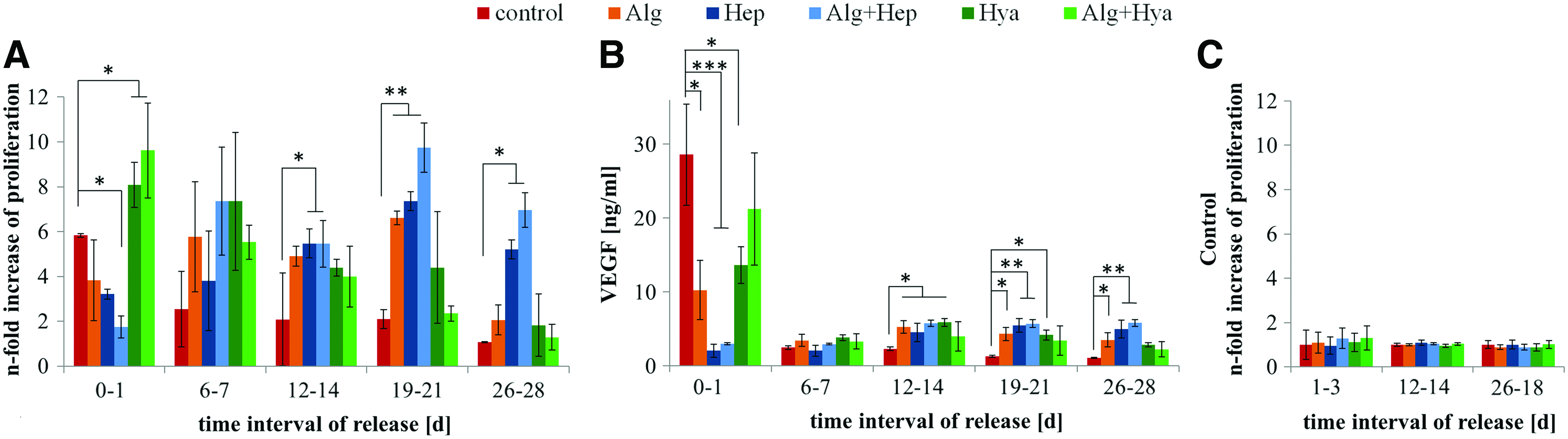
Maintenance of the biological activity of VEGF released from the various depot variants in different time intervals was proven in an endothelial cell proliferation assay (Fig. 5A). Compared to extracts from nonloaded scaffolds, which served as control and were set as 1, for all extracts collected from VEGF-loaded scaffolds, an increased cell proliferation was discernible. The differences in the increase of proliferation reflect the VEGF concentration in the different extracts. Depending on the respective time point and the depot composition, the depicted proliferation patterns correlated with the amount of released VEGF (Fig. 5B). The depot variants with a stronger initial VEGF release in the first days (Hya and Alg+Hya) revealed comparatively high proliferation rate at early time points. Hep-containing depot variants, which are characterized by a retarded and prolonged VEGF release, led to clearly higher proliferation rates at later time points. This result shows that scaffolds modified with a heparin containing depot still release biologically active VEGF after 28 days of cultivation. Supernatants taken from scaffolds, which were loaded with VEGF-free biopolymer depots, showed no promoting effect on HDMEC proliferation (Fig. 5C), indicating that the biopolymer components alone did not affect endothelial cells.
Biological activity of released VEGF. Scaffolds with a central biopolymer-based VEGF depot (400 ng) were incubated in 1 mL release medium under cell culture conditions (for the control, PBS was used instead of biopolymer). For each scaffold type, VEGF-loaded samples as well as nonloaded reference samples were analyzed. Release medium extracts collected within different time periods were tested in an endothelial cell proliferation assay: HDMEC were cultivated in the presence of the extracts for 72 h, cell numbers were determined by DNA quantification.
Cell migration toward the central depot
Based on the release profiles, the depot variants Alg, Alg+Hep, and Hya were selected for further characterization with focus on the induction of cell migration into the scaffolds. Figure 6 shows the number of cells detected in the several scaffold planes in relation to the total cell number counted for each variant (Fig. 6A) and in relation to nonloaded scaffolds serving as control (Fig. 6B). The results show significant differences between the scaffolds with different depot variants. While nonloaded scaffolds showed most cells in the first 250 μm, the cell numbers of all depot variants in the scaffold areas deeper than 250 μm were higher compared to the control. However, the depot variants affected the cell migration differently. Injected VEGF diluted in PBS showed significantly higher cell numbers in a depth of 500–1000 μm compared to nonfunctionalized scaffolds. When VEGF was released in a retarded manner from an Alg depot, significantly more cells were found in the depths of 500–1000 μm and 1000–1500 μm. The cell migration in scaffolds injected with a VEGF-loaded Hya depot showed a comparable cell distribution, with slightly more cells in deeper scaffold regions (1500–2000 μm). Interestingly, for Alg+Hep-depots, the detected cell numbers in 1500–2000 μm and 2000–2500 μm were higher than for every other depot variant. These results were highly significant compared to nonloaded scaffolds as well as for those loaded with VEGF in the absence of biopolymers.

HDMEC migration into scaffolds modified with a central VEGF-loaded depot. One hundred nanograms VEGF was injected into the scaffold center either alone or with Alg, Hya, or Alg+Hep. Cells were seeded on the scaffolds' surfaces and incubated for 3 days.
Discussion
The induction of vascularization in engineered tissues, which is defined by polarization and directional sprouting of endothelial cells, is highly dependent on the cellular response to diffusible and matrix-bound growth factors, and their local availability. 26 Due to its key role in angiogenic processes, many strategies focus on the functionalization of scaffolds with VEGF. Growth factors gradually released from scaffolds have already been shown to promote vascularization in vitro and in vivo. As one example, Nillesen et al. evaluated acellular collagen–heparin scaffolds functionalized with VEGF and basic fibroblast growth factor (bFGF) in vivo, and observed clearly enhanced formation of mature vessels capable of supplying the infiltrated cells with sufficient oxygen. 27 However, with enlarging the transplants, in terms of clinical translation, strategies are required, which not only promote vascularization but also direct the vascular organization toward deeper scaffold regions. Recent studies demonstrated that particularly, the presence of growth factor gradients controls the vascular migration, rather than their overall availability. 3 Odedra et al. showed, for instance, that by dropping VEGF on the center of a collagen sheet, a radial gradient is formed, which lead to a more centralized cell distribution. They hypothesized that generating a gradient of a growth/survival factor in the opposite direction to the oxygen gradient enables cell guidance into the interior of a scaffold. 28
In this study, VEGF was integrated as a depot in the scaffold center, forming a gradient during its release that should encourage a directed cell migration toward deeper scaffold regions. By using different biopolymers to retard the VEGF release from the scaffolds, different release kinetics were achieved, which sustained over 28 days (Fig. 4). In our former study, we have demonstrated that the uniform modification of the whole mineralized collagen scaffold with heparin enables a steady VEGF release from the loaded scaffolds 22 ; in this work, the application of heparin within a central depot goes a step beyond. Beside a continuous VEGF release in an almost linear manner over 28 days (Fig. 4), different concentrations in the scaffold center and the periphery were observed (Fig. 3), indicating the formation of a VEGF gradient. The controlled release of VEGF from the Hep-based depots is a result of the stability of the heparin integration (Fig. 2) and the high affinity of heparin to bind VEGF as one of the heparin binding growth factors. Strong electrostatic interactions of the highly anionic heparin with positively charged groups of collagen 29 and Ca2+ ions of the mineral phase 30 led to direct binding of heparin to the scaffold material at the site of injection. In case of scaffolds containing a depot, VEGF is concentrated in the center after injection. That seemed to additionally slow down the release due to the longer distance of diffusion from the scaffold center to the periphery. For example, after 7 days, the cumulative release of VEGF from scaffolds with a Hep-based depot was ∼10% of those of the control, whereas in case of the scaffolds uniformly modified with heparin, the cumulative release was between 13% and 25% (depending on the heparin content) of those of the heparin-free control. 23
In our previous work, we observed for the scaffolds completely modified with heparin (30, 75, and 150 mg per gram collagen corresponding to 0.3, 0.75, and 1.5 mg per scaffold, respectively) that the majority of the heparin molecules remained bound to the mineralized collagen matrix over 28 days of incubation under cell culture conditions. Moreover, we found that the lower the amount of integrated heparin, the higher the percentage of heparin remaining in the scaffolds. 23 Therefore, it can be assumed that in case of Hep-based depots, which contained only 0.015 mg heparin per scaffold, most of the heparin remained immobilized in the scaffold center and, if at all, only a very low amount (below the detection limit) was released. This is supported by the results displayed in Figure 5C showing that supernatants taken from depot scaffolds without VEGF functionalization had no promoting effect on cell proliferation. However, the biological activity of VEGF released from the scaffolds containing a Hep-based depot appeared rather high in relation to the released amounts (Fig. 5). Therefore, an enhancement of the bioactivity of released VEGF by heparin coreleased in very small quantities is conceivable. Such a synergistic effect was already supposed for VEGF released from mineralized collagen scaffolds completely modified with heparin and referred to the known effect of an enhanced heparin-mediated receptor binding to endothelial cells. 22
Scaffolds with an Hya-based depot showed a weaker retardation of VEGF release in comparison to the Hep-containing variants reflected by a significantly different release profile (Fig. 4). This also corresponds with the images of depots loaded with FITC-VEGF showing a more persistent VEGF depot for the biopolymer compositions Alg+Hep and Alg (Fig. 3). One reason for this might be the low stability of the depot consisting of noncrosslinked Hya (Fig. 2): only a weak interaction of hyaluronic acid with mineralized collagen can be assumed as data from the literature indicate no considerable binding of hyaluronic acid to collagen 29 and only low affinity binding to hydroxyapatite.31,32 On the other hand, the lower retention of VEGF in scaffolds with a Hya-based depot can be attributed to weak electrostatic interactions with VEGF due to the low negative charge density of the nonsulfated hyaluronic acid. 33 However, it is conceivable that Hya in the scaffold pores may also act as a physical barrier slowing down the diffusion of VEGF, as a result of its high molecular mass and specific properties to be highly viscous and behave as large coil structure entrapping a high amount of water. 34 As an ECM component, hyaluronic acid is degradable in vivo by hyaluronidases. Therefore, upon implantation, the Hya-containing scaffolds could additionally release shorter fragments as degradation products of the long chains, which in turn are highly bioactive and could potentiate the activity of the released angiogenic growth factors.16,19
Similar to the Hya variant, release of VEGF from scaffolds containing an Alg-based depot represented an intermediate state between the fast VEGF release in the absence of any biopolymer (control) and the continuous release from scaffolds with a Hep-based depot (Fig. 4). After injection of Alg, the scaffolds were incubated in CaCl2 solution to crosslink the alginate chains and form a hydrogel within the scaffold pores. Accordingly, staining of scaffolds with Alg depot visualized the central core (Fig. 2). Over the period of 28 days, the alginate core appeared to continuously diffuse. As the mineral phase of the scaffold material contains Ca2+ ions and is known to absorb further Ca2+ ions from its environment, 11 an interaction between the alginate gel and the scaffold material can be assumed, which might have caused a marginal weakening and leaking of the hydrogel within the scaffolds. Nevertheless, the alginate hydrogel with its dense network might have acted as a physical barrier, which is most probably responsible for the slowed diffusion of VEGF, rather than electrostatic interactions, as alginate is known for its poor binding capacity for proteins, including VEGF. 35
The cell migration experiments revealed significant differences between the scaffolds with different depot variants. Thereby, the migration distances of HDMEC appear to relate to the release profiles of VEGF from the various depot variants and, in particular, to the degree of VEGF retardation (Fig. 6). Thus, HDMEC migrated deepest in scaffolds containing an Alg+Hep depot, the depot variant that showed the strongest VEGF retardation by releasing VEGF in an almost linear manner without initial burst. This observation suggests that the more retarded the VEGF release and, consequently, the steeper the developed gradient, the deeper the cells migrated into the scaffold. Also, Baker et al. demonstrated that human umbilical vein endothelial cells (HUVEC), which colonized microfluidic gelatin channel templates, showed the most intense invasion and sprouting at locations, where the VEGF gradients were steepest. 36 Terranova et al. postulated a chemotactic response of endothelial cells, isolated from umbilical vein (HUVEC), to heparin and a synergistic effect in combination with endothelial cell growth factor. 37 However, the low concentrations of heparin we used for this study did not stimulate the migration of HDMEC (data not shown), indicating that increased migration distances in scaffolds equipped with Alg+Hep depots are predominantly determined by VEGF and its retarded release from the depot.
To further enhance the cell migration, a combination of synergistically acting factors can be promising, like VEGF and FGF 27 or BMP-2 and SDF-1α. 38 Due to the lack of clinical approval for many recombinant factors, the utilization of autologous factors or cell-conditioned media can be promising for application in a central depot. Focus of our ongoing work is on the integration of supernatants from hypoxia-treated hBMSC (hypoxia-conditioned media [HCM]) into the central depot, which have been found to be highly potent for the attraction of mesenchymal stromal cells. 39 HCM contain a number of angiogenic factors and are able to induce angiogenesis in vitro (article in preparation). Within an envisaged in vivo study, we intent to study vascularization and migration of hBMSC into these HCM depot-containing scaffolds.
Conclusion
A novel scaffold system containing a central depot loaded with chemotactic factors was developed and characterized using VEGF as model factor. The combination of VEGF with several biopolymers resulted, to different extent, in a retardation of its release that opens the possibility to adjust the release kinetic by varying the biopolymer composition of the depot. A high degree of retardation, achieved by Hep-based depots, seemed to be crucial for formation of a steep VEGF gradient within the scaffolds. The retention of VEGF can be supposed to be determined by (1) the stability of the biopolymer-based depots, (2) the binding affinity of the biopolymer for VEGF, and (3) the deceleration of VEGF diffusion by the biopolymer acting as a physical barrier. Migration experiments with endothelial cells as model cell type revealed a relationship between the degree of retardation and the migration distance by showing that cells invaded deepest in scaffolds with a Hep-containing depot, which developed the steepest VEGF gradient. Heparin also appeared to enhance the biological activity of the released VEGF, which may synergistically promote cellular migration. The novel scaffold system developed herein is a promising candidate for in situ tissue engineering approaches, which are of particular interest as they can offer potent bone grafts, which are easy to obtain, cost-effective, and circumvent complex regulatory effort favoring translation into the clinics.
Footnotes
Acknowledgments
The authors thank the German Research Society (DFG) for financial support. This study was performed as part of the Collaborative Research Centre/Transregio 79 (subproject M4). Collagen type I used in this study was kindly provided by Syntacoll (Saal/Donau, Germany). We thank Ortrud Zieschang for excellent technical assistance.
Disclosure Statement
No competing financial interests exist.