
Abstract
Regeneration of complex bone defects remains a significant clinical challenge. Multi-tool biofabrication has permitted the combination of various biomaterials to create multifaceted composites with tailorable mechanical properties and spatially controlled biological function. In this study we sought to use bioprinting to engineer nonviral gene activated constructs reinforced by polymeric micro-filaments. A gene activated bioink was developed using RGD-γ-irradiated alginate and nano-hydroxyapatite (nHA) complexed to plasmid DNA (pDNA). This ink was combined with bone marrow-derived mesenchymal stem cells (MSCs) and then co-printed with a polycaprolactone supporting mesh to provide mechanical stability to the construct. Reporter genes were first used to demonstrate successful cell transfection using this system, with sustained expression of the transgene detected over 14 days postbioprinting. Delivery of a combination of therapeutic genes encoding for bone morphogenic protein and transforming growth factor promoted robust osteogenesis of encapsulated MSCs in vitro, with enhanced levels of matrix deposition and mineralization observed following the incorporation of therapeutic pDNA. Gene activated MSC-laden constructs were then implanted subcutaneously, directly postfabrication, and were found to support superior levels of vascularization and mineralization compared to cell-free controls. These results validate the use of a gene activated bioink to impart biological functionality to three-dimensional bioprinted constructs.
Introduction
T
The degree of customized control offered by 3D bioprinting has enabled the production of scaled up, mechanically reinforced materials for musculoskeletal tissue engineering.14,15 Another attractive feature of this spatial control is the ability to deposit specific biological cues in relevant locations, to drive complex tissue formation. 16 An efficient gene activated bioink would be particularly beneficial in this regard as successful cell transfection could produce localized, sustained protein expression; something that is not as easily achieved through the use of growth factors as they can diffuse easily and cause nonlocalized effects. 17 Calcium phosphate has been successfully used as a delivery vector within a 3D bioprinted alginate hydrogel previously, leading to elevated BMP-2 expression and ALP production in vitro.18,19 However, no bone formation was observed after 6 weeks following subcutaneous implantation of this approach. In addition, more demanding defects such as load-bearing bone defects may require more mechanical integrity than can be provided by a gene activated hydrogel alone. 20 Hydrogels have previously been combined with various polymeric support structures to fabricate composite materials with both biological and mechanical functionality.21,22 These constructs are typically cell-laden and cultured in vitro to engineer a mature tissue that can promote bone repair following implantation.23,24 The inclusion of a gene activated bioink may permit the bioprinting of a material that can be implanted directly postfabrication, inducing sustained therapeutic protein expression in vivo and hence accelerating regeneration.
In this work we developed a gene activated bioink by combining a printable alginate hydrogel with nHA-pDNA complexes and co-printing this ink with a reinforcing polycaprolactone (PCL) scaffold to produce a gene activated 3D construct. Bone marrow-derived mesenchymal stem cells (MSCs) were combined with the bioink directly before printing. The capacity of this strategy to successfully transfect MSCs was first assessed using reporter genes, before utilizing a combination of therapeutic genes encoding for BMP2 and TGF-β3 in an attempt to induce osteogenesis of MSCs in vitro. The final phase of the study sought to examine whether a vascularized and mineralized tissue could be generated in vivo by implanting such MSC-laden gene activated constructs directly postbioprinting. If successful, such an approach could be potentially be used at the point of care to develop personalized gene activated implants for treating complex bone defects.
Materials and Methods
Plasmid propagation
Four different plasmids were used in this study: two plasmids encoding for the reporter genes red fluorescent protein (pRFP, also called pTomato, kind donation from Prof. Gerhart Ryffel through Addgene) and luciferase (pLUC, pGaussia luciferase; New England Biolabs, Massachusetts), and another two encoding for the therapeutic genes BMP2 (kind donation from Prof. Kazihusa Bessho, Kyoto University, Japan) and TGF-β3 (InvivoGen, Ireland). Plasmid amplification was performed by transforming chemically competent Escherichia-coli bacterial cells (One Shot TOP10; Biosciences, Ireland) according to the manufacturer's protocol. The transformed bacteria were cultured on LB plates with 100 mg/L ampicillin (Sigma-Aldrich, Ireland) as the selective antibiotic for the four plasmids. Bacterial colonies were harvested and inoculated in LB broth (Sigma-Aldrich, Ireland) and incubated overnight for further amplification. The harvested bacterial cells were then lysed, and the respective pDNA samples were purified using Qiagen plasmid kit (MaxiPrep Kit; Qiagen, Ireland). Nucleic acid concentration (ng/μL) was determined by analyzing the 260:280 ratio and 230 nm measurement using NanoDrop spectrophotometer (Labtech International, Uckfield, United Kingdom).
Preparation of nano hydroxyapatite nHA-pDNA complexes
The synthesis of the nHA particles was performed as previously described. 25 Briefly, a solution of 12 mM sodium phosphate (Sigma-Aldrich), containing 0.017% DARVAN 821A (RTVanderbilt, Norwalk) was added to an equal volume of a 20 mM chloride solution (Sigma-Aldrich) and filtered through a 0.2 mm filter. 8 nHA-pDNA complexes were prepared by adding 1845 μL of the nHA solution to 125 μg of pBMP2 (in 250 μL), and 125 μg of pTGF-β3 (in 105 μL) pretreated with 300 μl 250 mM CaCl2 (Sigma-Aldrich). This 2.5 mL solution was then added to 50 × 10^6 MSCs and alginate as described below.
Gene activated bioink
Low molecular weight sodium alginate (γ alginate, 58 000 g/mol) was prepared by irradiating sodium alginate (Protanal LF20/40, 196 000 g/mol, Pronova Biopolymers, Oslo, Norway) at a gamma dose of 5 Mrad, as previously described.26,27 RGD-modified (arginine-glycine aspartic acid) alginates were prepared by coupling the GGGGRGDSP to the alginate using standard carbodiimide chemistry. Briefly, 10 g alginate was dissolved in 1 L MES Buffer (0.1 M MES, 0.3 M NaCl, and pH 6.5). 274 mg sulfo-NHS (Pierce, Rockford, IL), 484 mg EDC (Sigma), and 100 mg GGGGRGDSP peptide (AIBioTech, Richmond, VA) were then added into alginate solution. The reaction was stopped by addition of hydroxylamine (0.18 mg/mL; Sigma), and the solution was purified by dialysis against ultrapure deionized water (MWCO 3500, Spectrum Laboratories, Inc., Rancho Dominguez, CA) at 4°C for 3 days, treated with activated charcoal (5 g/L, 50–200 mesh, Fisher Scientific, Inc., Pittsburgh, PA) for 30 min, filtered (0.2222 μm filter), and lyophilized. 27
Bone marrow-derived MSCs were isolated from the femoral shaft of 4 month old pigs and expanded to passage 2 in standard culture media (high glucose Dulbecco's modified eagle's medium GlutaMAX (hgDMEM), 10% (v/v) fetal bovine serum (FBS), and 100 U/mL penicillin per 100 μg/mL streptomycin) before transfection. The nHA-pDNA complexes (2.5 mL) were prepared immediately before transfection, and added to a suspension of MSCs in 1 mL of standard expansion media. After 1 h of incubation, alginate was added to the cells and nHA-pDNA complexes to yield a final volume of 5 mLs, containing a concentration of 10 million cells/mL in 1% alginate. Then, the solution was mixed until a homogenous mixture was obtained. 10
Bioprinting gene activated constructs
Gene activated polymer/bioink scaffolds were fabricated using the 3D Discovery multi-head bioprinting system (Regen HU, Switzerland). The 3D Discovery was set up to allow for co-printing of a pneumatic driven syringes containing the bioinks alongside a fused deposition modeler (FDM) allowing for deposition of molten polycaprolactone (PCL; Sigma, Mn 45 000). First, the RGD-γ alginate bioink was dissolved at 3.5 wt% and mixed thoroughly using a luer lock system with the MSCs in either nHA solution (nHA alone control) or the nHA-pDNA complexes (both containing 50 mM CaCl2) to yield a gene activated bioink with 1% final alginate concentration [41]. To ensure homogeneity the suspension was mixed between syringes 25 times. The gene activated bioink solution was loaded into the pressure driven piston system and co-printed alongside PCL melted at 60°C (Fig. 1). A pressure of 0.2 MPa and a 25 Gauge needle were used to deposit the bioink strands, using an orthogonal 90 degree angle print pattern to build the constructs to the predesigned height. Following this, the constructs were immersed in a 100 mM CaCl2 solution for 15 min to fully cross-link the bioink. The 3D Discovery was operated within a laminar flow hood to ensure sterility throughout the biofabrication process.
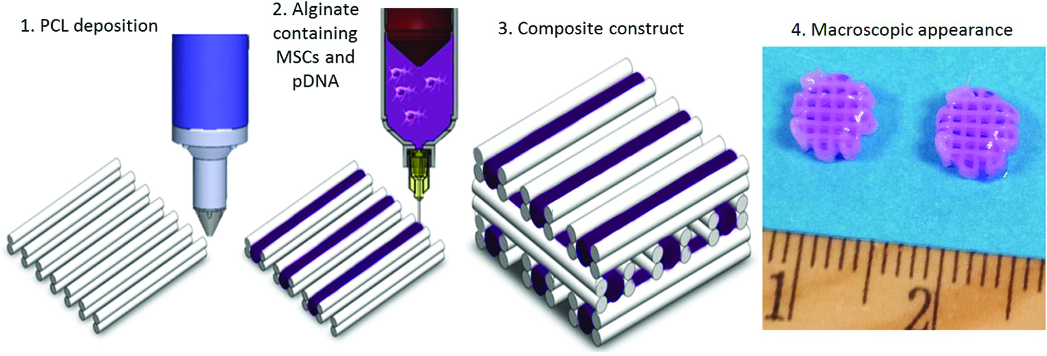
Schematic representation of the bioprinting process, with co-deposition of PCL and the gene activated bioink comprising of alginate, nHA-pDNA complexes and MSCs, and the macroscopic appearance of the constructs before implantation. MSCs, mesenchymal stem cells; PCL, polycaprolactone; pDNA, plasmid DNA. Color images available online at www.liebertpub.com/tea
Constructs of dimensions 10 × 2 mm (diameter × height) were printed for in vitro evaluation, while constructs 6 × 3 mm (diameter x height) were printed for subsequent in vivo implantation. In vitro analysis was conducted over 28 days in either control medium (high glucose Dulbecco's modified eagle's medium GlutaMAX [hgDMEM], 10% [v/v] FBS, and 100 U/mL penicillin per 100 μg/mL streptomycin) or osteogenic culture conditions (high glucose Dulbecco's modified eagle's medium GlutaMAX [hgDMEM], 100 nM dexamethasone, 10 mM β-glycerol phosphate, and 0.05 mM ascorbic acid [all from Sigma-Aldrich]) at 20% oxygen.
Live/dead confocal microscopy
Cell viability was assessed 24 h after bioprinting using a LIVE/DEAD viability/cytotoxicity assay kit (Invitrogen, Bio-science). Briefly, constructs were cut in half, washed in phosphate-buffered saline (PBS) followed by incubation for 1 h in PBS containing 2 μM calcein AM (green fluorescence of membrane for live cells) and 4 μM ethidium homodimer-1 (red fluorescence of DNA for dead cells). Sections were again washed in PBS, imaged at magnification ×10 with an Olympus FV-1000 Point-Scanning Confocal Microscope (Southend-on-Sea, United Kingdom) at 515 and 615 nm channels, and analyzed using FV10-ASW 2.0 Viewer software. Live/dead-based semi-quantification on n ≥ 4 separate regions, chosen at random, was carried out using Image J.
Biochemical analysis
To perform biochemical analysis, constructs were digested with papain (125 mg/mL, pH 6.5) in 0.1 M sodium acetate, 5 nM L-cysteine HCl, and 0.05 M ethylenediaminetetraacetic acid (all Sigma-Aldrich) at 60°C under constant rotation for 18 h. Calcium content was determined using a Sentinel Calcium Kit (Alpha Laboratories Ltd, United Kingdom) after digestion in 1 M HCl at 110°C for 48 h. Proteoglycan content was estimated by quantifying the amount of sulfated glycosaminoglycan (sGAG) in the constructs using the dimethylmethylene blue dye-binding assay (Blyscan, Biocolor Ltd., Northern Ireland), with a chondroitin sulfate standard. Total collagen content was determined by measuring the hydroxyproline content. Samples were hydrolyzed at 110°C for 18 h in concentrated HCl 38%, allowed to dry, and analyzed using a chloramine-T assay with a hydroxyproline-to-collagen ratio of 1:7.69. 28 Four samples per group were analyzed for each biochemical assay.
Reporter gene detection
Red fluorescent protein (RFP) expression was detected using Leica SP8 scanning confocal microscope (Leica Microsystems, Ireland) 24 h postbioprinting. Luciferase expression was imaged using a real-time bioluminescence imaging system (PhotonImager; Biospace lab, France) to visualize the spatial distribution of luminescence over time. Luciferase expression in the culture media was also quantified using a Pierce Gaussia Luciferase Flash Assay Kit (ThermoFisher, Ireland) at different time points up to 14 days.
Micro-computed tomography
Micro-computed tomography (μCT) scans were performed using a Scanco Medical 40 μCT system (Scanco Medical, Bassersdorf, Switzerland) with a 70 kVp X-ray source at 114 μA. Six constructs were analyzed per in vivo experimental group and quantification was performed by setting a threshold of 210 corresponding to a density of 432.58 mg hydroxyapatite/cm3) and recording the mineral volume (mm3). N = 3 samples were scanned and analyzed at a threshold of 100, corresponding to 120.81 mg hydroxyapatite/cm3 for the in vitro study. Reconstructed 3D images were generated from the scans and used to visualize mineral distribution throughout the constructs.
Subcutaneous Implantation
Gene activated constructs (n = 9) were implanted subcutaneously into the back of nude mice (Balb/c; Harlan, United Kingdom) as previously described with three samples inserted in each of two pockets. 29 Each mouse contained two separate pockets, with n = 3 constructs from a particular group implanted into either pocket. The constructs were harvested after 4 and 12 weeks. Mice were euthanized by CO2 inhalation and the animal protocol was reviewed and approved by the Ethics Committee of Trinity College Dublin and the Health Products Regulatory Authority (HPRA).
Statistical analysis
Statistical analysis was carried out using GraphPad software. The results are reported as means ± standard deviation and groups were analyzed using Student's two-tailed t-tests or by a general linear model for analysis of variance with groups of factors. Tukey's post hoc test was used to compare conditions. Significance was accepted at a level of p < 0.05.
Results
Gene activated bioinks support sustained expression of reporter genes following co-printing with PCL filaments
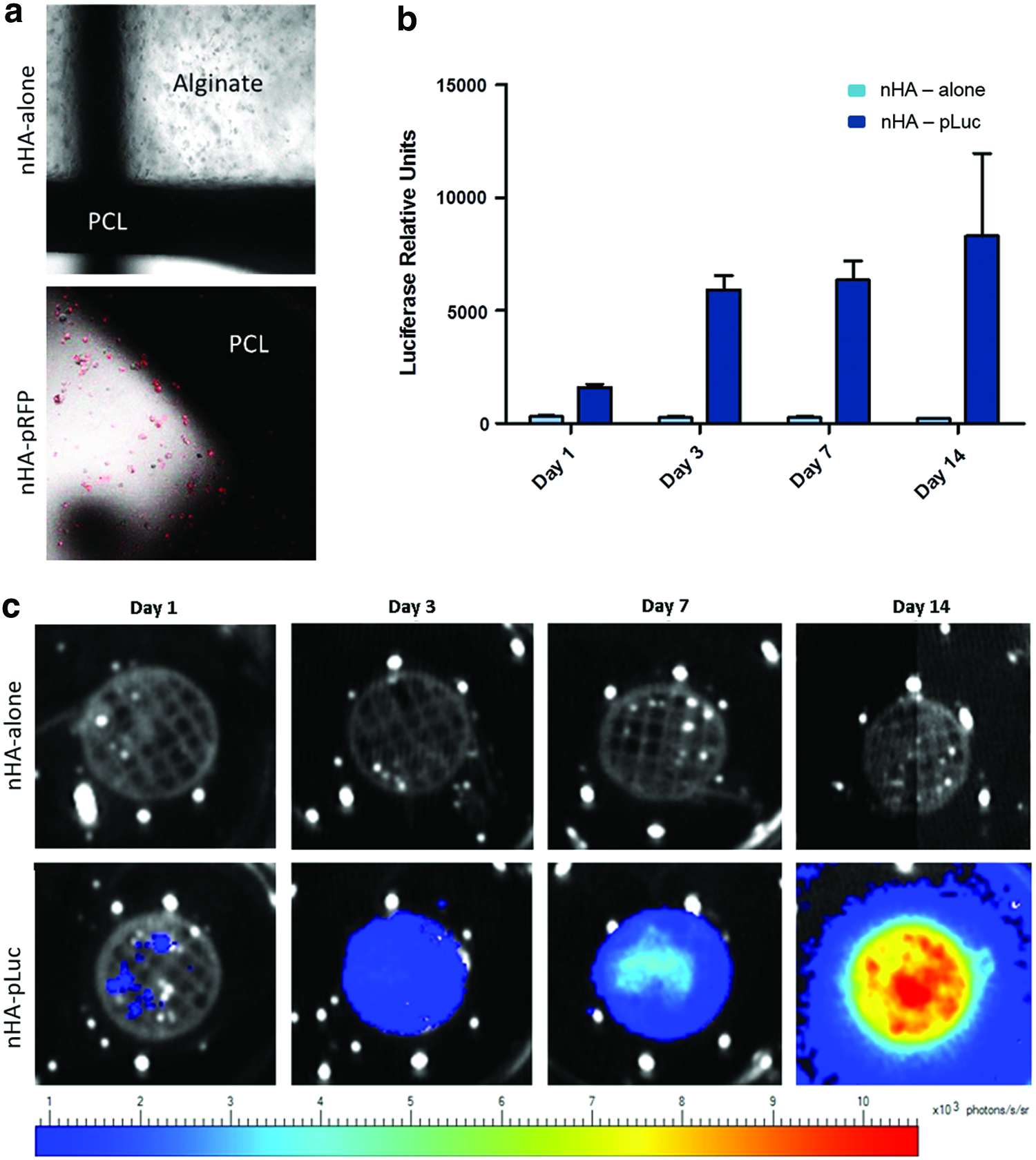
To establish that the gene activated bioink would remain functional following 3D bioprinting with a PCL support structure, reporter genes (pLUC and pRFP) were utilized to validate successful transfection of MSCs encapsulated within the bioinks at the time of bioprinting. The viability of MSCs printed within the gene activated bioink was not affected by the presence of the pDNA encoding for luciferase, however, some cell death was observed due to co-printing the cell-laden bioink with PCL (nHA-alone 64 ± 10%, nHA-pLUC 69 ± 2%, Fig. 2, Supplementary Fig. S1; Supplementary Data are available online at www.liebertpub.com/tea). By 14 days, the DNA content remained at the same level as that quantified at day 1 and almost 100% of cells within the construct were observed to be viable using live/dead staining.

Cell viability is maintained following pDNA incorporation. Live/dead images demonstrate the presence of viable cells (green) at both day 1 and day 14 postbioprinting, while quantification of DNA indicated no difference between groups cultured with or without pDNA encoding for luciferase. Color images available online at www.liebertpub.com/tea
Reporter gene analysis usingRFP and luciferase indicated that successful transfection of bioprinted MSCs was achieved within the gene activated bioink (Fig. 3). RFP was observed 24 h postbioprinting using fluorescent microscopy to provide an initial validation of successful pDNA uptake and protein expression. Luciferase was then employed to investigate temporal expression of a reporter protein. Luciferase was found to increase in expression over 14 days of culture, as assessed both by quantifying the luciferase expressed and released into the media (Fig. 3b) and qualitatively by imaging the protein remaining within the constructs (Fig. 3c).
Therapeutic gene delivery enhanced osteogenesis of MSCs in vitro
Following validation of successful transfection using reporter genes, a combination of therapeutic genes encoding for BMP2 and TGF-β3 was incorporated into the bioink system. These combinations of genes were chosen as delivery of recombinant BMP-2 and TGF-β protein from MSC-laden alginate hydrogels has previously been shown to promote bone formation in vivo. 30 Constructs were bioprinted and cultured for 28 days in either control medium or osteogenic culture conditions. Macroscopically, evidence of matrix deposition can be observed in all groups at this time point relative to constructs at day 0 (Fig. 4). Biochemical quantification indicated that significantly higher levels of DNA and deposition of GAG and collagen was achieved in both culture conditions following inclusion of pDNA within the bioink. Live/dead quantification at day 1 and day 14 for the constructs transfected with therapeutic genes (Supplementary Fig. S1) had not indicated any differential response in DNA content between the transfected and nontransfected control groups.
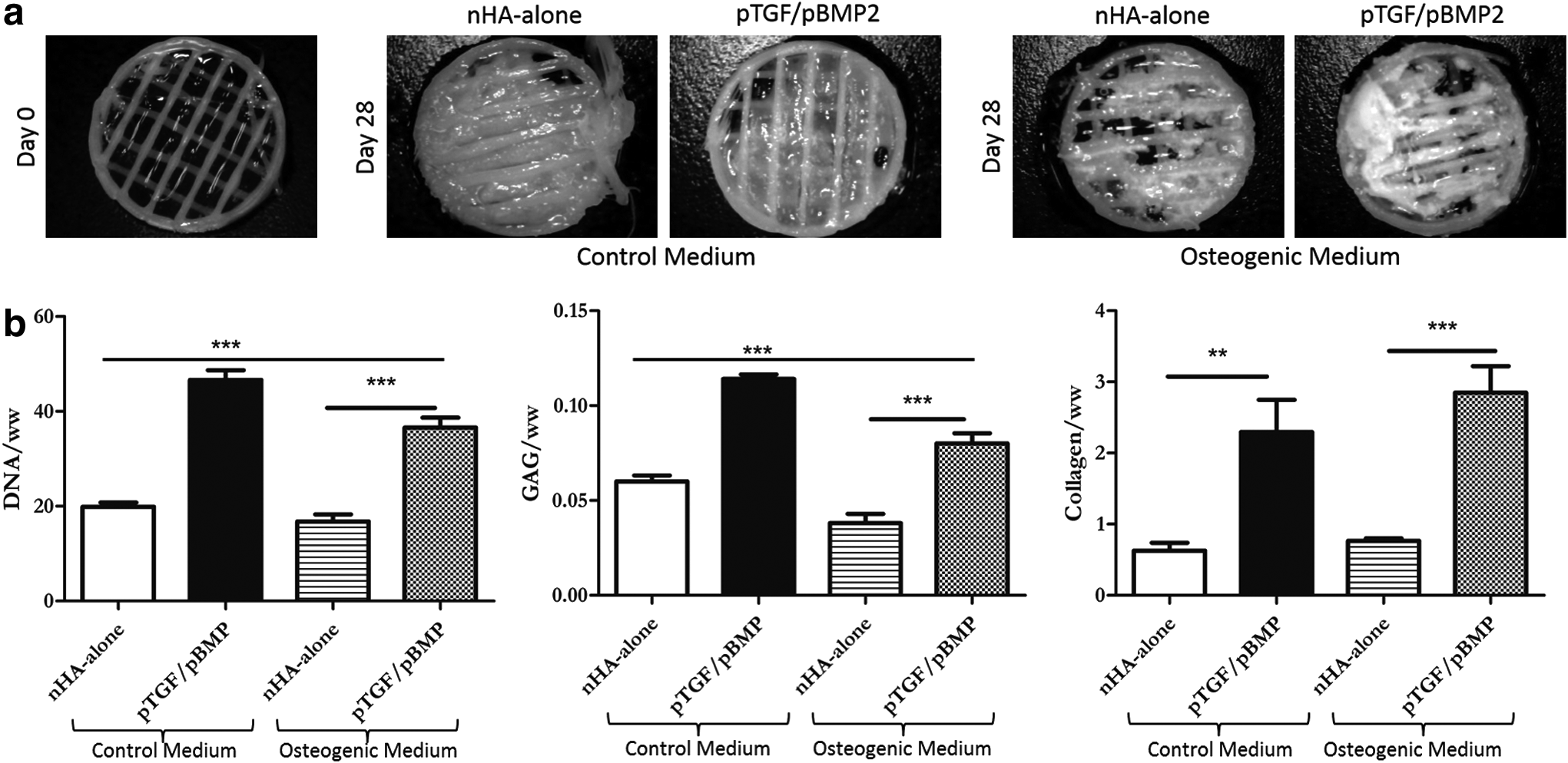
Upon quantification of calcium content, the matrix was found to be mineralized, indicating the onset of osteogenesis (Fig. 5). Significantly higher levels of mineral deposition were observed within the pDNA containing bioinks in control medium, and this effect was greatly amplified following culture in osteogenic supplemented medium. 3D reconstructed μCT images demonstrated the homogeneity of the mineral distribution throughout the cultured constructs.

Bioprinted gene activated constructs containing MSCs promote the development of vascularized and mineralized tissues in vivo
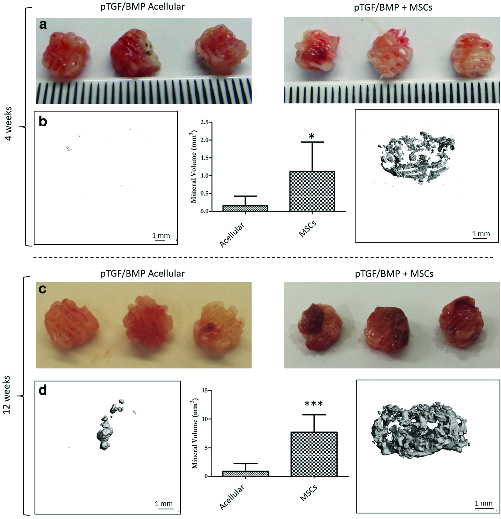
Bioprinted gene activated constructs were implanted directly postfabrication, and were compared to bioprinted acellular control constructs containing nHA-pDNA complexes only after 4 and 12 weeks in vivo (Fig. 6 a, c). Macroscopic evidence of vascular in-growth was observable at both time points, and verified using histological analysis (Supplementary Fig. S2). By 12 weeks, MSC-laden constructs appeared to be more vascularized. Regions of de novo bone formation and immature osteoid was also detected in the MSC-laden constructs. Mineral quantification at both time points indicated that the incorporation of MSCs resulted in significantly higher levels of mineral deposition compared to the acellular control, and that the deposition increased significantly with time (Fig. 6 b, d). Distribution of mineral can be observed homogeneously throughout the construct.
Discussion
This study describes the successful development of a gene activated bioink capable of transfecting mesenchymal stem cells post 3D bioprinting. These MSC-laden bioinks were co-deposited alongside a reinforcing PCL network to produce composite constructs suitable for bone tissue engineering applications. Reporter genes indicated that protein expression was detected after 24 h and that protein expression could be sustained, and in fact continued to increase, over 14 days of in vitro culture. Transfection with therapeutic genes encoding for BMP2 and TGF-β3 promoted enhanced osteogenesis in vitro compared to nontransfected controls containing only the nHA vector, implying that this gene activated bioink system could induce the expression of biologically functional proteins. Implantation of these gene activated MSC-laden constructs directly postfabrication was capable of driving vascularization and mineralization in a subcutaneous environment. These findings support the continued development of 3D printed gene activated scaffolds as putative “point-of-care” treatment options for a range of musculoskeletal defects.
The choice of material for the gene activated bioink was motivated by a number of factors, including the printability of the alginate hydrogel, the presence of the RGD ligand to allow cell spreading, the ability to facilitate calcium phosphate-based gene delivery, and established capacity to enable the osteogenic differentiation of MSCs.10,19,23,31–34 Previous studies from our lab have demonstrated that reinforcement of MSC-laden alginate hydrogels with printed PCL microfibers results in dramatic improvements in the mechanical properties of the construct. 23 Polymeric scaffolds are typically inert and may require supplementation with various factors to induce a favorable biological response, often provided through the addition of extracellular matrix components, or exogenous growth factors.35–38 A number of publications have also reported superior biological activity solely due to the addition of alginate hydrogel to PCL scaffolds.39,40 Furthermore, alginate can be modified to have tunable degradation rate and mechanical properties 27 and some formations already have FDA approval for clinical indications.
The temporal production of gene product observed over 14 days of culture clearly demonstrates the potential of this gene activated bioink approach for sustained therapeutic protein delivery, especially when compared to the burst release profiles typically observed with traditional growth factor delivery hydrogels. By employing the cells themselves to express the desired protein, limitations with protein delivery including rapid degradation of potentially supra-physiological, toxic doses, and dispersion of the drug to dangerous locations can be overcome. 41 The bioprinting process itself, or the fact that the bioinks were co-deposited alongside molten PCL, does not appear to detract from the ability of the nonviral delivery vector nHA to successfully transfect cells. In fact, the intensity of luciferase signal increased over 14 days of culture, suggesting sustained transfection of encapsulated MSCs following the bioprinting process.
Having demonstrated it was possible to bioprint gene activated constructs reinforced by a network of PCL micro-filaments, the capacity of this system to promote MSC differentiation along the osteogenic pathway was then tested. Alginate is commonly used as a biomaterial in bone regeneration strategies,19,26,31,42,43 and more recently has been used as a bioink for bone and cartilage bioprinting.18,34,41,44 In the absence of osteogenic supplements, the co-delivery of BMP-2 and TGFβ3 pDNA within these MSC-laden alginate bioinks resulted in the deposition of a mineralized matrix, with the differences compared to nontransfected controls becoming particularly apparent when cultured in osteogenic conditions. The nHA particles used to deliver the plasmids may be providing an osteogenic stimulus, although the concentration used to deliver pDNA is relatively low compared to that used previously to induce mineralization.45–47 Furthermore, it has previously been shown that while nHA transfects cells with lower efficacies to other vectors, its use still promotes higher overall levels of osteogenesis. 45 In a previous study, we observed that nHA-mediated delivery of TGF-β3 and BMP2 in an alginate hydrogel promoted a more chondrogenic rather than osteogenic stimulus. 10 This may be explained by the conditions (normoxia and osteogenic media) and the RGD modification of the alginate that we implemented in this study to promote direct osteogenic differentiation of encapsulated MSCs. Therefore, in the basal (control) medium, gene delivery may be promoting a more endochondral phenotype compared to a more intramembranous phenotype in the osteogenic media, although further studies are required to confirm this. Regardless, these findings support the use of alginate hydrogels containing nHA-pDNA complexes as gene activated bioinks for bone tissue engineering.
Bioprinted gene activated constructs became well vascularized in vivo, supporting the development of a mineralized bone-like tissue. These in vivo results point to the benefit of including mesenchymal stem cells when developing “point-of-care” bioprinted constructs for bone regeneration. Mineralization was considerably higher at both 4 and 12 weeks in the MSC-laden constructs compared to the acellular control. It could be argued, however, that the acellular, pDNA containing group may perform better upon implantation into an orthotopic defect site compared to the subcutaneous site due to the likelihood of greater infiltration of host osteo-progenitor cells. A study investigating the use of pDNA encoding for BMP2 and delivered using an alginate hydrogel-based nonviral approach also reported enhanced results when the gene activated biomaterial was combined with MSCs. 44 Histological evidence of blood vessels in-growth was detected in both acellular and MSC-laden constructs by 12 weeks, and mineralization was observed to increase with time in vivo corresponding with evidence of de novo immature bone formation at this later time point. This result agrees with previous in vivo studies delivering a combination of BMP2 and TGF-β3, either as recombinant proteins or through the use of gene delivery.30,48
Conclusion
The treatment of challenging fractures and large osseous defects presents a formidable clinical problem. Multi-tool biofabrication has permitted combination of various materials to create complex composite implants with tailorable mechanical properties and spatially controlled biological function. This study validated the efficiency of a gene activated bioink to induce cell transfection within a 3D bioprinted PCL-bioink composite construct. Sustained protein expression was achieved for up to 14 days postbioprinting, and the combined delivery of the therapeutic genes BMP2 and TGF-β3 led to enhanced osteogenesis of MSCs in vitro and formation of a vascularized and mineralized tissue upon subcutaneous implantation. These results demonstrate an effective platform technology to enrich biofabrication techniques with gene activated bioinks for musculoskeletal applications.
Disclosure Statement
No competing financial interests exist.
References
Supplementary Material
Please find the following supplemental material available below.
For Open Access articles published under a Creative Commons License, all supplemental material carries the same license as the article it is associated with.
For non-Open Access articles published, all supplemental material carries a non-exclusive license, and permission requests for re-use of supplemental material or any part of supplemental material shall be sent directly to the copyright owner as specified in the copyright notice associated with the article.