
Abstract
The wound healing process requires enough blood to supply nutrients and various growth factors to the wound area. However, chronic wounds such as diabetic skin ulcers have limited regeneration due to a lack of cellular and molecular signals because of a deficient blood flow. Mesenchymal stem cells (MSCs) are known to provide various factors, including growth factors, cytokines, and angiogenic mediators. Although MSCs have great therapeutic potential, their transplantation has many obstacles, including the time required to culture the cells, the invasiveness of the procedure, and limited stem cell sources. In this study, we induced a diabetic 1 model in rats aged 7 weeks by injecting streptozotocin and citrate buffer solution. After confirming that diabetes was induced in the rats, we created critical sized wounds on the dorsal area of the rats and then injected hydrogels. We performed the experiments with four groups (defect model for the control, self-assembled peptides (SAPs), SAP with soluble substance P, and SAP conjugated with substance P) to treat the wound defect. Tissues were harvested at 1, 2, and 3 weeks after injection and examined for the wound closure, histological analysis, quantitative real-time polymerase chain reaction analysis, and quantification of collagen deposits to investigate stem cell recruitment and full recovery of wounds at an accelerated time period. As our results show, the wounds treated with SAP and substance P exhibited significantly accelerated wound closure, enhanced collagen deposition, and increased angiogenesis. Furthermore, we confirmed the ability of SAP with substance P to promote the recruitment and homing of cells by immunofluorescence staining of a MSC marker. In addition, it was observed that substance P remained in the wound area up to 3 weeks after the injection of SAP with substance P. It is believed that the endogenous MSCs mobilized by substance P had therapeutic effects through their proper differentiation and release of paracrine factors into the wound sites. In conclusion, this study shows that SAP with substance P can promote wound healing to enhance skin regeneration without cell transplantation in a diabetic model.
Introduction
W
When the skin is damaged, lesions can heal rapidly and naturally within 1 or 2 weeks in healthy skin. 3 However, chronic wounds such as skin ulcers in patients with diabetic mellitus show delayed or incomplete progress in the healing process due to poor blood perfusion resulting in chronic inflammation and the formation of necrotic tissues.4,5 Notably, diabetic wounds are less able to repair tissues due to limited blood perfusion and consequently a lack of cellular and molecular signals required for the normal wound repair process. Moreover, the injured region has reduced levels of growth factors, including transforming growth factor-β (TGF-β), epithelial growth factor (EGF), vascular endothelial growth factor (VEGF), keratinocyte growth factor, platelet-derived growth factor, and basic fibroblast growth factor, which are essential factors leading to healing responses from the surrounding tissues.6,7 Especially, a decrease in VEGF levels results in a lack of endothelial progenitor cells, which are involved in angiogenic stimulation and, subsequently, provokes impaired angiogenesis. 7
It is well known that mesenchymal stem cells (MSCs) have therapeutic potential through the secretion of growth factors, cytokines, and antifibrotic or angiogenic mediators which have paracrine effects. 8 The protective biologically active factors secreted by MSCs have important roles in tissue regeneration.9,10 For example, VEGF secreted by MSCs can stimulate vascularization and angiogenesis. TGF may stimulate keratinocyte migration and wound reepithelialization, and interleukin (IL)-6 can promote dermal fibroblast and keratinocyte migration.11–13 MSCs also participate in anti-inflammatory responses through secreted soluble factors to decrease inflammatory and immune reactions and apoptosis. 14 Although inflammation is the first step in the wound healing mechanism when an injury occurs in the tissue, uncontrolled inflammation can provoke tissue fibrosis. Thus, the anti-inflammatory action of MSCs could contribute to wound healing and regeneration of the defect lesion.
Recently, substance P, which is an 11-amino acid neuropeptide that is secreted from the peripheral terminals of sensory nerve fibers as a neurotransmitter or hormone, has been reported to have anti-inflammatory functions by increasing the level of IL-10 and decreasing the level of TNF-α. 15 In addition, we previously reported that substance P decreased pro-inflammatory cytokines and increased anti-inflammatory cytokines, including IL-4. 16 Furthermore, a new role for SP was reported in which it accelerates wound healing by mobilizing endogenous stem cells from bone marrow to wound sites. 17 Based on the novel function of SP as a systemic injury-inducible wound messenger, mobilizing stem cells may have potential to overcome the limitations of stem cell transplantation in clinical applications in terms of the low cell survival, limited cell sources, long culture times, and high costs.18,19
Our previous work demonstrated that bioactive peptides, which are peptide hydrogels immobilized with substance P, are a good treatment for ischemia by supplying a sufficient blood flow by forming matured vessels, as well as reducing fibrosis formation and apoptosis.20,21 Like in the ischemia model, to treat full-thickness skin defects, it is important to supply a sufficient amount of blood into the wound area. Newly formed blood vessels deliver a sufficient amount of nutrients and trigger the desired cell recruitment for the recovery of the wound area.22,23
Therefore, we selected the bioactive peptides, which can recruit stem cells and enhance angiogenesis, as a treatment material in this study. Furthermore, self-assembled peptide (SAP) has the potential to form a network of stable β-sheet nanofibers, which assemble into a three-dimensional (3D) scaffold in physiological conditions. 24 This structure would closely mimic the porosity and gross structure that promote adhesion, proliferation, and differentiation of a variety of cells in a microenvironment. Furthermore, SAPs can be easily designed and modified through the peptide bonds to have biological activities.25–28 Bradshaw et al. reported that functionalized RADA16 influenced the proliferation rate of keratinocytes and dermal fibroblasts in skin wound healing. They used three kinds of peptides which were RADA16, RADA16 with a fibronectin motif, and RADA16 with a collagen type I motif. 25 Wang et al. reported that the modified RADA16 peptide hydrogel scaffold with a linked N peptide significantly promoted cell adhesion and upregulated the synthesis of chondrogenic proteins by providing an improved microenvironment. 29
In this study, we hypothesized that SAP and substance P might be helpful in promoting skin regeneration in a diabetic model. After creating a full-thickness skin wound on a diabetic rat, we loaded the site with SAP with substance P which is expected to recruit endogenous stem cells to the wound site. Then, skin regeneration was evaluated by measuring the wound closure and area to identify the degree of wound repair at the site. In addition, the wound-healing effects of the peptide hydrogel were evaluated by histological analysis of the skin structure, including the epidermal and dermal layers. We expected the bioactive peptides to increase the mobilization of MSCs which would consequently promote skin tissue regeneration in a type I diabetic model.
Materials and Methods
SAP hydrogel preparation and characterization
The peptides RADA16 (Ac-RARADADARARADADA-NH2) and RADA-SP (Ac- RARADADARARADADAGGRPKPQQFFGLM-NH2) were synthesized (Peptron) and dissolved in a 295 mM sucrose solution to prepare a 1% (w/v) peptide hydrogel. RADA16 and RADA16-SP were mixed with different concentrations for the experimental groups. The experimental groups were as follows: (1) PBS, (2) RADA, (3) RADA and soluble substance P (R+SP), and (4) RADA and RADA-SP (R+R-SP). RADA and RADA-SP were mixed at a ratio of 200:7 (v/v). The total amount of substance P (Substance P; Merck) was 35 μg in the R+SP and R+R-SP groups. Then, the peptide hydrogel was sonicated with an ultrasonic cleanser for 30 min.
Creation of the skin wounds and injection of the hydrogels into the diabetic rats
All animal experiments were approved by the committee on the safety and ethical handling of animals for laboratory experiments at the Korea Institute of Science and Technology in accordance with the recommendations for handling laboratory animals for biomedical research.
To induce type I diabetes in Sprague-Dawley rats (Nara Biotech) weighing 200–250 g, we used the streptozotocin (STZ) injection method. First, rats were fasted approximately for 16 h, but water was provided ad libitum. Then, the diabetic rats were induced with a single intraperitoneal injection of 50 mg/kg STZ dissolved in 0.1 M sodium citrate buffer. We stopped fasting the rats after the injection and checked their blood glucose 2 days later with a blood glucose monitoring meter. To create wound defects, the diabetic rats were anesthetized with an intraperitoneal injection of a 1:1 mixture of tiletamine and zolazepam (Zoletil 50; Virbac) with xylazine (Rompun; Bayer Vital GmbH) at a dose of 30 mg of a tiletamine and zolazepam mixture and 10 mg of xylazine per kg of body weight. Four wounds 1 cm in diameter on each side of the midline were created on the dorsal skin of the 1-week diabetic rats. The diabetic skin wounds received different treatments: PBS for the control group, RADA hydrogel (R), RADA and soluble substance P (R+SP), and RADA and substance P conjugated RADA (R+R-SP). The full-thickness wound area was filled with 150 μL of peptide hydrogel or PBS, and then, the wounds were guarded with silicon rings and dressed with petrolatum gauze (Tegaderm® 3 M). At 1 (n = 5), 2 (n = 3), and 3 (n = 5) weeks after the treatments, the rats were sacrificed, and the wound closure was quantified using photographic images and the ImageJ program.
After the wounds were isolated, the samples were fixed with 10% buffered formalin (Sigma-Aldrich) for 1 day and embedded in paraffin for histological and immunostaining evaluation.
Quantitative real-time polymerase chain reaction analysis
Skin regeneration was determined by the gene expression levels of type I and III collagen in cells present at the defect site. Furthermore, the gene expression of Ki-67, which is an indicator of cell proliferation in skin tissue, was also examined. Total RNA was purified with the RNeasy Mini Kit (QIAGEN), and 2 mg of each RNA sample was reverse transcribed with cDNA in a 20 mL reaction using the Omniscript System Kit (QIAGEN). The RNA of each sample was evaluated with the A260/280 absorbance before reverse transcription. Quantitative real-time polymerase chain reaction (qRT-PCR) was performed with an Applied Biosystems 7500 Real-Time PCR system with the Power SYBR Green PCR Master Mix (Applied Biosystems). The final volume of the sample was 25 μL, and the cDNA was amplified with 45 cycles of 95°C for 15 s and 55°C for 60 s (n = 3). The primer pairs were as follows: collagen I (forward: ATCCTGCCGATGTCGCTAT, reverse: CCACAAGCGTGCTGTAGGT), collagen III (forward: CTGGTCCTGTTGGTCCATCT, reverse: ACCTTTGTCACCTCGTGGAC), GAPDH (forward: ATGCTGGTGCTGAGTATGTC, reverse: AGTTGTCATATTTCTCGTGG), and Ki-67 (forward: AGGACTTTGTGCTCTGTAACC, reverse: CTCTTTTGGCTTCCATTTCTTC).
Histological and immunofluorescence analysis
The paraffin-embedded blocks were sectioned into 0.6 μm thicknesses. The sections were deparaffinized and hydrated with sequential ethanol solutions for sealing and microscopic observations. Then, the hydrated sections were stained with hematoxylin and eosin (H&E) to examine epithelial regeneration and integration with the original surrounding tissues. To examine the collagen compositions and structure of the epithelium, we also did Masson's trichrome staining with hematoxylin solution, acid fuchsin, phosphomolybdic acid solution, and methyl blue solution to stain the collagen, nuclei, and cytoplasm. After dehydrating all the sections again, they were scanned with microscope (Eclipse TE 2000U; Nikon) (n = 3).
For immunofluorescence staining, dorsal skin samples were fixed in 4% Paraformaldehyde overnight. Involucrin (Abcam), type I collagen (GeneTex), type III collagen (Abcam), and cytokeratin 14 (Abcam) were detected by immunofluorescence staining to identify keratinocyte differentiation and the composition of collagen. In addition, the samples were stained with alpha smooth muscle actin (α-SMA; Abcam) and von Willebrand factor (vWF; Abcam) antibodies to confirm angiogenesis. Alexa fluor 488 goat anti rabbit IgG (Invitrogen), Alexa fluor 594 rabbit anti mouse IgG (Invitrogen), and Alexa fluor 488 goat anti mouse IgG (Invitrogen) were used as the secondary antibodies, and nuclei were counterstained with 4′,6-diamidino-2-phenylindole (DAPI; Molecular Probes). All images were acquired with fluorescence microscopy.
Collagen assay
Total soluble collagen was measured colorimetrically with the Sircol Collagen Assay Kit (Biocolor) following the manufacturer's instructions. Briefly, 30 μg of tissues were chopped with 1 mL of acetic acid-pepsin solution and shook overnight at 4°C. To each sample, 100 μL of acid neutralizing reagent was added, and 200 μL of cooled collagen isolation and concentration reagent were added with vortexing for 24 h at 4°C. Later, all the samples were centrifuged for 10 min, and the supernatant was gently removed and transferred to a new tube. Then, 1.0 mL of Sircol Dye Reagent was added to each of the sample supernatants and mixed. During the reaction time, a collagen-dye complex formed and precipitated out from the soluble unbound dye. Then, the tubes with the collagen-dye complex were transferred to a microcentrifuge and spun at 12,000 rpm for 10 min. After the supernatant was removed, 750 μL of ice-cold acid-salt wash reagent was added gently to the collagen dye to remove any unbound dye from the surface of the pellet and from the inside surface of the tube. Then, we centrifuged the tubes at 12,000 rpm for 10 min. After the supernatant was removed, we added 250 μL of an alkali reagent, and 200 μL of each sample was transferred to the individual wells of a 96 micro well plate. Afterward, the plate was set into a microplate reader and read at 555 nm.
Examination of the recruitment of intrinsic MSCs
To investigate the recruitment of MSCs to the injury site during wound healing, sections were double stained with MSC markers, CD90 (Thy-1 antibody; Santa Cruz Biotechnology) and CD105 (Anti-Endoglin monoclonal antibody; Millipore). Alexa Fluor 594 donkey anti rabbit IgG (Millipore) and Alexa Fluor 488 goat anti mouse IgG (Millipore) were used as the secondary antibodies. In addition, to assess the retained substance P in the skin wounds, the sections were stained with the substance P antibody (Santa Cruz Biotechnology). Alexa Fluor 488 chicken anti goat IgG (Invitrogen) was used as the secondary antibody. In addition, a blinded rater observed the substance P stained sections in four random fields at 200 × magnification in the border zone, and a positive stained area was quantified as the mean per unit area (1 mm2) with the ImageJ program (NIH).
Statistical analysis
Quantitative data are expressed as the mean ± standard deviation with the GraphPad Prism software (GraphPad Prism, version 7; GraphPad Software, Inc.). Statistical analysis was performed either with one-way analysis of variance (ANOVA) with Tukey's posttests or two-way ANOVA with Bonferroni posttests. Significance was determined with the posttests between the various treatments and experimental results. The p-value for statistical significance was set at p < 0.05.
Results
The quantification of the retained substance P
In this study, we hypothesized that the recruitment of MSCs, which promotes skin regeneration, was due to the remaining substance P in the injured skin tissue up to 3 weeks after the treatments.
The immunofluorescence analysis was done with anti-substance P antibody, and the positive area was quantitatively analyzed at 1 and 3 weeks after the treatments to identify the retention time of substance P (Fig. 1). The substance P expression area gradually decreased in all the groups. The R+SP group had the highest expression of substance P at 1 week (702.4 ± 123.8 μm2 in the control group, 1395.9 ± 165.1 μm2 in the R group, 3434.1 ± 779.3 μm2 in the R+SP group, and 3214.9 ± 381.1 μm2 in the R+R-SP group), whereas the R+R-SP group had the largest positively stained area at 3 weeks after the treatments (225.1 ± 62.4 μm2 in the control group, 738.5 ± 15.6 μm2 in the R group, 1035.6 ± 296.4 μm2 in the R+SP group, and 1918.2 ± 424.6 μm2 in the R+R-SP group).

Immunofluorescence analysis of substance P. The expressed positive area of substance P means that substance P was retained in wound sites at 1
These results may be related to the release pattern of substance P, which is different between the R+SP and R+R-SP groups. Because substance P exists as a soluble factor, the burst effect occurred in the R+SP group initially. In addition, substance P was covalently bound with the SAPs in the R+R-SP group; therefore, it could remain for a longer time compared with the other treatment groups up to 3 weeks.
Macroscopic observation of the wound healing process
The effects of the substance P conjugated SAP on wound healing were evaluated in a full-thickness skin wound model for up to 21 days. We induced type I diabetes in rats aged 7 weeks by injecting a STZ and citrate buffer solution. After confirming that type I diabetes was induced in the rats, we created a round wound with a 10-mm diameter on the dorsal area of the rats followed by the injection of the treatments.
Figure 2 shows a gross view of the dorsal skin wound and the wound healing process after treatment with PBS, RADA (R), RADA and substance P (R+SP), and RADA and RADA-SP (R+R-SP), respectively. The photographic evaluation clearly shows that the wound area decreased as time passed in all the experimental groups. However, a remarkable difference was observed between each group. The R+SP and R+R-SP groups had clearly a smaller wound area compared with the other two groups at 2 weeks after treatment. Similarly, treatment with SAP and substance P (the R+SP and R+R-SP groups) gradually decreased the wound area which was mostly filled with regenerated tissue.

These results show that there was accelerated wound healing induced by substance P at 3 weeks after the treatments. In addition, these results indicate that substance P contributes to not only accelerating wound healing but also to decreasing the size of the wound area by recruiting MSCs which are involved in the early events of wound healing.
Histological analysis
To observe tissue ingrowth and skin regeneration, tissues from the wound site and the surrounding vicinity were explanted from the diabetic rats (Fig. 3). H&E stained images of tissues at 1 week after treatment showed enhanced granulation in the tissues and infiltrated inflammatory cells from the foreign body reaction; however, the wound defect surface was not fully covered by the reepithelialized fraction in all the treatment groups. However, the wound defect site of all the groups except for the PBS group was mostly filled with a newly epithelialized layer at 3 weeks after treatment. Especially, the R+SP and R+R-SP group had formed a sebaceous gland which is found in normal skin tissue at the adjacent boundary of native tissue. In the R+SP group, the dermal–epidermal junction was well defined, which provides support for the epidermis and serves as a partial barrier between the epidermis and the dermis. Moreover, newly generated epidermis was uniformly formed, and the thickness of the epidermis layer was thicker compared with the other groups except for the R+SP group. In contrast, a loose-organized structure was seen only at the wound site of the PBS treated group.
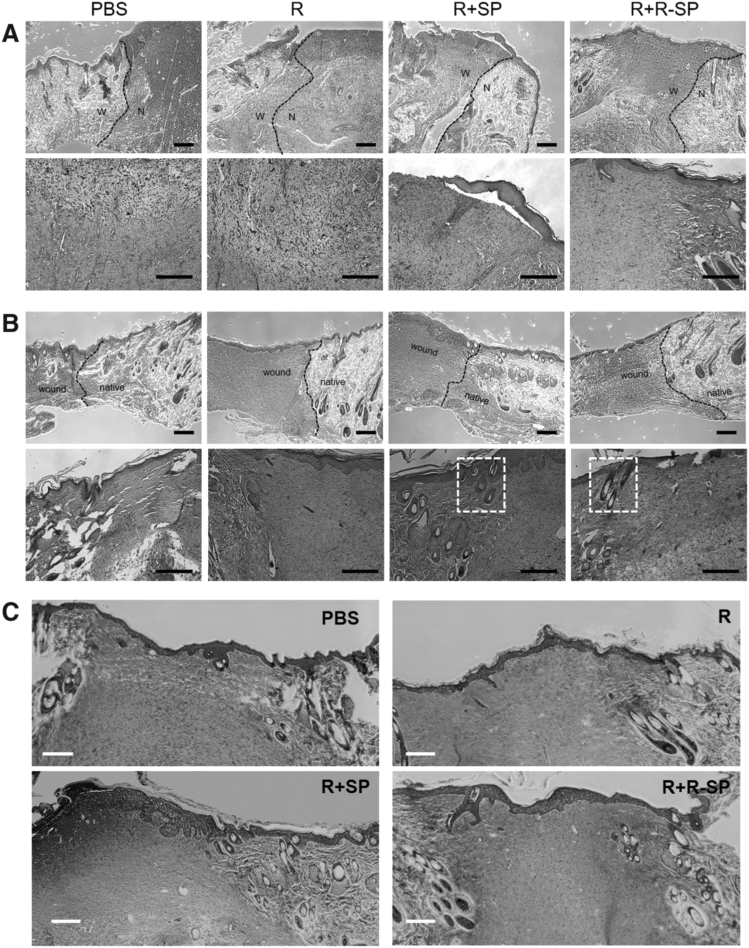
Histological analysis of injured tissues. The R+SP and R+R-SP treatment groups show accelerated wound healing by the filling of a newly epithelialized layer and increased collagen organization, respectively (W: wound area, N: native area).
Subsequently, the levels of collagen deposited in the tissue were examined by Masson's trichrome staining which shows the accumulation of collagen (blue color) and keratin (red color). The stratum corneum, which is the outermost layer of the epidermis, was clearly seen in the R, R+SP, and R+R-SP groups, but not in the control group. In addition, collagen deposition was evenly distributed in all the groups except for the control group.
This result indicates that SAP and substance P can accelerate collagen deposition during wound healing in damaged skin tissue.
Identification of collagen production
The immunofluorescence staining results of collagen deposition is shown in Figure 4A. Collagen is a structural protein and the major component in skin tissue, especially type I and type III collagen which are found in all dermal layers and within the adventitial dermis layer, respectively. In this study, sections were double stained for type I and type III collagen, and then, the collagen staining images were merged with DAPI staining images. When comparing each treatment group, type I and III collagen was strongly expressed in the R+SP group. Likewise, the R+R-SP group also had a positively stained section for type I and III collagen; however, the control and R groups had the smallest positively stained areas for type I and III collagen.
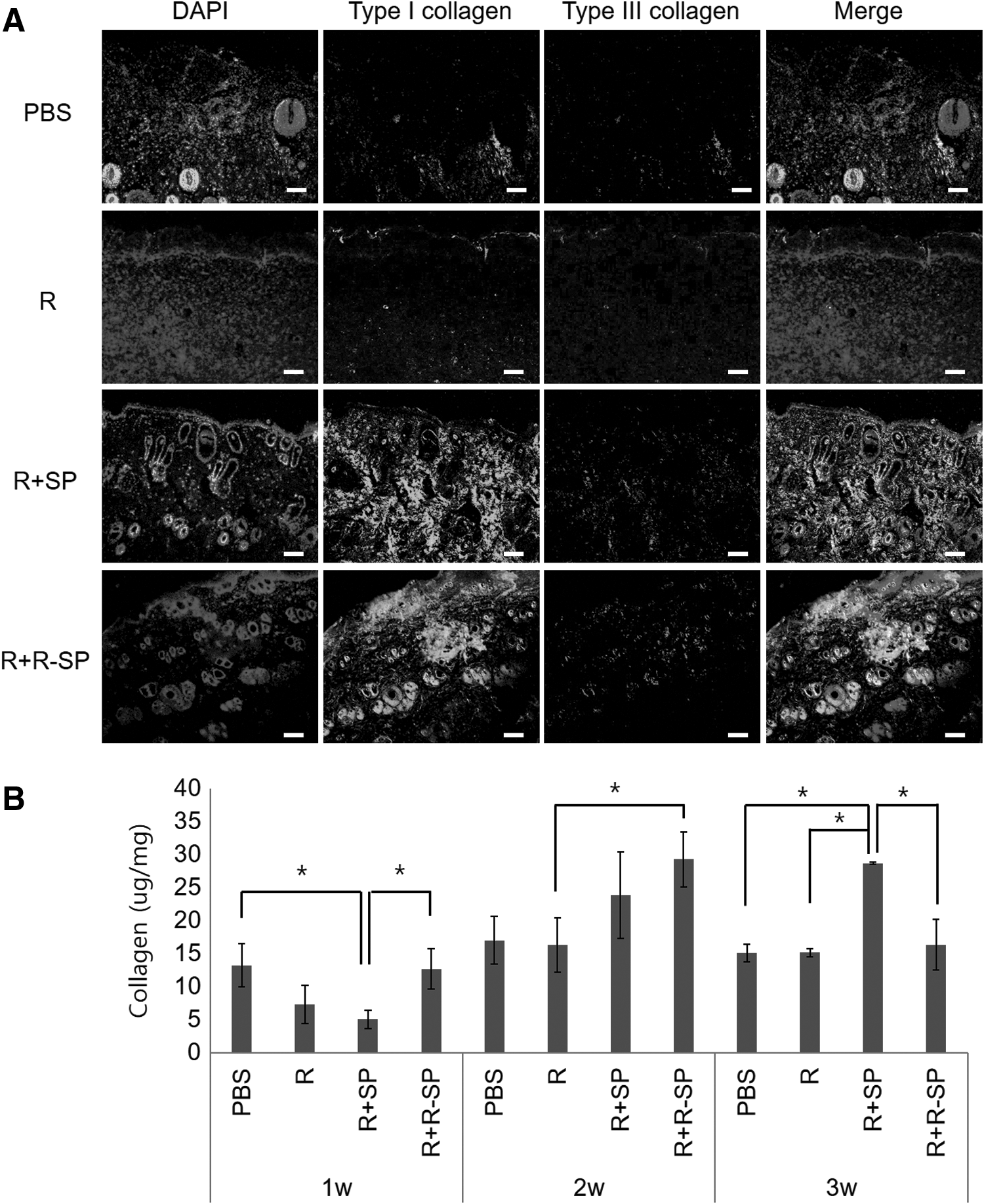
Identification of collagen production using immunofluorescence analysis and Collagen Assay Kit.
To quantitatively investigate collagen production and deposition, a collagen assay was done that measured the total soluble collagen for types I–IV in a chronic wound healing model. As shown in Figure 4B, the collagen contents for the R group at 3 weeks after the operation only gradually increased compared to the early stage at 1 week. The R+R-SP group had the highest total collagen among all the treatment groups at 2 weeks, although this was not statistically significant compared with the R+SP group. Furthermore, the control group had no significant change in the collagen contents. Based on this result, substance P promotes the synthesis of collagen, and treatment with RADA is the most effective way to promote the wound healing process.
Quantitative real-time polymerase chain reaction
To assess the gene expression at the wound site according to each treatment in the chronic skin wound model, qRT-PCR was done at 1, 2, and 3 weeks after the operation.
Markers related to skin regeneration (collagen types I and III) and the proliferation of cells (Ki-67) were identified at the mRNA level. The expression levels of the target genes were normalized relative to that of GAPDH.
In the early stage, the R+R-SP group had the highest gene expression for type III collagen among the treatment groups (Fig. 5). However, although the gene level for type III collagen in the R+R-SP group was more compared with the R+SP group, there was no significant difference between the groups, which is related to the formation of less granulation/fibrotic tissue. At 3 weeks, the gene level for type III collagen in the R+SP group was 24.9, 25.6, and 1.6 times higher compared to the control, R, and R+R-SP groups, respectively. In addition, the difference between the type III and type I collagen levels is important in the healing time of injured skin.
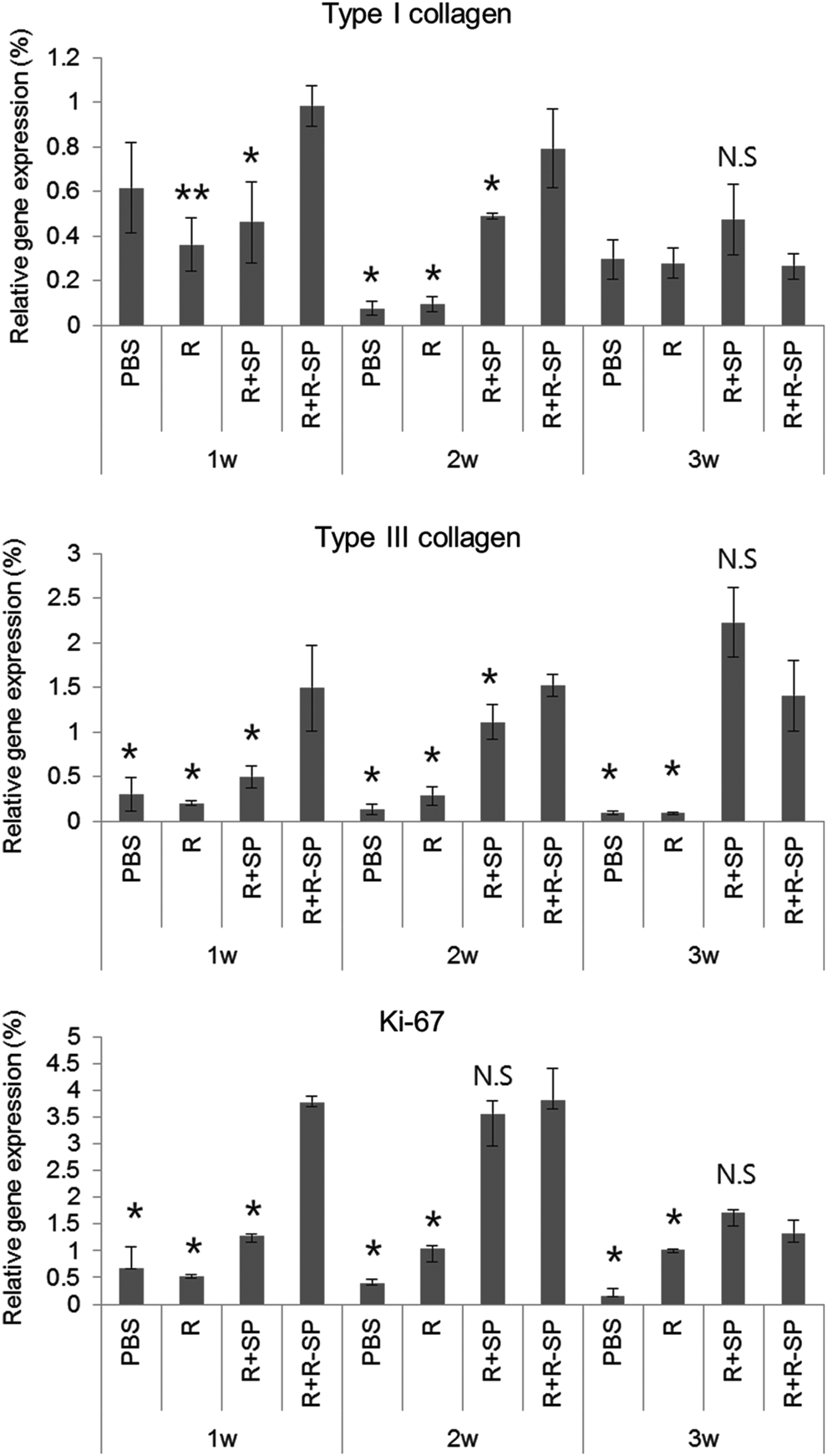
Quantitative real-time PCR analysis of type I and III collagen and cell proliferation marker (Ki-67) expression in the treated wound sites at 1, 2, and 3 weeks. The gene expression was normalized to the housekeeping gene, GAPDH. *p < 0.05 compared to the R+R-SP group. **p < 0.01 compared to the R+R-SP group. N.S., not significant with R+R-SP group.
Our data show that the expression of type I collagen was higher in the R+SP group than in the other groups at 3 weeks after the operation. Furthermore, the marker Ki-67 was increased from week 1 to 2; however, the level decreased at week 3 in all the treatment groups. These results are explained as follows: the cells of the regenerated skin tissues start to move from the proliferation stage to the differentiation stage. Ki-67 was highly expressed in the R+R-SP group; however, the R+SP and R+R-SP groups had a similar gene level at 2 and 3 weeks after the operation with no statistical difference.
Keratinocyte differentiation
To observe keratinocyte differentiation in injured tissue during the wound healing process, immunofluorescence was done for keratinocyte marker genes, including involucrin and cytokeratin 14. At 1 week, cytokeratin 14 and involucrin were slightly expressed in the dermis adjacent to the epidermis in all the treatment groups except for the control group (Fig. 6). The keratinocyte differentiation markers, including cytokeratin 14 and involucrin, were not detected at any specific sites in the control group, which means that the immunostaining had no specificity. In the wound after 3 weeks, cytokeratin 14 and involucrin positive cells were strongly expressed in the R+SP group; however, the other treatment groups had smaller positively stained areas on the section.
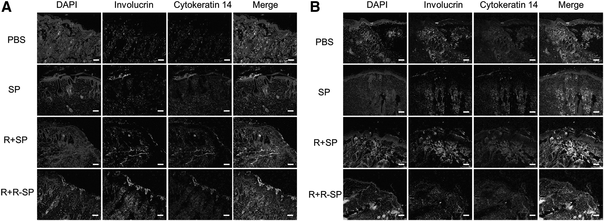
Immunofluorescence analysis of keratinocyte differentiation markers. Cytokeratin 14 and involucrin markers were expressed in the dermis adjacent to the epidermis 1
These results indicate that the R+SP group contributes more to the differentiation of keratinocytes in injured skin. This seems to be the therapeutic effect of differentiation and release of paracrine factors by the recruitment of MSCs from early stimulation by substance P.
Angiogenesis
Angiogenesis is crucial in the wound healing process. It provides sufficient nutrients and the appropriate conditions for the reconstruction of skin tissue. To examine angiogenesis in the injured tissue, the vWF+ cell density (μm2) and α-SMA+ cell density (μm2) of all the treatment groups were analyzed in the injured tissues at 1 and 3 weeks after the treatments. The vWF+ cell density of the R+SP and R+R-SP groups increased to 8.2 and 5.5 times that of the control group at 1 week, respectively, (8234.4 ± 494.2 μm2 in the R+SP group, 5559.6 ± 889.9 μm2 in the R+R-SP group, and 1006.8 ± 332.5 μm2 in the control group). Similar to the control group, a few vWF positive vessels were found in the wound site of the R group (1337.8 ± 387.9 μm2 in the R group). At 3 weeks, the values were similar to the values at 1 week in all the groups except for the R+R-SP group (1330.9 ± 92.2 μm2 in the control group, 2385.8 ± 134.9 μm2 in the R group, and 8584.3 ± 550.4 μm2 in the R+SP group).
The vWF+ cell density of the R+R-SP group was remarkably 1.7 times higher than that at 1 week; this effect may be a result of the sustained delivery of substance P from the peptide hydrogel (7707.5 ± 657.0 μm2 in the R group).
The α-SMA vessel density of the R+SP group was the highest among all the groups which increased to 16.2, 5.4, and 1.5 times that of the control, R, and R+R-SP group, respectively; however, there was no significant difference between the R+SP and R+R-SP groups at 1 week (10,443.1 ± 1983.5 μm2 in the R+SP group, 644.1 ± 283.6 μm2 in the control group, 1928.4 ± 451.8 μm2 in the R group, and 4026.5 ± 1437.5 μm2 in the R+R-SP group). At 3 weeks, the α-SMA vessel density of the R+SP group also was the highest among all the groups; however, the values were slightly decreased in the R+SP and R+R-SP groups compared to that of 1 week (6977.4 ± 1350.8 μm2 in the R+SP group and 4026.5 ± 631.6 μm2 in the R+R-SP group). As seen in Figure 7, the R+SP group had ingrowth and small-sized vessels; however, vessel formation changed to mature and large-sized vessels as vascularization continued. Furthermore, the R+R-SP group had less mature vessels compared with the R+SP group although the R+R-SP group did have remarkably enhanced positively stained vessel formation compared to the control and R groups.

Representative images of the ECs and SMCs by immune staining. ECs were stained with vWF (red) and SMCs with α-SMA (green)
Based on this result, SAP with substance P was effective in promoting the formation of mature vascular structures in the wound healing process, which is essential for the new formation of granulation tissue.
Recruitment of intrinsic MSCs
To determine whether skin regeneration, which is related to keratinocyte differentiation, angiogenesis, and new granulation tissue formation, occurred due to the recruitment of MSCs from the treatment of substance P, immunofluorescence analysis was performed for CD90 and CD105, which are MSC specific markers, at 3 weeks after the operation. The specimens retrieved 3 weeks after the operation were stained with anti-CD90 and anti-CD105 antibody, and the resulting images were quantitatively analyzed (μm2/μm2) by the ImageJ program.
As shown in Figure 8, the groups with SP (R+SP and R+R-SP group) had a significantly higher recruitment of intrinsic MSCs compared with the control group. In addition, there was no significant difference between the R+SP and R+R-SP groups. In contrast, only a few MSCs were observed at 3 weeks after the treatments in the R group. These results indicate that substance P contained in SAPs is effective in wound healing by recruiting MSCs; therefore, homing MSCs will contribute to keratinocyte differentiation, as well as skin regeneration at wound sites.
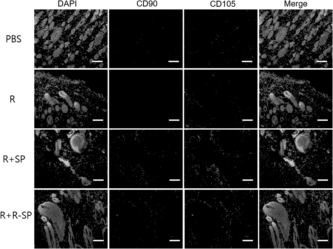
Immunostained images of MSC markers. To study the recruitment of MSCs, the sections were stained with CD90 (red), CD105 (green), and DAPI (blue) 3 weeks after injection. Representative images of the wound site from each group. Scale bars 100 μm. The R+SP and R+R-SP treatment groups were effective in recruiting MSCs to full-thickness wound tissues in the diabetic wound model. MSC, mesenchymal stem cell.
Discussion
Various factors such as cell migration, production of growth factors, collagen synthesis, and vascular biology, including stimulation of blood vessels and enhancing vascularization, will improve the wound healing process.
Most of all, MSCs are an attractive source as regenerative cellular therapeutic materials because they have the potential to secrete essential cytokines for successful wound repair. MSCs have the ability to differentiate into various cell types, including endothelial cells (ECs),30,31 neural cells,32,33 hepatocytes,34,35 and a variety of tissue-specific cells.36,37 Furthermore, it has been reported that MSCs contribute to wound repair by differentiating into keratinocytes resulting in accelerated skin wound healing. 38 Keratinocytes are known to reepithelialize and restore the epidermal barrier during wound healing. 39 For these reasons, MSC therapy has already improved the treatment for wound recovery and the regeneration of skin recently. Kwon et al. investigated the treatment effect of bone marrow-derived mesenchymal stromal cells (BMSCs) by systemic and local injections into a diabetic wound rat model. 6 Chen et al. reported that BMSCs within a cell-laden biocompatible hydrogel can inhibit chronic inflammation at the wound sites of diabetic ulcers and contribute to the rapid healing of the wound sites. 40
However, the focus of these studies has been on directly injecting MSCs for clinical trials to regenerate damaged tissue; whereas, in this study, we focused on an endogenous regeneration system by increasing the number of circulating MSCs without exogenous cell transplantation. Therefore, we expected that the MSCs mobilized to the injured defect site would improve the therapeutic effect through differentiation into cell types associated with skin regeneration without the addition of exogenous MSCs.
It was reported that substance P can mobilize CD29+ stromal-like cells from the periphery to the site of injury as a systemically acting wound messenger. 17 In addition, β1-integrins (CD29) are related to keratinocyte migration during wound reepithelization. 41 Furthermore, keratinocyte migration was studied according to the concentration of substance P; thus, the cell migration speed was highly detected at 10−7 M substance P compared with the control group (no addition of substance P). Therefore, these studies prove that substance P has the potential to accelerate wound healing and tissue regeneration.
As shown by our results, keratinocyte differentiation markers, involucrin and cytokeratin14, were expressed higher in the substance P containing groups (R+SP and R+R-SP groups). As such, the higher levels of substance P affected keratinocyte migration and differentiation by recruiting CD29+ stromal-like cells. We confirmed that the MSC specific markers (CD90 and CD105) were higher in the substance P containing groups (R+SP and R+R-SP groups) than in the control and R groups. Although we did not confirm whether stem cells were recruited to the injured lesion by direct analysis such as cell tagging or labeling, it was speculated that the R+SP and R+R-SP groups have many mobilized MSCs because the quantity of positive expression for the MSC specific markers was lower in the control group and the RADA group without substance P. Thus, these mobilized MSCs may differentiate into a skin cell type through local interaction with skin cells in native tissues adjacent to the wounded site.
Collagen is an essential component which can provide strength, form the structure of normal tissues, and engage in repairing the defects created by injuries; thus, it facilitates the restoration of tissue structure and function.6,42 In this study, we measured the total amount of collagen with the Sircol Collagen Assay; thus, it does not measure specifically type I and III collagen critical to the formation of the ECM in healing wounds but the amount of total collagen. Type I and III collagen are the main collagen types as a structural and functional scaffold in healthy skin and predominantly expressed during the repair process. For these reasons, we additionally focused on the organization of collagen, the mRNA levels for type I and III collagen produced by cells, and the biological factors in tissues adjacent to the wound site. Type III collagen is important in establishing a scar-free wound with less granulation/fibrosis tissue and provides a basic lattice for subsequent healing events when wound healing occurs in the early phase. Type I collagen is known to have a role as a main constituent in regenerated skin.5,43
As shown by our results, although the expression of type I and type III collagen was higher in the R+SP group than in the other groups, there was no statistically significant difference with the R+R-SP group. This tendency is believed to be due to the stimulation by substance P from the injected mixture. This substance P stimulation results in the recruitment of MSCs, and subsequently, their interactions between the defect site and adjacent native tissues facilitate the upregulation of collagen expression.
In addition, substance P was reported to not only stimulate collagenous healing of the skin but also influence collagen organization particularly after injury. MSCs also are known to contribute to enhancing collagen deposition; thus, endogenously mobilized MSCs were an efficient stimulant in this study because both the R+SP and R+R-SP groups have a similar quantity of collagen expression at the gene level.
To efficiently and successfully form new granulation tissue, neovascularization and the formation of new blood vessels are important in skin tissue regeneration. 23 While most skin wounds can heal naturally, chronic wounds such as diabetic wounds lead to prolonged and insufficient wound healing due to impaired neovascularization. In our immunofluorescence results, treatment with R+SP resulted in fine vessel structures and a greater vWF+ cell density, as well as an α-SMA+ vessel density, although it was not significantly different from that of the treatment with the R+R-SP.
The SAPs have nanofibrous structures and form 3D microenvironments; thus, they can promote cell attachment, proliferation, migration, and differentiation for various cellular functions and inhibit scar tissue formation. Among the various types of SAPs, RADA16 has superior characteristics enhancing angiogenesis as a result of infiltrating angiogenic cells such as ECs and smooth muscle cells inhibiting the apoptosis of ECs because then it forms nanofibers and assembles into a 3D scaffold. Therefore, we anticipated that the 3D microenvironment provided by the RADA conjugated peptide hydrogel could supply oxygen without prolonging neovascularization in this study. In addition, substance P is known to stimulate angiogenesis, epidermal cell proliferation, and capillary and fibroblast proliferation. Kohara et al. reported that substance P induces the recruitment of cells and increases angiogenesis and, thus, has new therapeutic potential as factor-induced host cell recruitment for ischemic diseases. 18
Overall, our findings show that both the R+SP and R+R-SP treatment groups induced angiogenesis as follows: (1) secretion of angiogenic factors from endogenously mobilized MSCs stimulated by substance P, (2) biological regulation of angiogenesis by substance P's own potential, and (3) the synergetic effect of the fibrous structure provided by the SAPs and the beneficial environment provided by these factors described above.
We evaluated the efficacy of the substance P conjugated peptide hydrogels in a chronic wound rat model by inducing type I diabetes mellitus. Diabetic ulcers are characterized by a chronic inflammation state, which causes imbalances in proteases and their inhibitors, as well as in pro- and anti-inflammatory cytokines, leading to the inhibition of the healing process resulting in wounds that do not heal. Chen et al. developed a biodegradable and biocompatible hydrogel that inhibits pro-inflammatory M1 macrophage expression and then used it in a diabetic wound healing model. The result was not only inhibiting chronic inflammation but also rapid wound healing at the wound sites. 40 Considering these results, we anticipated that the treatment groups with substance P would be beneficial for wound healing because substance P has an anti-inflammatory function in a chronic inflammatory microenvironment.
In our previous study, we used SAPs conjugated with substance P in various disease models such as osteoarthritis, 16 calvarial defect, 44 and hind limb ischemia.20,21 The results showed that it had therapeutic effects as a remedy and in regeneration at each defect site compared to the treatment group which only had a simple mixture of SAPs and substance P (not conjugated to the SAPs).
Substance P is known to induce the recruitment and migration of MSCs; thus, regeneration will be accelerated in a defect or wound model. However, in our previous study, we promoted tissue regeneration by orchestrating stem cell homing by providing a suitable 3D microenvironment and promoting stem cell recruitment in the substance P conjugated SAP treated group at the defect site compared to only the substance P treated group. 20 The SP, R+SP, and R+R-SP groups were more effective in recruiting MSCs compared to the control group (nontreated group). Especially, the R+R-SP group was the most powerful based on the sustained delivery of substance P.
These results mean that the retention and sustained delivery of substance P contributed to tissue regeneration by physically adsorbing and covalently binding to the SAP (RADA). Furthermore, we confirmed that the group with varying concentrations of substance P conjugated SAPs exhibited higher regeneration factors compared with the nontreated control group. 16 It shows that substance P stimulation through its sustained release from the substance P conjugated SAP not only facilitated continuous recruitment of MSCs but also provided a proper microenvironment for tissue regeneration. For these reasons, we determined that the experimental groups should be R, R+SP, and R+R-SP and PBS as the control group except only substance P treated group in this study. As shown by the results in this study, both the R+SP and R+R-SP treatment groups efficiently improved wound healing in this study.
The reason for these results could be that the rate of recovery and regeneration is relatively faster than for other wound healing models for other diseases. Furthermore, it is generally known that sufficient recruitment of host stem cells or progenitor cells into an implanted scaffold in the initial stage is important for in situ tissue regeneration. 45
In a chronic wound, pro-inflammatory cytokines such as TNF-α and IL-β are elevated, and they synergistically cause MMPs to increase. These increasing active factors result in the degradation of the ECM inhibiting cell migration and collagen deposition. 46 In our previous work, we identified that substance P, SAP, and substance P conjugated SAP inhibited the expressions of pro-inflammatory cytokines, including GM-CSF, IL-2, IL-17A, IFN-γ, TNF-α, MIP, and MIP-1α, β. Furthermore, they improved the secretion of anti-inflammatory factor IL-4. Therefore, we assumed that substance P may have contributed to tissue remodeling due to the burst effect from the SAPs during the wound healing process which acts on the initial point, namely in this study, the inflammation stage. In addition, the SAP-SP conjugate group (R+R-SP group) may induce tissue regeneration because of its sustained release effect during the healing process. Therefore, the action of the mixed substance P group (R+SP group) is powerful during the recovery and regeneration of tissue in the initial stage, whereas the covalently bonded substance P group (R+R-SP group) has improved anti-inflammation and mobilization of MSCs due to the sustained release, and finally, efficacy of wound healing will be equivalent between the two groups.
Our results also confirmed that the quantity of remaining substance P in the R+SP and R+R-SP groups was 4.6 and 8.52 times greater compared with the control group. This result suggests that substance P was continuously released regardless of whether it was conjugated with RADA, which resulted in enhanced recruitment of MSCs and more effective interactions between the cells adjacent to the wound site.
However, it is unclear whether the stained substance P is the administered substance P or endogenous substance P by only the immunofluorescence analysis. When we previously studied whether SAP-SP conjugates have different therapeutic potentials according to the ratio of SAP to substance P, the quantity of detected substance P increased as the substance P amount increased. 16 Therefore, we speculated that some of the stained area was from endogenously expressed substance P by mobilization to the defect site during the wound healing process, while another stained area is possibly due to the administered substance P with the SAPs which was detected by attachment at the tissue surface.
Although there are no data for the residual amount of administered substance P, we investigated the peptide disappearance in the synovial joint of the rat model; the SAP concentration was quantitatively analyzed in a previous work. 47 The tagging SAP was detected until 6 weeks after the injection in the rat animal model. From the results of the SAP disappearance analysis in vivo, it is possible that R+SP or R+R-SP could have remained up to 4 weeks in this study. In a future study, we will study the retention of the peptide hydrogel (SAP and substance P) by tagging with an immunofluorescence substrate to observe the stability of the peptide in vivo.
In summary, we improved wound healing by effectively mobilizing endogenous stem cells without using exogenous MSCs in a diabetic rat model. This study showed that SAP with substance P provides a microenvironment that is appropriate for the differentiation of homing MSCs through the secretion of cytokines and growth factors required for efficient tissue regeneration.
Conclusion
In this study, SAP with substance P was used in easy injectable treatments in a diabetic wound model. SAP with substance P can mobilize circulating cells due to homing effect of substance P and thus could possibly 1 day eliminate the need for exogenous cell transplantation. Therefore, SAP with substance P recruited endogenous MSCs, leading to accelerating tissue regeneration by wound closure, deposition of collagen, and increased neovascularization. These results show that the therapeutic effect is due to keratinocyte differentiation and the release of paracrine factors by the mobilized MSCs. Furthermore, our results suggest that SAP with substance P could efficiently restore the structure and function of injured skin. Consequently, SAP with substance P is capable of recruiting MSCs and thus can be used in skin regeneration as a potential treatment.
Footnotes
Acknowledgments
This work was supported by a grant of the Korea Health technology R&D Project through the Korea Health Industry Development Institute (KHIDI), funded by the Ministry of Health & Welfare (HI15C3060-010115), and by a grant of Basic Science Research Program through the National Research Foundation of Korea (NRF) funded by the Ministry of Science, ICT and future Planning (2016R1A2B2009550), Republic of Korea.
Disclosure Statement
No competing financial interests exist.