
Abstract
In vitro generated human skin equivalents are generating interest as promising tools in basic study, as alternatives to animal testing, and for clinical applications in regenerative medicine. For prediction of skin irritation and corrosion, three-dimensional human skin equivalents consisting of differentiated human keratinocytes (KCs) have been developed and some models have been internationally accepted. However, more delicate assessments using full-thickness skin models, such as skin sensitization tests, cannot be performed due to the lack of a dermis containing fibroblasts or appendages. In a previous study, we developed dermo–epidermal human skin equivalents (DESEs) using a cell coating technique, which employs cell surface coating by layer-by-layer assembled extracellular matrix (ECM) films. The DESEs with dermis consisting of normal human dermal fibroblasts (NHDFs) and epidermis consisting of human KCs were easily fabricated by using this technology. In this study, the constructed DESEs were evaluated as an alternative skin for skin permeation and irritation tests. A good relationship of permeability coefficient of chemicals was observed between the DESEs and human skin data. We investigated whether the DESEs, a new in vitro skin model, are capable of identifying skin irritant and nonirritant substances among 20 reference chemicals. It was confirmed that the DESEs are applicable to skin irritation testing as defined in the European Centre for the Validation of Alternative Methods (ECVAM) Performance Standard (OECD Test Guideline 439). We further studied the construction of DESEs with density-controlled blood capillary networks using human umbilical vein endothelial cells (HUVECs). The results suggest that DESEs allowing incorporation of skin appendages are more promising alternatives to animal testing and can be applied to the design of physiologically relevant in vitro skin models.
Introduction
I
The appropriate construction technique for a 3D tissue is required to enhance the cell functions to fabricate engineered tissues. Various techniques have recently been developed to construct 3D stereoscopic tissues, including cell sheet engineering, 5 multilayer scaffolds, 6 and 3D printing technologies. 7 Although these methods are able to construct cells into 3D composites, they have limitations due to the complicated handling of the fragile cell sheet or residual scaffolds in the composites. Moreover, it is difficult to precisely control the thickness and components of an extracellular matrix (ECM) in constructed tissues. We recently reported a simple and unique bottom-up approach, called cell accumulation technique, to develop 3D multilayered cell composites with the desired layer number and location by the fabrication of thin films consisting of nanometer-sized layer-by-layer (LbL) fibronectin (FN)–gelatin (G) (FN-G) onto the single cell surfaces. 8 ECM films comprising FN-G with a thickness of less than 10 nm allowed all cells to adhere to each other through interactions between the FN-G nanofilms and the cell membrane proteins to create various types of tissues, such as liver 9 and heart. 10 Using this technique and a sandwich culture, highly dense and homogeneous endothelial tubular networks were formed in fibroblast tissues. 11
Three-dimensional tissue-engineered constructs primarily developed for clinical application are now supporting the design of physiologically relevant in vitro models as alternatives to animal testing. This is particularly important for tissue-engineered skin substitute as the European Commission (EC) announced that cosmetic products tested by animal experiments could not be marketed in the European Union from 2013. The animal experiments traditionally used for irritation, corrosiveness, and phototoxicity tests must be replaced by alternative methods.12,13 Using animal models in the cosmetic and pharmaceutical research fields has become more difficult because of ethical concerns about animal welfare and the species differences between animals and humans in drug metabolism and pharmacokinetics. For skin irritation and corrosion testing in vitro, various types of human skin equivalents have been researched and some of these are available commercially, such as Skinethic™ RHE, Episkin™ (SkinEthic/L'Oreal), Epiderm™ (MatTek), and LabCyte EPI-MODEL (J-TEC).14–16 These skin equivalents can be divided into two classes. The first class consists of human differentiated keratinocytes (KCs) seeded on a substrate mimicking the epidermis (epidermal model). The second class consists additionally of a dermis comprising human skin fibroblasts embedded in various kinds of matrices (full-thickness skin model). The epidermal models show a major advance over 2D cell culture models and are mainly used for skin irritation studies according to the OECD Test Guideline 439 (OECD TG439) as alternatives to the rabbit Draize skin irritation test. 17 However, more delicate assessments using full-thickness skin models such as skin sensitization tests cannot be performed due to the lack of a dermis containing fibroblasts, immune cells, or appendages. Many skin irritants induce inflammatory reactions without direct cytotoxicity, 18 suggesting the need to develop physiologically relevant skin equivalents.
In a previous study, we developed a full-thickness human skin equivalent using a cell coating technique, which is a rapid fabrication method of 3D cellular composites by cell coating using LbL assembled FN-G thin films. 19 The 3D human skin equivalents with dermis consisting of normal human dermal fibroblasts (NHDFs) and epidermis consisting of human KCs were easily fabricated by using this technology. In this study, the constructed dermo–epidermal human skin equivalents (DESEs) were evaluated as an alternative skin for skin permeation and irritation tests. Four chemical compounds with different lipophilicities were used as test penetrants. We evaluated whether the DESEs, a new in vitro skin model, react to skin irritant and nonirritant chemicals in a manner similar to in vivo among the 20 reference chemicals defined in the European Centre for the Validation of Alternative Methods (ECVAM) Performance Standard. We further studied the construction of DESEs with density- and size-controlled blood capillary networks using human umbilical vein endothelial cells (HUVECs). In the human dermis, blood and lymphatic vessels play major roles in tissue fluid homeostasis and immune cell trafficking. 20 The tissue engineering of capillary networks not only generates valuable in vitro/ex vivo research models but is also useful for regenerative medicine as in skin tissue regeneration. 21
Materials and Methods
Materials
All chemicals were used as received without further purification. FN from human plasma (Mw = 4.6 × 10⁵ Da) and collagen type IV (Col IV) from human placenta were purchased from Sigma-Aldrich (MO, USA). Gelatin (G) (Mw = 1.0 × 10⁵ Da), phosphate-buffered saline (PBS), Dulbecco's modified Eagle's medium (DMEM), 10% formalin neutral buffer solution, 4% paraformaldehyde phosphate buffer solution, and 3-(4,5-dimethylthiazol-2-yl)-2,5-diphenyl tetrazolium bromide (MTT) were purchased from Wako Pure Chemical Industries (Osaka, Japan). The mouse monoclonal anti-human CD31 antibody was purchased from Dako (Glostrup, Denmark). Goat anti-mouse IgG secondary antibody (Alexa Fluor 488 or 546 conjugate), 4,6-diamidino-2-phenylindole dihydrochloride (DAPI), and fetal bovine serum (FBS) were purchased from Life Technologies. The 24-well cell culture inserts (culture area 0.33 cm2) with 0.4 μm pore size were purchased from Corning.
Cell culture
NHDFs from neonatal tissue, HUVECs, and EGM™-2MV BulletKit™ Medium were purchased from Lonza. Human epidermal KCs from neonatal tissue and the HuMedia-KG growth factor set were purchased from KURABO (Osaka, Japan). EpiLife® Medium with 60 μM calcium was purchased from Life Technologies. The NHDFs (passage was <8) were cultured in DMEM containing 5% FBS, 100 units/mL penicillin, 100 μg/mL streptomycin, and 0.25 μg/mL amphotericin B. The cells were maintained in 5% CO2 at 37°C and subcultured with 0.05% trypsin +0.02% EDTA in PBS. HUVECs (passage was <8) were cultured in EGM-2MV medium and subcultured with 0.05% trypsin +0.02% EDTA in PBS. KCs (passage was <3) were cultured in EpiLife Medium supplemented with HuMedia-KG growth factor and subcultured with pronase (KYOKUTO, Tokyo, Japan).
Construction of a dermis consisting of NHDFs using a cell coating technique
NHDFs were suspended with 0.04 mg/mL FN and G in PBS and alternately incubated for 1 min at 12 rpm using a microtube rotator (MTR-103; AS ONE). After each procedure, the cells were washed with PBS using centrifugation at 400 g for 1 min to remove uncoated FN and G. After nine steps of coating, FN-G nanofilms were covered onto single cell surfaces. The cells coated with FN-G nanofilms (1 × 10⁶ cells/300 μL) were seeded into the cell culture insert (0.33 cm2) coated with an FN, and 2 mL of DMEM containing 5% FBS was added into the microplates. The cells were then incubated in 5% CO2 at 37°C. After 1 day of incubation, the NHDF multilayered tissues with the desired thickness were constructed on the insert membrane. These tissues as a dermis were used for further experiments.
Construction of dermo–epidermal composites and KC differentiation by lifting to air–liquid interface
The epidermal layers were constructed on the surface of the prepared NHDF dermis. First, the outer layer of the NHDF dermis was coated with 100 μL of Col IV (0.2 mg/mL in PBS) for 30 min. KCs (6 × 10⁵ cells/300 μL) were then seeded onto the surface of the dermis, and 2 mL of DMEM/EpiLife mixed medium (1:1 v/v) was added into the microplates. After 1 day of incubation, the NHDF-KC constructs were lifted to the air–liquid interface and 500 μL of cornification medium (DMEM/EpiLife mixed medium [1:1 v/v] supplemented with 25 μg/mL ascorbic acid) was added into the 24-well plate (of culture insert) (airlift culture). 22 The medium was changed every day for 7 or 14 days. Paraffin-embedded formalin-fixed DESEs were sectioned at 5 μm and stained with hematoxylin–eosin (HE). The tissue morphology and behavior of KC differentiation were estimated by HE and immunohistochemical-stained images of the histological sections.
Fabrication of DESEs containing blood capillary networks
FN-G nanofilm-coated NHDFs (5 × 10⁵ cells) suspended in DMEM containing 5% FBS were seeded into the cell culture insert to construct the five-layered (5L) NHDF tissues. After 1-day incubation at 37°C, HUVECs (0.5, 1, 2, or 4 × 10⁴ cells) were seeded onto the surface of the Col IV-coated NHDF dermis and incubated in DMEM containing 5% FBS at 37°C for 6 h to construct the 1L–5L tissues. Then, FN-G nanofilm-coated NHDFs (5 × 10⁵ cells) were seeded onto the 1L–5L tissues to construct the 5L-1L-5L heterocellular tissues, which sandwiched the HUVECs between the 5L-NHDF dermises. 11 After 1-day incubation, the epidermis layers were constructed using the same procedure (airlift culture) as described above. After 7 or 14 days of incubation, the DESEs with widespread and dense blood capillaries were fabricated. The HUVECs were immunostained using an anti-CD31 antibody after construction of DESEs. Fluorescently stained vascularized DESEs were observed using a confocal laser scanning microscope (CLSM; FLUOVIEW FV10i; Olympus). For quantitative analysis of capillary network formation in the DESEs, the % area of the capillary networks and vessel size was measured by ImageJ 1.45I software (National Institutes of Health).
Transepithelial electrical resistance profiles of DESEs
The transepithelial electrical resistance (TEER) values were measured during airlift culture for 1 week using a Millicell ERS-2 (Millipore Co. Ltd.). Before measurements, the DESEs were washed with PBS, and 300 μL (inner) and 1 mL (outer) of PBS were added to the cell culture insert, respectively. TEER values were measured for three times per sample and average values (Ω × cm2) were used.
Permeation experiments
The constructed skin model (DESEs) on the insert membrane was set in a diffusion cell (effective diffusion area, 0.24 cm2) and the receiver chamber was incubated at 32°C (close to normal skin temperature). Frozen, excised human abdominal skin was purchased from Biopredic International (Rennes, France). Four kinds of chemicals, antipyrine (ANP, Mw = 188, log Ko/w = −1.55), isosorbide 5-mononitrate (ISMN, Mw = 191, log Ko/w = −0.15), aminopyrine (AMP, Mw = 231, log Ko/w = 0.50), and flurbiprofen (FP, Mw = 244, log Ko/w = 1.61), were used for permeation experiments. 23 Saturated chemical compounds were dissolved in PBS (ANP and ISMN) or 40% polyethylene glycol (PEG) 400/PBS (AMP and FP). The chemicals (250 μL) were added on the stratum corneum (SC) side. The receiver solution (5.75 mL of PBS) was mixed using a magnetic stirrer during the experiments. At predetermined intervals, an aliquot was collected from the receiver chamber and the same volume of fresh buffer was added to the chamber (TransView® Automated Sampling System; CosMED Pharmaceutical Co. Ltd., Kyoto, Japan). The concentration of chemical compounds in the receiver chamber was measured by high-performance liquid chromatography (HPLC). The permeation parameters of 4 kinds of chemicals were calculated from the time course of the cumulative amount of chemicals that penetrated through the skin models. 24
Skin irritation tests
The 20 reference test chemicals (10 nonirritants and 10 irritants) shown in Table 1 were selected from the list in OECD TG439. The DESEs on a culture insert were set into a 24-well plate with 500 μL DMEM/EpiLife mixed medium (1:1) and were then exposed to the test chemicals. Liquid samples (25 μL) were added to the surface of the DESEs with a micropipette, and solid samples (25 mg) were applied from microtubes to the DESEs moistened with 25 μL sterile water. Each test chemical was added to three samples. In addition, samples serving as negative controls and positive controls were treated with 25 μL distilled water and 5% sodium dodecyl sulfate (SDS), respectively. After 15 min of exposure at room temperature, to remove any residual test chemicals, each tissue was rinsed in a washing procedure where PBS was spritzed on the tissues (filling and emptying the insert cup) with a washing bottle for 2 min (about 50 mL PBS used) and then residual PBS on the tissue was completely wiped off with a cotton bud. The washed tissues were then transferred to new wells on a 24-well plate containing 1 mL of DMEM/EpiLife mixed medium (1:1). The treated tissues were postincubated for 42 h at 37°C. After incubation, the tissues were transferred to new wells on a 24-well plate containing 500 μL of MTT medium (0.3 mg/mL) for cell viability assay. The tissues were incubated for 3 h at 37°C and the cut out membranes from the inserts were then put into microtubes containing 500 μL isopropanol for overnight at 4°C. After extraction reaction, 200 μL extracts were added to a 96-well plate. The optical density (OD) was measured at 570 nm wavelength and at 650 nm (reference wavelength), with isopropanol as a blank. The negative control OD value of the test methods was greater than 1.0 (standard deviation <18%). A chemical was judged as an irritant when the mean cell viability of the tested tissues was reduced to <50% of the negative control and judged as a nonirritant when the cell viability remained above 50%. 15
I, irritant; NI, nonirritant; SD, standard deviation; SDS, sodium dodecyl sulfate.
Results and Discussion
Construction of DESEs
The airlift culture of KCs on NHDF layers was carried out for KC differentiation of epidermis layers. Figure 1 shows HE-stained images of KC+NHDF constructs after differentiation for 7 days of airlift culture. The DESEs were successfully generated and were histologically comparable with human skin. The homogeneous formation of a cornified and basal layer at the epidermis was clearly observed during the process of KC differentiation by airlift culture, and the thickness of the cornified layer increased during the airlift culture. HE images of cross section clearly indicated the formation of the cornified, granular, spinous, and basal layer as an epidermis equivalent (Fig. 2a). The thickness of dermis and cornified layer was controlled by changing the seeding number of NHDFs (Fig. 2b) and period of airlift culture (Fig. 2c). To estimate the barrier functions of the epidermal layer in the DESEs, TEER values were measured during the 7 days of differentiation. The TEER values of DESEs at days 0, 1, 2, 3, 5, and 7 were 4, 40, 560, 770, 1130, and 1080 (Ω × cm2). The values increased with increasing duration of differentiation and reached a plateau (over 1000 Ω × cm2) at 5 days.
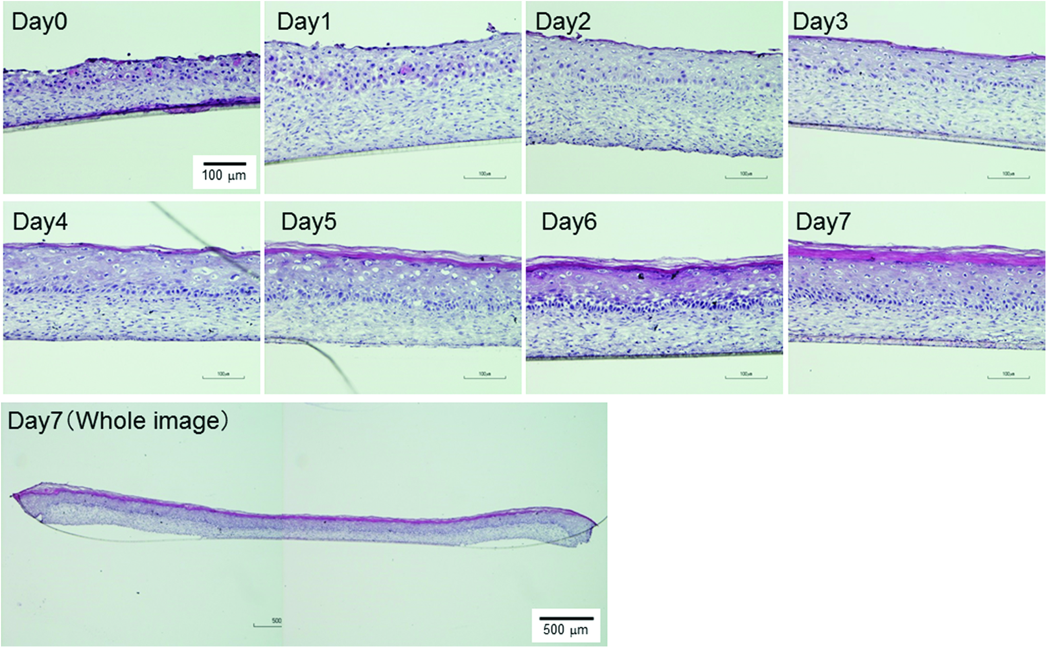
Time course of HE-stained images of DESEs with NHDF and KC layers after differentiation for 7 days of airlift culture. DESEs, dermo–epidermal human skin equivalents; HE, hematoxylin–eosin; KCs, keratinocytes; NHDF, normal human dermal fibroblast. Color images available online at www.liebertpub.com/tea
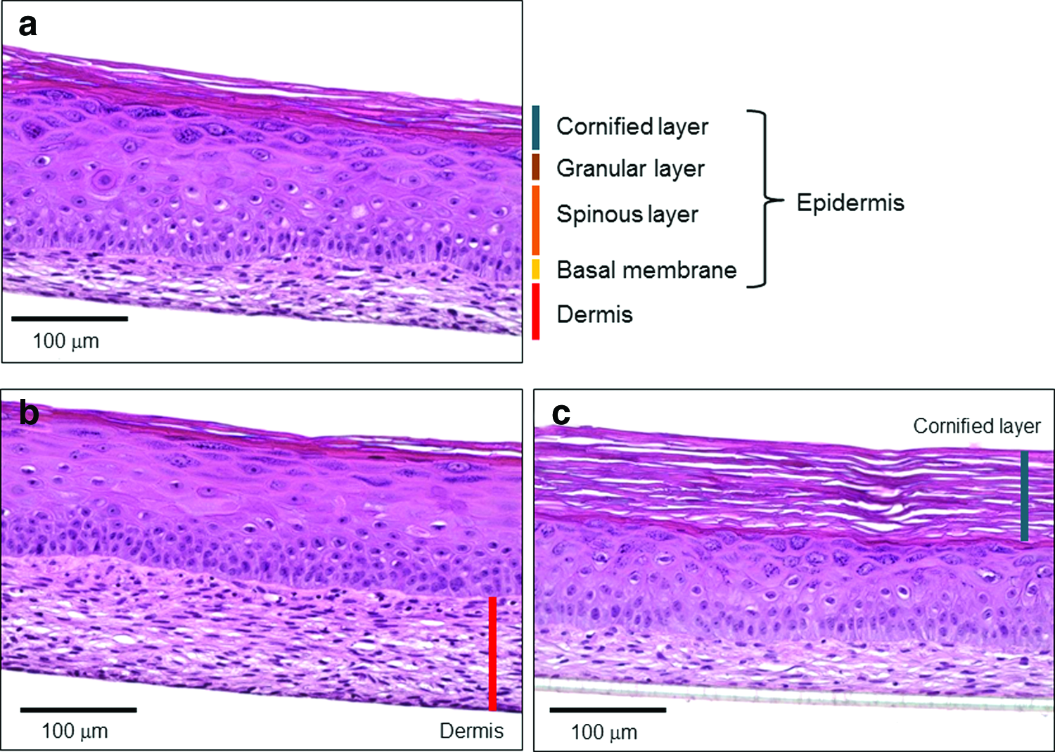
HE-stained images of DESEs consisting of
In a previous study, we used KCs isolated by ourselves from human skin tissues. The collected primary cells were used in their fourth passage. For construction of skin models, a total of 1.8 × 10⁵ KCs were seeded onto the surface of the NHDF dermis. 19 In this study, commercially available KCs were used in their third passage, and a total of 4 × 10⁵ KCs were used for construction of the skin models. The tissue structure of DESEs was significantly improved in comparison with a previous report. We found that the passage number of KCs and the number of KCs seeded onto the surface of the NHDF dermis affect the KC differentiation in the skin models because KC native function gradually decreases with increasing culture passage number (population doubling).
Skin permeation experiments
For evaluation of the skin permeation profiles and skin concentrations of test chemicals, permeation experiments in vitro using native animal and human skin are very helpful. However, in vitro experiments using these skins are limited because of the low availability of samples, ethical issues, and low reproducibility of obtained data.
The permeation experiments were performed on both DESEs after KC differentiation for 7 days and excised human skin to obtain P (permeability coefficient of chemicals), DL−2 (diffusion parameter), and KL (partition parameter) of the applied chemicals from an aqueous solution. Four chemical compounds with lipophilicities (log Ko/w) of −1.55 to 1.61 (ANP, −1.55; ISMN, −0.15; AMP, 0.50; and FP, 1.61) were selected in this study. Figure 3a shows the relationships between log Ko/w and log P obtained from permeation experiments through the DESEs and excised human skin. The log P values of DESEs were increased with increasing the lipophilicity of the applied chemicals, and a similar tendency was observed with the native human skin. Figure 3b and c shows the relationships between log Ko/w and log DL−2 or log KL in the DESEs. Log KL in the DESEs was increased with an increase of log Ko/w of the chemicals, whereas log DL−2 remained almost constant despite the different log Ko/w of the chemicals, suggesting that the solubility parameters or lipophilicity characteristics of the DESEs were similar to those of human skin. These results indicated that skin permeation of chemicals might be predicted according to their permeability coefficients through the DESEs. However, the log P and log KL values of high lipophilic chemicals (FP) in the DESEs showed a relatively low tendency. In general, human skin equivalents were more permeable than human skin due to a low barrier function. 23 The outer layer of skin, the SC, is a highly lipophilic layer consisting of free fatty acids, cholesterol, and ceramides. 25 It has been reported that the lipid amount, composition, and structure in the SC are important parameters for the barrier property of skin.26,27 The reason for the log P and log KL difference of high lipophilic chemicals in the DESEs is not yet fully understood. Further studies of SC lipid analysis in the DESEs will be needed to clarify the difference between the DESEs and native human skin.

Relationship between
Skin irritation tests
Four reconstructed human skin models, Episkin (SM), Epiderm SIT (EPI-200), SkinEthic RHE, and LabCyte EPI-MODEL24 SIT, are being estimated in an ECVAM Skin Irritation Validation Study.28–30 However, although commercially available 3D skin models exist, they need to be upgraded to include fibroblasts in the dermis or appendages for reproduction of skin microenvironments and functions. We performed several examinations of the DESEs and confirmed that they are applicable to skin irritation testing as defined in the ECVAM Performance Standards (OECD TG439).
First, to estimate the barrier function of DESEs after KC differentiation for 14 days, the DESEs were exposed to SDS (from 0.05% to 1%) for 18 h, and the SDS concentration required to decrease cell viability by 50% (IC50) as an indicator of barrier function was measured using an MTT assay. From Figure 4a, the mean of the IC50 of the DESEs was 0.29% (2.9 mg/mL). It has been reported that the IC50 value of human epidermal models, Episkin model and LabCyte EPI-MODEL, was 2.32 and 2.57 ± 0.27 mg/mL, respectively. 15 The result indicated that DESEs have a skin barrier function that resists quick penetration of the test chemicals.

Cell viability of DESEs in skin irritation tests.
Next, to optimize the protocol of skin irritation tests for the DESEs, the exposure time of test chemicals and washing time in which chemicals on the DESEs could be almost completely removed were investigated using test chemicals, allyl phenoxy-acetate and heptyl butyrate, which are nonirritant chemicals in the UN-GHS (United Nations-Globally Harmonized System of Classification and Labelling of Chemicals) classification. First, the exposure periods for the test chemicals were investigated. Allyl phenoxy-acetate, which is a nonirritant chemical, showed a false positive at the exposure periods of 30 min or more. It was judged as a skin nonirritant at the exposure time of 15 min (Fig. 4b). Next, we examined the washing time in which chemicals on the DESEs could be almost completely removed. When the washing time was 2 min or more, in vitro results of heptyl butyrate corresponded to the UN-GHS classification of the chemical; however, the 1-min washing time returned a false-positive result (Fig. 4c). From these results, the optimal exposure time and washing time for our developed DESEs were determined as 15 min for exposure and 2 min for washing.
The 20 reference test chemicals (10 nonirritants and 10 irritants) shown in Table 1 were used to evaluate whether the DESEs as in vitro skin model are able to judge skin irritant and nonirritant chemicals. Of the 10 irritant chemicals (No. 11–20) in vivo, the results for all test chemicals were consistent with those of the classification (Table 1). On the other hand, for the 10 nonirritants (No. 1–10), 3 chemicals, 1-bromo-4-chlorobutane (No. 1), 4-methyl-thio-benzaldehyde (No. 6), and cinnamaldehyde (No. 10), were judged as positive when using DESEs. Based on these results, 7 of 10 UN-GHS nonirritants and 10 of 10 UN-GHS irritants were classified correctly when using DESEs. Regarding the predictive potency of the skin irritation tests, the sensitivity, specificity, and overall accuracy were 100%, 70%, and 85%, respectively. These values were sufficient according to the criteria in the OECD performance standards (80% sensitivity, 70% specificity, and 75% accuracy). These results suggest that the DESEs are applicable to the skin irritation tests. We presented the DESE results of prediction for the 20 reference chemicals described in OECD TG439. Although 1-bromo-4-chlorobutane, 4-methylthio-benzaldehyde, and cinnamaldehyde were judged as false positive in the DESEs (Table 1), similar results were obtained with the LabCyte EPI-MODEL24, Episkin, and Epiderm test method.28,31,32 These different predictions may be due to different properties of the SC between these in vitro skin models and native skin.
Fabrication of DESEs with blood capillary networks as appendages
To fabricate advanced human skin equivalents with blood capillary networks, we performed a sandwich culture of HUVECs based on our previous reports where we constructed blood capillary networks in NHDF layers.8,11 After the sandwich culture of HUVECs with NHDFs, KCs were seeded onto the top of sandwich cultured constructs and lifted to the air–liquid interface for KC differentiation. After 7 and 14 days of incubation, obvious network structures of HUVECs in the dermis and epidermal layers consisting of differentiated KCs on the dermis were observed (Fig. 5). In addition, CLSM images immunostained with anti-CD31 antibody clearly showed tubular structures existing inside the NHDF dermis after 7 and 14 days of KC differentiation (Fig. 6). The percent area of the capillary networks in the DESEs increased with increasing the seeding number of the HUVECs. These results indicate that the density of capillary networks could be controlled by adjusting the seeding number of the sandwiched HUVECs. We have previously reported the construction of coindividual networks of blood and lymph-like capillaries in the dermis.19,33 In addition, it has been demonstrated that the implanted tissue with blood capillary networks was connected with the host circulatory system by anastomosis. 34 This method is an attractive technique for engineering prevascularized tissues for transplantation. The DESEs with circulatory system such as blood capillary have great potential for applications in drug secretion tests and regenerative medicines. In particular, high transplantation efficiency in clinical study using human skin equivalents with blood capillary is expected because of faster nutrient and oxygen supplies from the host blood capillary through the connected blood capillaries. Further experiments using the DESEs with blood capillary networks are now in progress for in vitro and in vivo.

HE-stained images of DESEs with blood capillaries constructed by a sandwich culture of
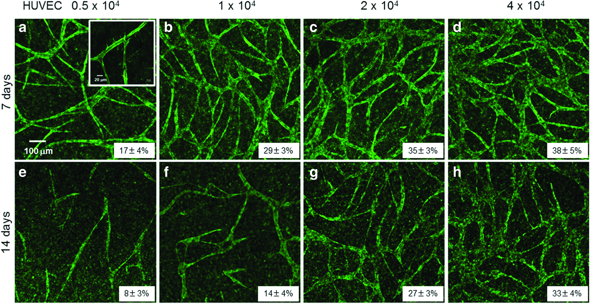
CLSM images of DESEs with blood capillaries constructed by a sandwich culture of
Although the majority of human skin equivalents used in drug development only comprise an epidermis, these skin models could be further improved by the addition of a dermis and subcutis containing appendages, such as fibroblasts, 35 endothelial cells,20,36,37 melanocytes, 38 dendritic cells, 39 Langerhans cells,40–42 hair follicles, 43 and stem cells.44,45 As one of the essential roles of the skin is to provide a barrier against foreign materials, it is evident that dermal and epidermal layers are highly immunogenic compartments. Therefore, the development of full-thickness skin equivalents with immune cells is important for the reproduction of skin microenvironments so that these models are suitable for skin sensitization tests. Although some skin models containing immune cells have been reported,39–42 at present, there is no full-thickness skin equivalent that is capable of accurately estimating skin sensitization. The construction method of DESEs shown in this study would allow for incorporation of various types of cells in the dermis part. It is expected that the DESEs with blood capillary networks and immune cells, such as mast cells, Langerhans cells, or dendritic cells, will be applicable to new in vitro models for skin sensitization tests.
In conclusion, we constructed a 3D dermo–epidermal skin equivalent using cell coating technology. Histological analysis of the constructed DESEs revealed four distinct layers, basal, spinous, granular, and cornified layer in the epidermis and dermis. The blood capillary networks were successfully incorporated into the dermis part of the DESEs. For functional characterization, the DESEs were evaluated for skin permeation and irritation tests. It was confirmed that the DESEs are applicable to skin permeation and irritation testing as defined in the ECVAM Performance Standard. It is thought that the results of the skin irritation tests meet the acceptance criteria described in the OECD TG439. The DESEs with blood capillary networks have potential for various applications, such as drug effect testing, toxicology, and tissue engineering. In particular, it is expected that they will be applied as an alternative method to animal testing.
Footnotes
Acknowledgments
The authors are grateful to H. Taguchi Senior Manager (Kao Corporation), Associate Prof. M. Matsusaki (Osaka University), Prof. K. Sugibayashi, and Associate Prof. H. Todo (Josai University) for technical advice on the experiments and helpful discussion.
Disclosure Statement
No competing financial interests exist.