
Abstract
The aim of this study was to assess the efficacy of a self-assembling peptide hydrogel as a scaffold for bone regeneration. We used a neutral and injectable self-assembling peptide hydrogel, SPG-178-Gel. Bone defects (5 mm in diameter) in rat calvarial bones were filled with a mixture of alpha-modified Eagle's medium and peptide hydrogel. Three weeks after surgery, soft X-ray and microcomputed tomography (micro-CT) images of the gel-treated bones showed new bone formations in the periphery and in central areas of the defects. Next, we evaluated the three-dimensional osteogenic induction of dental pulp stem cells (DPSCs), a type of mesenchymal stem cell, in SPG-178-Gel. We first confirmed that the osteogenic differentiation of DPSCs was significantly promoted by osteogenic induction medium containing recombinant human bone morphogenetic protein-4 (rhBMP-4) in a two-dimensional cell culture. Then, we verified DPSC proliferation and osteogenic differentiation in a three-dimensional cell culture using SPG-178-Gel. The gene expression levels of osteopontin, osteocalcin, and collagen type I were significantly increased when DPSCs were cultured in SPG-178-Gel with the osteogenic induction medium. Micro-CT observations showed the formation of widespread calcium deposition. In conclusion, SPG-178-Gel was adequately effective as a scaffold and can be a suitable tool for bone formation in vivo and in vitro. These findings suggest that the self-assembling peptide hydrogel, SPG-178-Gel, could be a promising tool for bone tissue engineering.
Introduction
I
In general, to succeed in regenerative medicine, three components, that is, cells, scaffolds, and signals, are indispensable. 2 Bone marrow-derived mesenchymal stem cells (BM-MSCs) have been investigated for their potential use as multipotent stem cells and have the ability to differentiate into osteoblasts. 3 However, the harvesting of these cells requires invasive surgery. Furthermore, the maximal proliferative life span, number, and proliferation rate of BM-MSCs exhibit an age-related decline.4–6 As an alternative cell source, dental pulp stem cells (DPSCs) have received recent attention. DPSCs are mesenchymal stem cells with high potential for proliferation and differentiation into odontoblasts, adipocytes, neuronal cells, and osteoblasts.7,8 DPSCs can also be easily extracted from third molar teeth and stored for long periods using cryopreservation.9–11 Furthermore, DPSCs have shown high proliferation rates and high potential of osteogenic differentiation when compared with BM-MSCs. 12 In bone regeneration, several types of scaffolds have been developed based on polymer materials, animal collagen, and ceramic materials, such as hydroxyapatite among others. However, these materials have certain deficiencies. Ceramic materials show low biodegradability. Polymer materials, such as poly(lactic-co-glycolic) acid (PLGA) copolymers, have been reported to release acidic components13,14 and shown to have poor osteoinductivity. 14 Few reports have indicated that animal-derived materials cause allergic reactions15,16 and can carry dangerous pathogens, including prions, causing a variety of neurodegenerative diseases in humans and other animals.17,18
Alternatively, self-assembling peptide hydrogels could be used as scaffolds for bone regeneration. The complete sequence of a self-assembling peptide, EAK16 (AEAEAKAKAEAEAKAK; A = alanine, E = glutamic acid, and K = lysine), was originally found in a region of alternating hydrophobic and hydrophilic residues in zuotin. 19 EAK-16 was later renamed as EAK16-II because it had a modulus of two based on the formula of (AEAEKAKA)n. Several self-assembling peptides, including RAD16-I (RADARADARADARADA, R = arginine, A = alanine, and D = aspartic acid), which had a modulus of one based on the formula of (RADA)n and RAD16-II (RARADADARARADADA), were derived from EAK16-II. 20 The peptide strands undergo self-assembly into nanofibers. The nanofibers then form interwoven matrices that further form a hydrogel scaffold.21,22 The self-assembling peptides minimize the risk of biocontamination due to their chemical-based synthesis. The hydrogel system with RAD16-I is well characterized and has already been used in a variety of tissue engineering studies.20,23,24 However, RAD16-I has a very low pH (∼3–4), thereby retaining a potential to harm inner cells and host tissues.
To avoid cell necrosis and receive the full benefit of this technology, the preneutralization procedure was often used.25–27 In one particular case, a gel required one week to gradually change its pH from acidic to neutral by a solvent substitution. 28
In this study, we selected a neutral self-assembling peptide hydrogel, SPG-178-Gel 29 (Fig. 1). The SPG-178 peptide consists of 13 amino acids, RLDLRLALRLDLR; R = arginine, L = leucine, D = aspartic acid, and A = alanine. Because of its high isoelectric point (pI = 11.5), the peptide can form a stable hydrogel (SPG-178-Gel) at neutral pH. The biocompatibility of the hydrogel was already confirmed by culturing rat skeletal muscle cells in the hydrogel. We first found the efficacy of the self-assembling peptide hydrogel as a scaffold for bone regeneration in the rat calvarial defect model. Then, we further investigated the proliferation and osteogenic induction of cultured DPSCs in this artificial scaffold. Radiographic examinations confirmed the efficacy of the peptide hydrogel scaffold in bone regeneration.
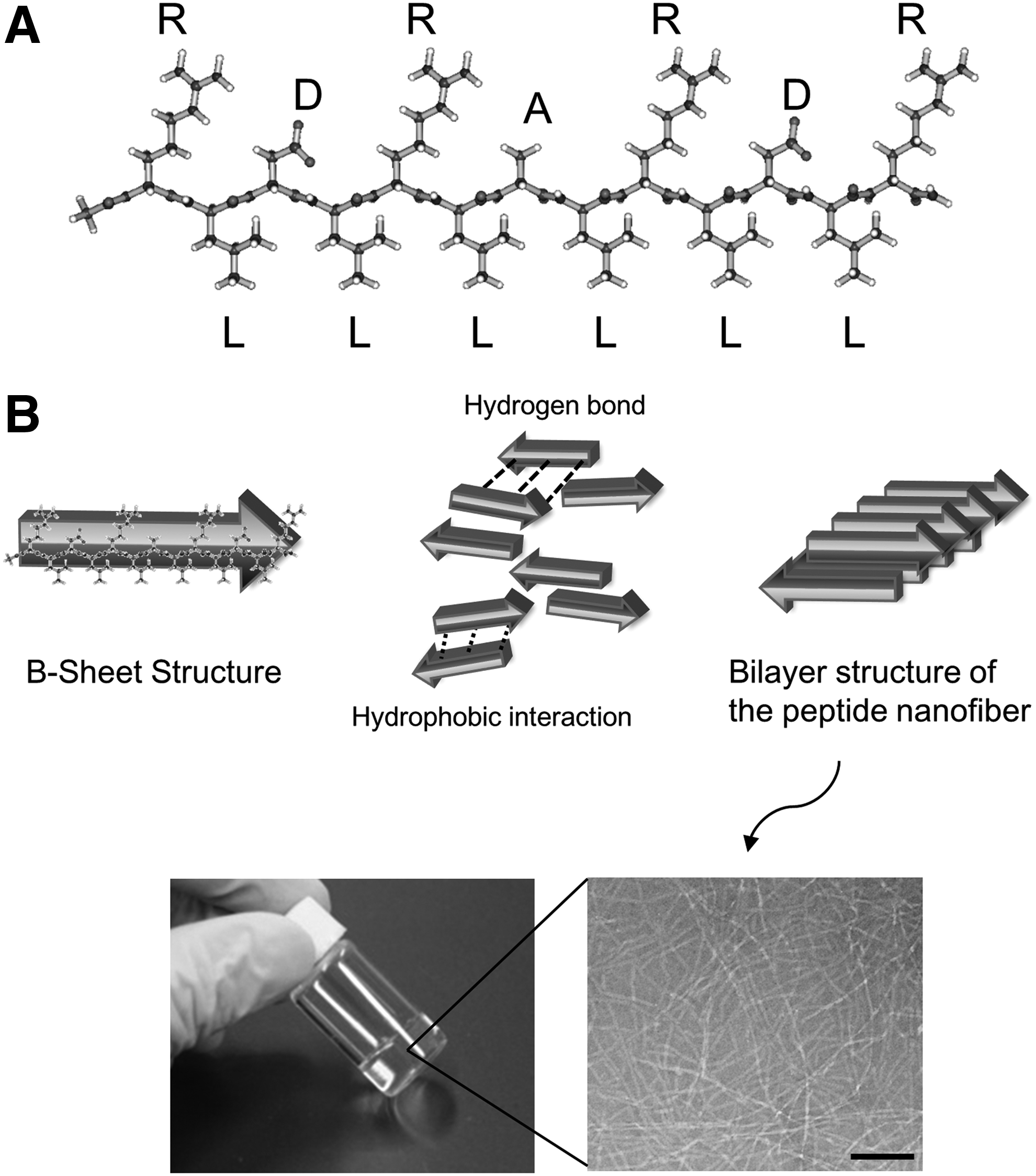
Self-assembling peptide SPG-178 and hydrogel formation.
Materials and Methods
Animals
Four-week-old male Sprague-Dawley rats for the hydrogel implantation in the calvarial defect model and six-week-old male Sprague-Dawley rats for DPSC isolation were obtained from Chubu Kagakushizai (Nagoya, Japan). These studies were carried out in strict accordance with the recommendations in the Guide for the Care and Use of Laboratory Animals of the National Institutes of Health. These studies were approved by the Institutional Animal Care and Use Committees of Aichi Gakuin University (AGUD 189). All rats were housed in cages (260 × 382 × 200 mm, three rats in each cage) under controlled temperatures (24°C ± 1.0°C) with a 12-h light/12-h dark cycle and were provided with standard laboratory rat chow and water ad libitum. Acclimatization period was four days. The surgeries for the hydrogel implantation in the calvarial defect model were performed using isoflurane anesthesia. Penicillin and potassium (200,000 UI/kg; Meiji Seika Pharma Co., Ltd.) were administered before the surgeries. All efforts were made to minimize suffering.
Surgical procedure
The rats were anesthetized with isoflurane (Intervet, Tokyo, Japan), and surgical defects were created in the calvarium. A critical-sized bone defect 5 mm in diameter was created between two bregmas on the midline with trephine (Implatex Co., Ltd., Tokyo, Japan) under running physiological salt solution. 30 The rats were divided into two groups: a Control group (n = 12), where the defects were not treated; and a Gel group (n = 8), where the defects were filled with ∼70 μL of 0.2% peptide hydrogel prepared by mixing alpha-modified Eagle's medium (α-MEM; GIBCO, Billings, MT) and 0.4% SPG-178-Gel (Menicon Co., Ltd., Aichi, Japan) at a 1:1 ratio. The rats were euthanized by CO2 3 weeks after surgery. The peripheral bones of the defects were excised, washed with water, and fixed in 10% formalin.
Radiographic examinations
Soft X-ray (SRO-M50; Sofron, Tokyo, Japan) and microcomputed tomography (micro-CT: R_mCT; Rigaku, Tokyo, Japan) observations were performed to assess the formation of new bone in the defects. The soft X-ray observations were carried out at 40-s exposure, 30 kV, and 4 mA. Micro-CT scans were performed at 2-min exposure, 90 kV, 88 μA, and 10-fold magnification, and at a cubic voxel size of 18 × 18 × 18 μm3. A region of interest (ROI) with a diameter of 5 mm and a height of 0.5 mm encompassing the original bone defect was chosen. Reconstructions of the three-dimensional cranial bone and calculations of new bone volumes were performed using TRI/3DBONE (RATOC System Engineering Co., Ltd., Tokyo, Japan). The volume of the new bone (n = 8–12) was expressed as total bone volume in ROI. The evaluation was performed by two distinct investigators in a blinded manner. Micro-CT images of angled top views, top views, and angled back views were obtained. Micro-CT scans with the same conditions as described above were also performed to observe three-dimensional calcification in SPG-178-Gel after osteogenic differentiation of cultured DPSCs.
Isolation and culture of DPSCs
Rats were euthanized by an overdose of pentobarbital. The incisors were surgically extracted and dissected. Dental pulp tissue was collected, and DPSCs were isolated as previously described. 11 DPSCs were cultured in growth medium (α-MEM+), which consisted of α-MEM with 20% fetal bovine serum (FBS; GIBCO), on plastic dishes at 37°C in 5% humidified CO2. Nonadherent cells were washed off after 3 days of the culture, and adherent cells were continuously expanded until passage 3. Subculturing was performed by detaching the primary culture cells with 0.05% trypsin and resuspending the harvested cells in alpha-MEM+ before centrifugation. Primary culture cells were trypsinized and harvested with α-MEM+ by centrifugation before subculturing. DPSCs were cultured from passages 3 to 6 and used for all experiments.
Comparison of osteogenic induction medium
We compared two different media for their ability to differentiate DPSCs into osteoblasts. One medium was a commercially available kit (osteogenic medium) and consisted of 0.5% dexamethasone, 2%
Proliferation assay in three-dimensional culture of DPSCs
To evaluate the three-dimensional proliferation ability of DPSCs in SPG-178-Gel, we used the Cell Counting Kit-8 (CCK-8; Dojindo, Kumamoto, Japan) with slight modifications to the manufacturer's instructions. DPSCs resuspended in α-MEM at 4.0 × 10⁶ cells/mL were mixed with 0.4% SPG-178-Gel at a 1:1 ratio. Then, 50 μL of these suspensions was placed into a 24-well cell culture insert (pore size: 8 μm, Becton Dickinson). Aliquots of 300 and 700 μL of α-MEM+ were placed inside and outside of the insert, respectively. The cells were cultured for up to 11 days at 37°C, in 5% CO2. Media were replaced every 2 to 3 days. Cell proliferation assays were carried out at each time point (0, 4, 8, and 11 days of culture). After culture, the medium was replaced with 1 mL of α-MEM+ containing 10 μL of CCK-8 solution. After 4 h of incubation, the cell/SPG-178-Gel mixture was transferred from the cell culture insert to the well and dispersed in media. An aliquot of 100 μL of each dispersed cell/SPG-178-Gel mixture solution was then transferred to a 96-well plate. Then, the amount of the formazan dye generated by living cells was determined by absorbance measurements at 450 nm using a spectrophotometer (1420 ARVO™ MX/Light; PerkinElmer, MA).
Three-dimensional osteogenic differentiation of DPSCs
DPSCs grown on culture dishes until 80% confluence were collected by trypsinization. The trypsin was blocked with α-MEM+, which contained 20% FBS. Then, the cells were washed twice with α-MEM. The cells were suspended with α-MEM containing 1.0 μg/mL rhBMP-4 and 0.4% SPG-178-Gel at a 1:1 volume ratio. The final concentration of the cells and the SPG-178-Gel were 2.0 × 10⁶ cells/mL and 0.2%, respectively. A total volume of 100 μL cell/SPG-178-Gel mixture was applied to a 24-well culture plate insert. The inserts were set in the wells of a 24-well culture plate with aliquots of 300 and 700 μL of α-MEM+ placed inside and outside of the insert, respectively. The cells were cultured for one day at 37°C, in 5% CO2. Then, the media were replaced with BMP-4+ for osteogenic differentiation or with α-MEM+ as controls. Media were added on top of the cell/SPG-178-Gel mixture layer in the insert. Media were replaced every 2 to 3 days.
Real-time polymerase chain reaction
Total RNA was extracted by the TRIzol® Reagent (Invitrogen, San Diego, CA) and transcribed into cDNA using ReverTra Ace® (TOYOBO, Osaka, Japan). Real-time polymerase chain reaction (PCR) was carried out using the ABI PRISM7000® Sequence Detection System (Applied Biosystems) with TaqMan® Gene Expression Master Mix (Applied Biosystems). First-strand cDNA was mixed with TaqMan Gene Expression Master Mix, TaqMan probes of the target genes, and endogenous control gene. We used collagen type I (Rn01463848_m1), osteopontin (Rn00563571_m1), osteocalcin (Rn00566386_g1), Osterix (Rn01761789_m1), and Runx2 (Rn01512298_m1) for probing, and β2-microglobulin (β2m) (Rn00560865_m1) as the endogenous control (all reagents were supplied by Applied Biosystems). The β2m RNA levels were referenced when the gene expressions of specific osteoblast markers were determined. The relative quantity was calculated by the ΔΔCt method.
Immunohistochemistry
To investigate the osteogenic differentiation of DPSCs, osteopontin was visualized by immunofluorescence. After fixing with 4% paraformaldehyde, the samples were washed with 1% bovine serum albumin (BSA) in phosphate-buffered saline (PBS), permeabilized, and blocked with 0.3% Triton X-100 (SIGMA-ALDRICH) and 1% BSA in PBS. Then, samples were stained with the primary antibody overnight. The primary antibody was mouse anti-mouse osteopontin (1:50, sc-21742; Santa Cruz Biotechnology). The samples were then incubated with secondary antibodies, Alexa Fluor® 488 rabbit anti-goat IgG or Alexa Fluor® 568 rabbit anti-mouse IgG (A-11078, A11061; Invitrogen, Carlsbad, CA), which were diluted 1:200 in PBS containing 1% BSA, and 4′-6-diamidino-2-phenylindole (DAPI; SIGMA-ALDRICH). The samples were observed using fluorescence microscopy (AF6000LX; Leica Microsystems, Wetzlar, Germany).
Statistical analyses
Results are expressed as mean ± SD. Statistical analyses were made by t-test for two samples, and one-way analysis of variance with the Bonferroni correction for multiple comparisons. The differences were considered to be significant at p < 0.05.
Results
Efficacy of a self-assembling peptide hydrogel as a scaffold for bone regeneration
To evaluate bone regeneration at the calvarial defect sites using the peptide hydrogel, radiographic examinations were carried out 3 weeks after scaffold implantation. As shown in Figure 2A, the soft X-ray images revealed that the calcified tissue covered the peripheral and central parts of the bone defects in the Gel group. The radiopaque intensity of the calcified tissue tended to be lower compared with that of the peripheral bone. Similarly, the micro-CT images showed regenerating bone-like structure covering the defect sites (Fig. 2B). The surface texture and the thickness of the structures were similar to those of the peripheral bone. Quantitative analysis using micro-CT demonstrated that bone regeneration was significantly accelerated in the Gel group by 2.5 ± 1.2-fold when compared with the Control group (p < 0.05) (Fig. 2C). The post hoc statistical power and effect size were calculated as 75.5% and d = 1.26, respectively.
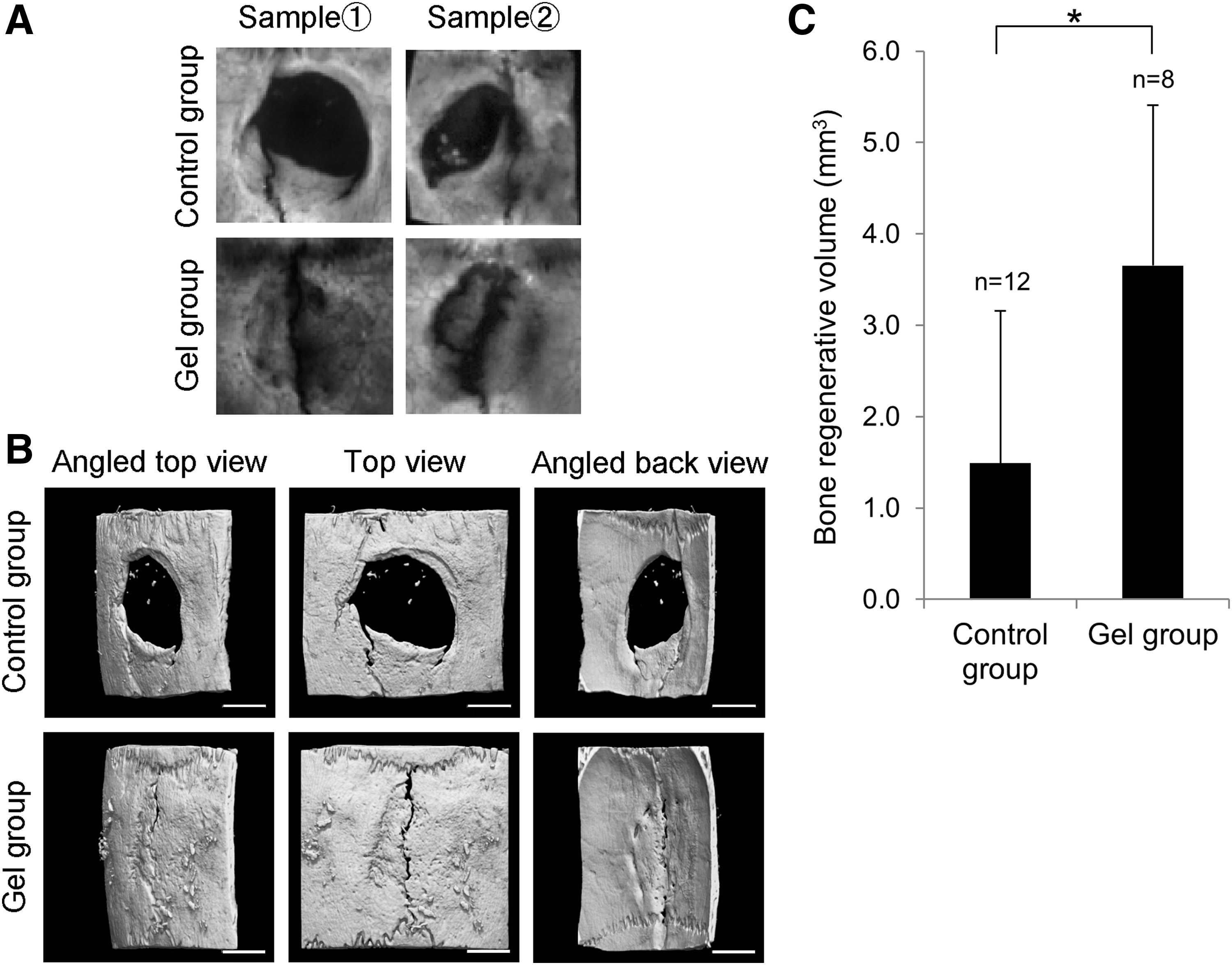
Evaluation of bone regeneration at calvarial defect sites using radiographic analysis. Typical images of a defect site at 3 weeks after surgery by
Comparison of the osteogenesis efficacy of DPSCs by different induction media
Two weeks after culturing with osteogenic medium (n = 4), BMP-4+ (n = 3), or α-MEM+ control (n = 4), the osteogenic differentiation of DPSCs was assessed by real-time PCR for osteogenic gene expression. The mRNA levels of osteopontin in BMP-4+ increased by 37.7 ± 25.3-fold compared with α-MEM+ and by 10.0 ± 6.7-fold compared with osteogenic medium (p < 0.05) (Fig. 3A). The osteocalcin expression level in BMP-4+ also significantly increased by 8352.1 ± 932.4-fold compared with α-MEM+ and by 106.5 ± 11.9-fold compared with osteogenic medium (p < 0.001) (Fig. 3B). In the immunofluorescence assay, we further confirmed the expression of osteopontin. It was observed in DPSCs cultured with BMP-4+ for 3 weeks (Fig. 3C). The fluorescent signal of osteopontin was relatively low in the DPSCs cultured with osteogenic medium.

Osteogenic gene and protein expressions of DPSCs cultured in three different media. After 2 weeks of culture, the osteogenic gene expression levels of
DPSC proliferation in three-dimensional culture using SPG-178-Gel
The proliferation of DPSCs in SPG-178-Gel with α-MEM+ was monitored by measuring the absorbance at 450 nm to quantify the amount of formazan dye generated by dehydrogenases in the cells. DPSCs gradually proliferated through 11 days of culture in 0.2% SPG-178-Gel (Fig. 4B).

Proliferation potency of DPSCs in SPG-178-Gel.
Osteogenic mRNA expressions of DPSCs in three-dimensional culture using SPG-178-Gel
The osteogenic gene expressions of DPSCs after 2 (n = 5) and 3 weeks (n = 6) of culture in the SPG-178-Gel with BMP-4+ or α-MEM+ and the levels of mRNA expressions of collagen type I, osteopontin, and osteocalcin were quantified (Fig. 5A). After 2 weeks of culture, the expression levels of osteopontin, osteocalcin, and collagen type I increased by 1.6 ± 0.6-fold, 1.2 ± 0.4-fold, and 1.4 ± 0.2-fold, respectively, in BMP-4+ compared with those of α-MEM+; these differences were not significant (Fig. 5B–D). The expression levels of each gene were significantly upregulated in BMP-4+ after 3 weeks of culture compared with the expression levels at 2 weeks (osteopontin; p < 0.01, osteocalcin; p < 0.001, collagen type1; p < 0.001). In addition, when compared with α-MEM+ at 3 weeks, osteopontin, osteocalcin, and collagen type I significantly increased by 18.5 ± 14.0-fold, 13.2 ± 2.2-fold, and 20.9 ± 12.7-fold, respectively (osteopontin; p < 0.01, osteocalcin; p < 0.001, collagen type1; p < 0.001).

Osteogenic differentiation of the three-dimensional DPSCs/SPG-178-Gel cultures.
We further investigated the expressions of key transcription factors associated with osteoblast differentiation, Osterix and Runx2. In comparison with α-MEM+, the expression level of Osterix in BMP-4+ significantly increased by 1.5 ± 0.2-fold after 2 weeks of culture and by 1.2 ± 0.1-fold after 3 weeks of culture (2 weeks; p < 0.001, 3 weeks; p < 0.05). The average expression level of Runx2 seemed to be increased in BMP-4+ compared with that in α-MEM+ (at 2 weeks; 1.6 ± 0.2-fold, at 3 weeks; 1.4 ± 0.5-fold), although there was no significant difference between BMP-4+ group and α-MEM+ group (Supplementary Fig. S1; Supplementary Data; Supplementary Data are available online at www.liebertpub.com/tea).
Micro-CT observations of three-dimensional calcification by the osteogenic differentiation of DPSCs in SPG-178-Gel
At 3 weeks after starting differentiation, three-dimensional images were reconstructed using micro-CT data (n = 2). These images revealed widespread three-dimensional calcification in DPSCs/SPG-178-Gel cultured with BMP-4+ (Fig. 5E). No calcification was observed in the control group (with α-MEM+) or in the BMP-4+ culture without cells (data of BMP-4+ without cells not shown). These results indicated that the osteogenic induction of DPSCs by rhBMP-4 with SPG-178-Gel triggered the calcification in the three-dimensional culture.
Discussion
This study aimed to determine a useful approach for bone regeneration using a self-assembling peptide hydrogel, SPG-178-Gel. In recent studies, DPSCs have been shown to have high proliferation potency and pluripotency in their differentiation into odontoblasts, adipocytes, neuronal cells, and osteoblasts.7,8 In this study, we successfully demonstrated the high efficacy of SPG-178-Gel for in vivo bone regeneration and for the in vitro three-dimensional osteogenic induction of DPSCs in SPG-178-Gel.
We showed that SPG-178-Gel contributed toward bone regeneration in a rat calvarial defect model. The new bone formation covered most of the defect sites as observed in the Gel group. The bone formation in the Gel group was significantly greater compared with that in the control group. Past studies have reported that scaffolds are required for bone regeneration to provide void space for cells until mature bone could be formed.14,31 In case of hydroxyapatite, a report found that the highest amount of bone was produced in the ceramic implants with pore size of 300–400 μm, which was much larger than that of the peptide hydrogel (∼10 μm). 32 On the contrary, another report found that optimal pore sizes were different and not necessarily around 300 μm. 33 Thus, pore sizes of the peptide hydrogel could be further optimized.
DPSCs can be used for tissue regeneration. DPSCs are mesenchymal stem cells that can be isolated from extracted teeth without requiring invasive surgical techniques. We and others have previously reported that DPSCs maintain their ability to proliferate and differentiate even after cryopreservation.9,11 In the three-dimensional culture using SPG-178-Gel as a scaffold, DPSCs proliferated successfully for more than ten days. However, while DPSCs were shown to have exponential growth in two-dimensional cultures, a limited, gradual proliferation was recognized in three-dimensional culture. In a previous report, murine C2C12 myoblasts showed a similar proliferation behavior when cultured in SPG-178-Gel. 29 The two-dimensional or three-dimensional environment seemingly affected cellular behavior in several ways.
For three-dimensional osteogenic induction, DPSCs successfully differentiated into osteoblast-like cells in SPG-178-Gel when the SPG-178-Gel was premixed with rhBMP-4 and the cell/SPG-178-Gel mixture was cultured in osteoinduction medium. These three-dimensional osteogenic differentiations of DPSCs seemed to be mineralized, which may interfere with the fluorescence image of the nuclear (DAPI). These results were confirmed by real-time PCR and by micro-CT observations of calcification in the cell/SPG-178-Gel mixture. The upregulation of the osteogenic markers was clearly observed after 3 weeks of incubation. Micro-CT imaging further confirmed the secretion of extracellular matrix, including calcium deposition, from the three-dimensionally cultured cells. This delayed induction may be due to the mild proliferation ratio of the cells in the hydrogel.
On the contrary, the gene expression of the osteogenic transcriptional factor, Osterix, was decreased in 3 weeks of osteogenic induction compared with 2 weeks. The mineralization in the three-dimensional culture, which was gradually increased by the osteogenic differentiation, may interrupt the nutrition supply and suppress further new bone formation. A recent in vivo study of bone regeneration using DPSCs and a poly(lactide-co-glycolide)-co-ɛ-caprolactone (PLGC) scaffold revealed that lower rates of new bone formation occurred in the inner region of the scaffold than in the bottom region. This suggested a limited diffusion of nutrition and blood in the internal part of the scaffold. 34 In future studies, the shape of the cell/SPG-178-Gel mixture, especially the thickness and the surface area exposed to media, should be addressed to optimize the proliferation ratio and differentiation efficiency.
In the present study, the high efficacy of new bone formation with the SPG-178-Gel peptide hydrogel scaffold in the rat calvarial defect model was demonstrated. The SPG-178-Gel showed biocompatibility and osteoconductive activity. Furthermore, the SPG-178-Gel was revealed to adequately act as a scaffold suitable for bone formation in vitro. In three-dimensional culture, DPSCs proliferated in SPG-178-Gel with α-MEM+. Osteogenic differentiation was successfully induced in the hydrogel. This study is the first demonstration that DPSCs could be cultured and differentiated into osteoblast-like cells in a self-assembling peptide hydrogel, as verified by gene expression levels and observed calcification. These findings suggest that the self-assembling peptide hydrogel, SPG-178-Gel, could be a promising tool for bone tissue engineering.
Footnotes
Acknowledgments
We thank Mr. Brent Bell for reading the manuscript. This research was supported, in part, by the Adaptable and Seamless Technology Transfer Program through Target-driven R&D (A-STEP) from the Japan Science and Technology Agency (JST).
Authors' Contributions
Conceived and designed the experiments: K.N. and T.M. Performed the experiments: J.T., S.K., N.N., M.H., and M.O. Analyzed the data: J.T., Y.N., T.H., and T.K. Contributed reagents/materials/analysis tools: J.T. and N.N. Wrote the article: J.T., K.N., and Y.N. Interpreted results of experiments: K.N., T.H., and T.K.
Disclosure Statement
J.T., Y.N., and S.K. are the employees of Menicon Co. None of the other authors has financial interests in the subject of this article.
References
Supplementary Material
Please find the following supplemental material available below.
For Open Access articles published under a Creative Commons License, all supplemental material carries the same license as the article it is associated with.
For non-Open Access articles published, all supplemental material carries a non-exclusive license, and permission requests for re-use of supplemental material or any part of supplemental material shall be sent directly to the copyright owner as specified in the copyright notice associated with the article.