
Abstract
The morbidity of bone fractures and defects is steadily increasing due to changes in the age pyramid. As such, novel biomaterials that are able to promote the healing and regeneration of injured bones are needed to overcome the limitations of auto-, allo-, and xenografts, while providing a ready-to-use product that may help to minimize surgical invasiveness and duration. In this regard, recombinant biomaterials, such as elastin-like recombinamers (ELRs), are very promising as their design can be tailored by genetic engineering, thus allowing scalable production and batch-to-batch consistency, among others. Furthermore, they can self-assemble into physically crosslinked hydrogels above a certain transition temperature, in this case body temperature, but are injectable below this temperature, thereby markedly reducing surgical invasiveness. In this study, we have developed two bioactive hydrogel-forming ELRs, one including the osteogenic and osteoinductive bone morphogenetic protein-2 (BMP-2) and the other the Arg-Gly-Asp (RGD) cell adhesion motif. The combination of these two novel ELRs results in a BMP-2-loaded extracellular matrix-like hydrogel. Moreover, elastase-sensitive domains were included in both ELR molecules, thereby conferring biodegradation as a result of enzymatic cleavage and avoiding the need for scaffold removal after bone regeneration. Both ELRs and their combination showed excellent cytocompatibility, and the culture of cells on RGD-containing ELRs resulted in optimal cell adhesion. In addition, hydrogels based on a mixture of both ELRs were implanted in a pilot study involving a femoral bone injury model in New Zealand white rabbits, showing complete regeneration in six out of seven cases, with the other showing partial closure of the defect. Moreover, bone neoformation was confirmed using different techniques, such as radiography, computed tomography, and histology. This hydrogel system therefore displays significant potential in the regeneration of bone defects, promoting self-regeneration by the surrounding tissue with no involvement of stem cells or osteogenic factors other than BMP-2, which is released in a controlled manner by elastase-mediated cleavage from the ELR backbone.
Introduction
I
Engineered biomaterials, in combination with growth factors, have been shown to be an effective approach in bone tissue engineering since they can act both as a scaffold and as a drug-delivery system to promote bone repair and regeneration.18,19 For instance, the osteoinductive bone morphogenetic protein-2 (BMP-2)20,21 has been shown to enhance the formation of bone tissue in situations that lead to bone degradation, such as alcohol dependence 22 and osteometabolic diseases. 23 Due to the high cost and rapid release of BMP-2 when placed at the site of injury, it is often associated with carrier matrices that act as drug-delivery systems to increase its half-life and to avoid the adverse effects associated with high doses of BMP-2.24–27
On the contrary, protein-based recombinant biomaterials, such as resilin-, silk-, collagen-, and elastin-like polypeptides, have been developed over the last few decades with the aim of improving the features of traditional biomaterials in terms of ease of design and synthesis, biocompatibility, and bioactivity. 28 As an example, elastin-like recombinamers (ELRs), thus named due to their polymeric and recombinant nature, 29 have been shown to be a potential tool for the development of biomedical devices for regenerative medicine due to their thermosensitivity. This smart behavior is a result of their composition, which is based on repetitions of the Val-Pro-Gly-X-Gly pentapeptide, in which X (guest residue) is any amino acid except L-proline. Moreover, it is characterized by a transition temperature (Tt), which itself depends on the polarity of the side chain in the guest residue. Thus, in an aqueous medium, the ELR chains remain soluble below their Tt while above that Tt (e.g., physiologic temperature), the ELR self-assembles hydrophobically, undergoing a phase transition. 30 In this study, two different ELRs have been developed, based on a previously described hydrogel-forming ELR. 31 Taking advantage of the recombinant nature of these biomolecules, one of the novel ELRs designed in this work has been genetically engineered to include Arg-Gly-Asp (RGD) motifs to enhance cell adhesion via cell membrane integrins, 32 whereas the other ELR was designed to include BMP-2. Both ELRs also contain elastase-sensitive domains resulting from repetition of the Val-Gly-Val-Ala-Pro-Gly hexapeptide 33 to improve the enzymatic biodegradability of the biomaterial (see Supplementary Fig. S1 for a schematic representation of both ELRs; Supplementary Data are available online at www.liebertpub.com/tea).
To study the potential of these novel ELRs in bone regeneration, we have used a previously developed model of femoral bone injury (FBI) in New Zealand white rabbits. This involves the creation of a defect 6 mm in diameter in a femoral condyle 8 mm in diameter.34,35 This animal model allows the study of the defect by computed tomography (CT) and by radiological studies given the size of the bone.
The aim of this work was to evaluate whether novel bioactive ELRs are cytocompatible and degradable, while being able to form extracellular matrix (ECM)-like hydrogels and promoting bone regeneration after implantation into an FBI in rabbits, as a preliminary step for their use in humans. For this purpose, the cytocompatibility and biodegradation ability were assessed in vitro, and a highly reproducible model was subsequently used to carry out a pilot in vivo study.
Materials and Methods
Ethical approval
Experimental procedures regarding the use of animals were approved by the Bioethics Committee of Rosario National University (Resolution No. 150/2015). Its regulations include well-established guidelines for animal care and manipulation to decrease pain and suffering of the animal, according to the 3Rs (replacement, reduction, and refinement), and are in accordance with international laws concerning the use of animals.
ELR biosynthesis and characterization
The genetic construction of the ELRs used in this work was performed as described elsewhere. 36 Briefly, their DNA sequences were obtained by genetic engineering techniques and cloned into a pET-25b(+) vector for expression in Escherichia coli. ELRs were biosynthesized in a 15-L bioreactor and purified by several cooling and heating purification cycles (inverse transition cycling) taking advantage of the ability of these recombinamers to precipitate above their Tt. Further centrifugation steps led to a pure product, which was dialyzed against ultrapure water, filtered through 0.22-μm filters (Nalgene; Thermo Fisher, USA) to obtain a sterile solution, and freeze-dried before storage. The ELRs were found to contain <2 endotoxin units per milligram of ELR, as determined using the limulus amebocyte lysate assay with the Endosafe®-PTS system (Charles River Laboratories). This process allowed the production of two different ELRs, both of which were derived from a previously synthesized block corecombinamer. 31 Further information can be obtained in Supplementary Methods.
The characterization techniques used included sodium dodecyl sulfate–polyacrylamide gel electrophoresis (SDS-PAGE) and matrix-assisted laser desorption/ionization time-of-flight (MALDI-TOF) spectrometry for purity and molecular weight (Mw) evaluation compared to the theoretical values of 113,556 Da for ELR-Elastase-RGD (ELR-E-RGD) and 107,752 Da for ELR-Elastase-BMP-2 (ELR-E-BMP-2); differential scanning calorimetry (DSC) to determine the Tt (Supplementary Fig. S2); high-performance liquid chromatography (HPLC) to determine the amino acid composition of both ELRs (Supplementary Tables S1 and S2); and nuclear magnetic resonance to provide recombinamer fingerprint data (Supplementary Figs. S3 and S4; Supplementary Tables S3 and S4). The procedure for the measurement of the mechanical properties of ELR-based hydrogels is described in Supplementary Methods.
Elastase-mediated cleavage of the ELR in solution
Different quantities (1.2, 1.8, and 2.4 U) of porcine pancreas elastase (4 mg/mL, 6.8 U/mg) (Sigma-Aldrich, USA) were added to solutions of the mixture of both ELRs [98% (w/w) ELR-E-RGD and 2% (w/w) ELR-E-BMP-2] at a final concentration of 1 mg/mL dissolved in ultrapure water to evaluate the biodegradation rate for each quantity of enzyme. The quantity of elastase used was 2000-, 3000-, and 4000-times the amount needed to cleave the mixture of ELRs used as substrate in 30 min, since preliminary experiments showed that larger quantities than those calculated are required to observe an actual effect of the enzyme in vitro. Samples were incubated at 37°C, collected at various time points (10, 20, 30, 45, 60, 90, and 120 min), and then stored frozen at –20°C until further use. A negative control, namely an ELR molecule lacking elastase-sensitive sequences but with the same elastin-like structure as the two ELRs designed for this work, 31 was also treated with 1.2 U of elastase for further comparisons. Methods concerning the evaluation of the biodegradation are explained in Supplementary Methods.
In vitro cell culture
Bone marrow-derived human mesenchymal stem cells (hMSCs) were extracted and isolated as described elsewhere 37 and were generously provided as a gift by Citospin S.L. (Spain). They were cultured for expansion in Dulbecco's modified Eagle's medium (DMEM) low glucose (1 g/L) (Gibco, USA) supplemented with 10% fetal bovine serum (Gibco) and 1% penicillin/streptomycin (Gibco).
All cells were used at passage 3–5 in subsequent experiments. They were detached from the wells using a trypsin-ethylenediaminetetraacetic acid (EDTA) solution (0.25%; Gibco) and counted using a hematocytometer.
Cell viability
hMSCs were used to determine the in vitro viability using the calcein AM assay (Molecular Probes, USA) when cultured in DMEM supplemented with 10 mg/mL of the different ELRs or the mixture of them [98% (w/w) ELR-E-RGD and 2% (w/w) ELR-E-BMP-2] for 3 days. This assay was performed in a black, 96-well plate with clear bottom (Greiner Bio One, USA) according to the manufacturer's instructions and the fluorescence intensity measured at 530 nm using a plate reader (SpectraMax M2e; Molecular Devices, USA). The intensity measured at this wavelength, corresponding to live cells, was then used to calculate cell numbers by using calibration curves obtained with different known quantities of cells (from 1000 to 10,000 cells per well) seeded on 96-well plates 24 h before the measurement. Each condition was performed in triplicate, with four experiments for each (n = 4).
Cell adhesion on ELR-coated tissue culture plates
Ninety-six-well plates were used for the coating of different wells with both ELRs separately and combined [98% (w/w) ELR-E-RGD and 2% (w/w) ELR-E-BMP-2]. Briefly, a 5 mg/mL solution of the recombinamers in ultrapure water was placed in the well and allowed to adsorb to the surface for 24 h at 4°C. The wells were washed twice with 1× phosphate-buffered saline (PBS; Gibco), blocked with 1% bovine serum albumin for 2 h at 37°C, then rinsed again and, finally, 3000 cells per well were seeded onto the modified surfaces to study cell adhesion after 24 h. The number of cells in each well was determined using the calcein AM assay as described above.
Dissolution of the ELRs for the in vivo experiments
A mixture of both ELRs [98% (w/w) ELR-E-RGD and 2% (w/w) ELR-E-BMP-2] was prepared and dissolved in sterile tubes (1 per animal) at 300 mg/mL with 1× sterile PBS (Gibco) by incubation at 4°C for 24 h. A 2% (w/w) ELR-E-BMP-2 solution at 300 mg/mL gives a similar amount of BMP-2 in our device (5.57 · 10−5 M) as in INFUSE® Bone Graft (5.77 · 10−5 M; Medtronic, USA). 38 This solution was kept in an ice bath during surgery until implantation.
In vivo experiments
Adult female New Zealand white rabbits (n = 7) with an average weight of 3.5 kg were used for the creation and treatment of bone defects. These animals were kept in individual cages with food (ACA Cooperativas, Argentina) and water ad libitum.
Antibiotic prophylaxis, anesthetic treatment, and surgical techniques were performed according to a previously described procedure.34,35 Further details regarding the surgical procedure can be found in Supplementary Methods. Three months postsurgery, the animals were euthanized using three doses of anesthesia, as previously described.34,39 The femora were then collected to perform different experiments to assess bone regeneration (see Multislice computed tomography and Bone histopathology).
Multislice computed tomography
Multislice computed tomography (MSCT) was performed on the seven right femurs of the rabbits using a Toshiba Alexion apparatus with 16 detectors and a thickness of 0.5 mm. Coronal, sagittal, and axial slices were obtained and the images were processed using Alexion Advance Edition software with the adaptive iterative dose reduction (AIDR 3D) algorithm, thus obtaining the 3D reconstruction for every sample. All images were analyzed together for an optimal comparison.
Bone histopathology
Femoral bone samples were evaluated by way of radiographic studies using a conventional dental X-ray machine with dental occlusal films (Eastman Kodak, USA) to determine the implant position to guide the histological procedures. The femoral epiphysis was cut 4 cm below the metaphysis using a carborundum disk cutter (Dochem, China) attached to a dental drill under irrigation with distilled water. The implanted area was marked with Indian ink. Two samples were selected for decalcification using modified Morse solution (Okayama University Dental School) and embedded in paraffin following well-established protocols. The samples were serially cut (7 μm thick) using a manual rotary microtome (Micron-Zeiss, Germany), and stained with hematoxylin and eosin. All specimens were examined by light microscopy and evaluated by a single pathologist. Subsequently, another pathologist (certified by the Argentinean Ministry of Health No. 31455) performed an independent review to verify microscopic observations. The reported results reflect the mutually-agreed-upon diagnoses by both pathologists. Photomicrographs were taken from slides of each specimen using a Sony digital camera fitted to an Olympus CH30 microscope with an Olympus stereo zoom SZ51.
Statistical analysis
Data for the in vitro experiments are reported as mean ± standard deviation (n = 4). Statistical analysis of data following a normal distribution was performed using a one-way analysis of variance and the Holm–Sidak method. A p < 0.05 was considered to be statistically significant, while p > 0.05 indicates no significant differences (n.s.d.). *p < 0.05, **p < 0.01.
Results
ELR biosynthesis and characterization
Both ELRs were obtained as a lyophilized product in a yield of ∼200 mg/L (ELR/culture volume). Their Mws and purities were confirmed as satisfactory by SDS-PAGE and MALDI-TOF (Fig. 1), while the Tt calculated by DSC for the ELRs dissolved in PBS (pH 7.4) was found to be 15.8°C and 15.3°C for ELR-E-RGD and ELR-E-BMP-2, respectively (Supplementary Fig. S2).
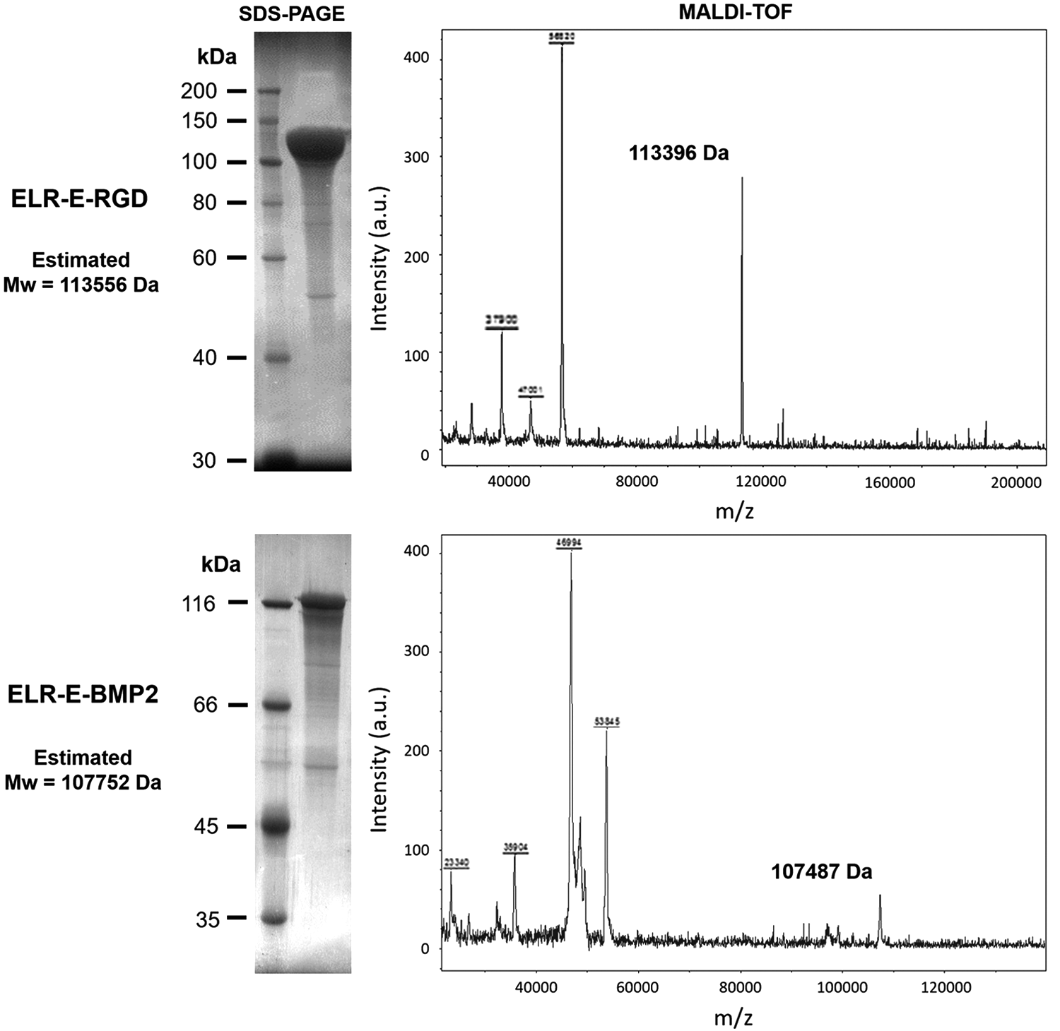
Molecular weight and purity assessment by SDS-PAGE and MALDI-TOF mass spectrometry for ELR-E-RGD and ELR-E-BMP-2. MALDI-TOF spectra represent nonquantitative intensity (a.u.) against m/z (mass divided by net charge of the molecule) of the ELRs. BMP-2, bone morphogenetic protein-2; ELRs, elastin-like recombinamers; ELR-E-BMP-2, ELR-Elastase-BMP-2; ELR-E-RGD, ELR-Elastase-RGD; MALDI-TOF, matrix-assisted laser desorption/ionization time-of-flight; Mw, molecular weight; RGD, Arg-Gly-Asp; SDS-PAGE, sodium dodecyl sulfate–polyacrylamide gel electrophoresis.
Regarding mechanical characterization, the storage modulus (G′) of the ELR-based hydrogel at a concentration of 300 mg/mL (98% ELR-E-RGD and 2% ELR-E-BMP-2) was found to be ∼1600 Pa at 37°C (Supplementary Fig. S5). In addition, hydrogels were formed above the Tt, as observed macroscopically (Supplementary Fig. S6).
Enzymatic cleavage of ELR molecules by elastase digestion
Due to the incorporation of elastase-sensitive domains in the ELR molecules designed for this work, we aimed to verify whether elastase was able to cleave these ELRs. Hence, a mixture of them (98% ELR-E-RGD and 2% ELR-E-BMP-2) was dissolved at 1 mg/mL, and ELRs were found to be cleaved in vitro in solution when different quantities of elastase were added. As observed in Figure 2, and as expected, the ELRs are sensitive to the quantity of elastase, and therefore, biodegradation was slower when only 1.2 U of elastase was added to the ELR solution (Fig. 2a), whereas an increase in the biodegradation rate was observed if 1.8 U (Fig. 2b) or 2.4 U (Fig. 2c) of elastase was supplemented. In contrast, no elastase-mediated cleavage was observed in the negative control at any sample collection time (Fig. 2d).
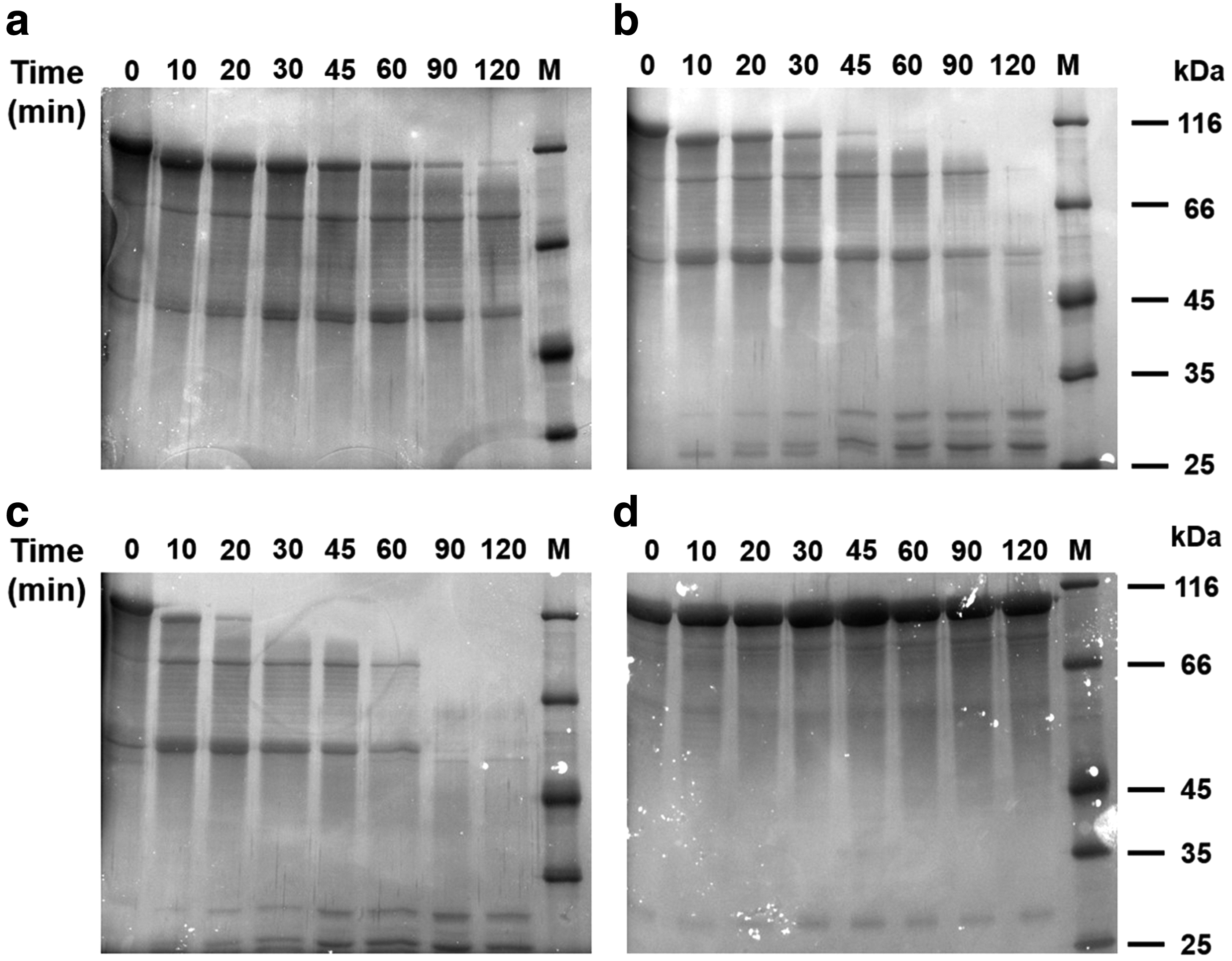
SDS-PAGE images showing the biodegradation of the mixture of ELR-E-RGD (98%) and ELR-E-BMP-2 (2%) in solution at 1 mg/mL mediated by
The disappearance of the larger bands at 113.6 and 107.8 kDa was further studied by image analysis, and the results are summarized in Figure 3. This figure clearly reinforces the statement made above regarding the biodegradation rate, namely that biodegradation is faster as more elastase is added to the solution.
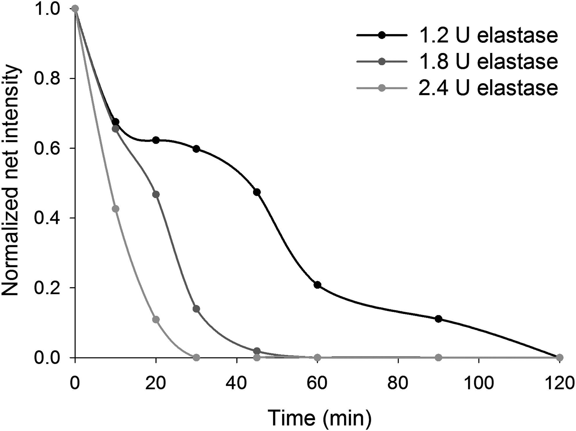
Graph showing the elastase-mediated cleavage rate of the highest molecular weight band with data obtained from analysis of the SDS-PAGE gels from Figure 2. The net intensity of this double band at 113.6 and 107.8 kDa is represented at different sampling times.
Regarding the nascent bands observed by SDS-PAGE (Fig. 2), we expected to obtain bands in three different Mw ranges, namely 65.5–66.5, 46.7–48.2, and 12–12.9 kDa, as by-products of ELR-E-RGD/BMP-2 digestion since there are two different elastase-sensitive domains at different points in the ELR-E-BMP-2 molecule. However, the Mw of the higher bands was found to be 80.8 and 54.2 kDa, respectively, while the band at 12–12.9 kDa could not be observed due to the limitations of SDS-PAGE in terms of resolution. Nevertheless, these results correlate well with previous studies that reported a 20% increase in the apparent Mw for different ELRs.40,41 As such, we estimated that the Mw plus 20% and the values showed good agreement with those found empirically, with the experimental values for the nascent bands being 80.8 and 54.2 kDa, while the expected values of Mw +20% were 78.6–79.8 and 56.0–57.8, respectively (Supplementary Table S5).
hMSC viability and integrin-mediated cell adhesion
The viability of the cells after culture for 3 days in media supplemented with the ELRs was found to be similar to that for the negative control, that is, medium without supplementation, as can be observed in Figure 4. Since no significant differences were observed, we can conclude that the ELRs alone, or the mixture thereof, do not affect cell viability.
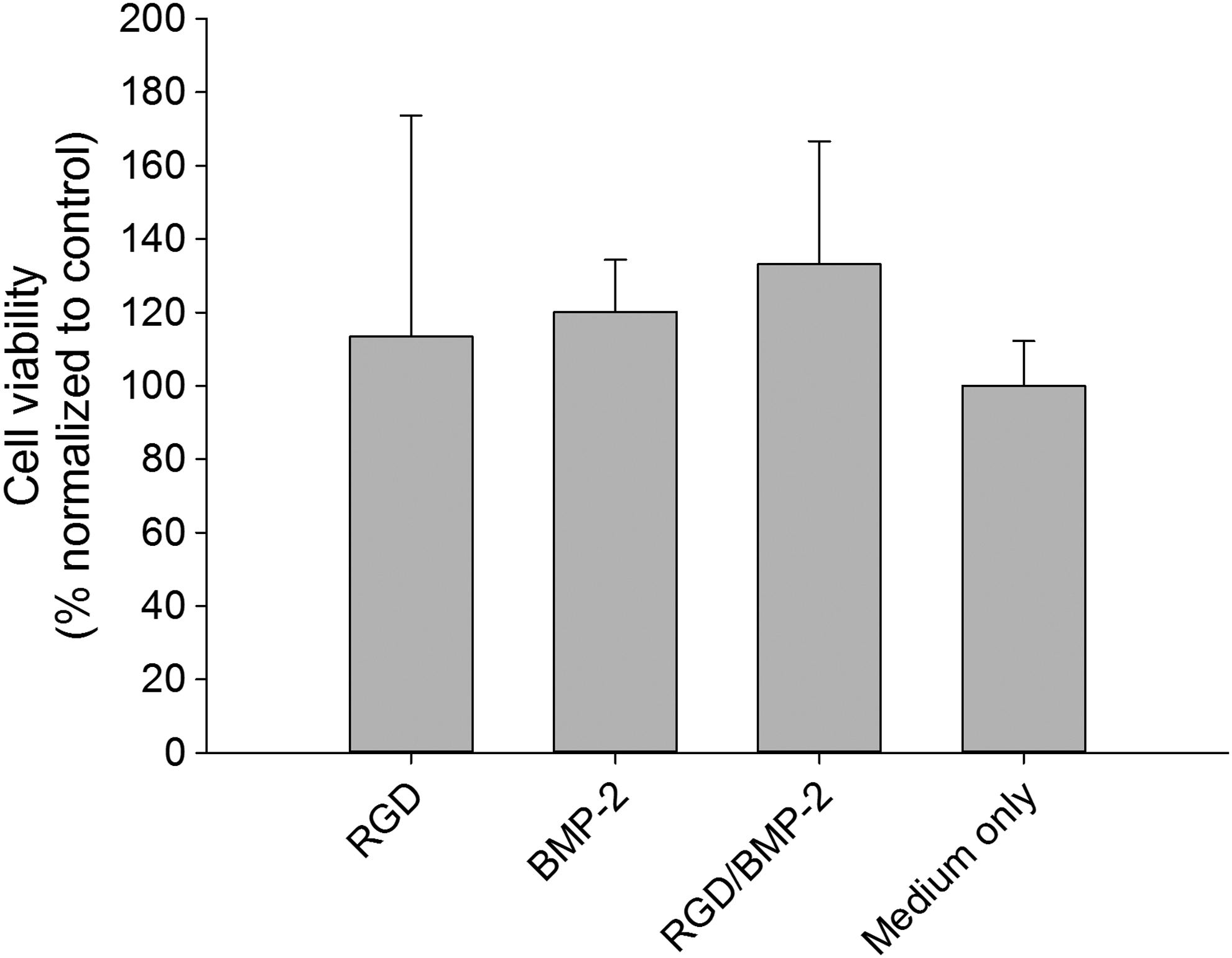
Graph showing hMSC viability results after 3 days of culture in terms of cell number as measured using the calcein AM assay for different ELR supplements in medium at 10 mg/mL: ELR-E-RGD (represented as RGD), ELR-E-BMP-2 (BMP-2), the mixture of both [98% (w/w) ELR-E-RGD and 2% (w/w) ELR-E-BMP-2, RGD/BMP-2], and supplement-free medium (medium only). No significant differences (p > 0.05) were found in any case. hMSCs, human mesenchymal stem cells.
Furthermore, the evaluation of cell adhesion in ELR-coated tissue culture plates showed good results in the case of ELR-E-RGD and the mixture of both. This finding was in agreement with our expectations since the mixture contains 98% ELR-E-RGD. However, coating only with ELR-E-BMP-2 led to statistically significantly lower levels of attachment due to the lack of cell adhesion domains in the recombinamer (Fig. 5).

Graph showing the number of hMSCs attached to the ELR-coated well plates as measured using the calcein AM assay for different ELR coatings absorbed at 5 mg/mL: ELR-E-RGD (represented as RGD), ELR-E-BMP-2 (BMP-2), the mixture of both [98% (w/w) ELR-E-RGD and 2% (w/w) ELR-E-BMP-2, RGD/BMP-2], and non-coated TCP (n = 4). *p < 0.05, **p < 0.01. TCP, tissue culture plates.
Biochemical and clinical results
The welfare of the animals in the first 2 days postimplantation was slightly affected, with disrupted walking, as expected. After 7 days, treated animals behaved similarly to their nonoperated control counterparts. The temperature values, food intake, and all the biochemical parameters measured were similar between animals from control groups at every time point studied (n.s.d., p > 0.05).
MSCT studies
MSCT studies showed bone healing in the defect area. A closer examination of the distal metaepiphysis region, in the medial cortical plane, showed total closure of the defect in most of the coronal and sagittal slices for all of the samples analyzed. However, it was possible to identify the persistence of a small defect with a diameter of 1 mm in the medial cortical plane of the lesion site in one of the femora extracted, although only in one coronal slice and two axial ones (Fig. 6a, b, white arrow). These results were confirmed by radiological studies (Supplementary Fig. S7).
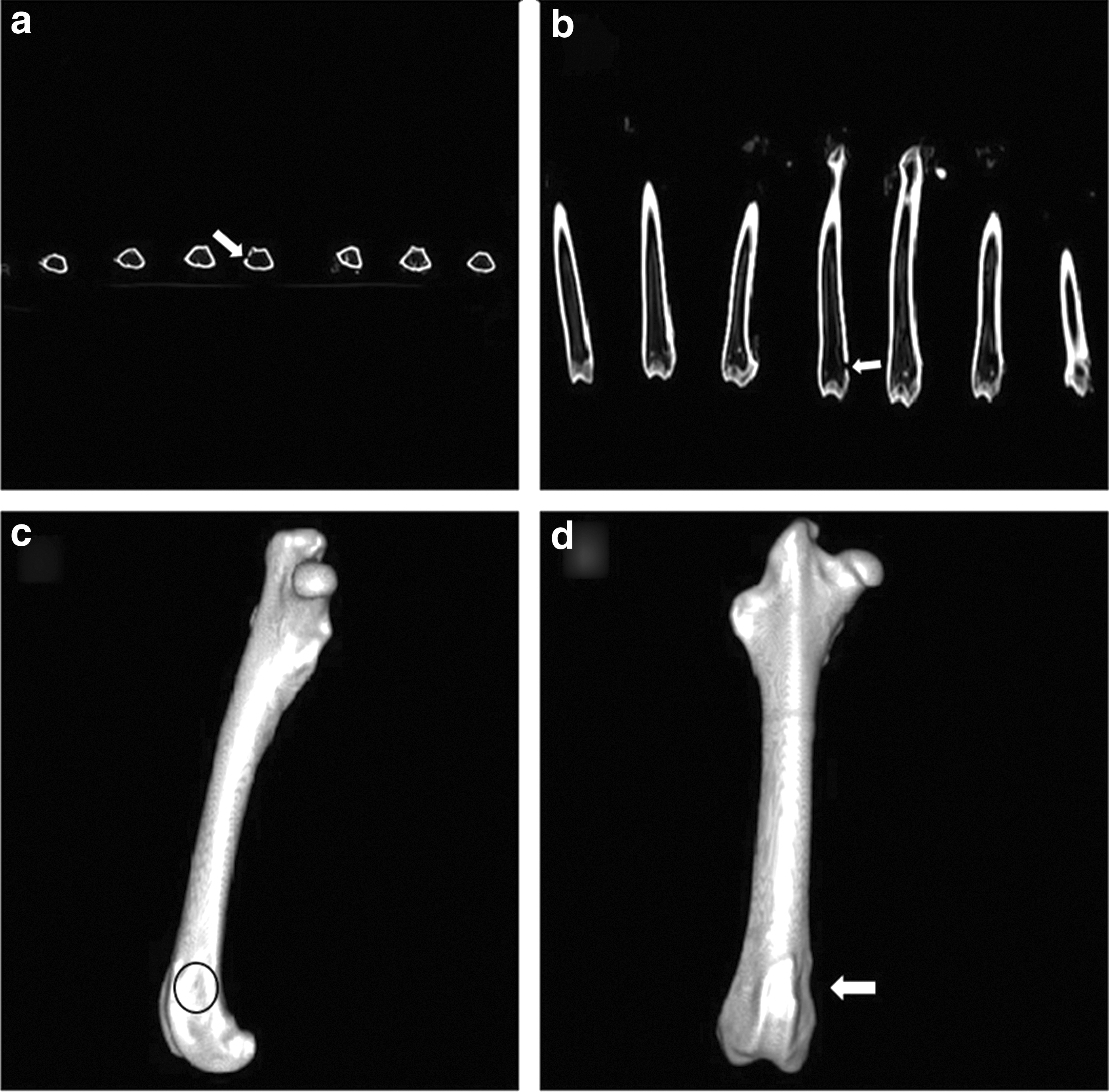
Bone restitution in the distal femoral metaphysis was also observed in the 3D reconstructions of all samples, and the created defect could not be detected (injury site indicated with a black circle; Fig. 6c), even in the case of the sample that showed a small defect remaining in the axial and coronal slices. This 3D reconstruction showed a tiny hollow (white arrow), but the processed signal correlates to cortical bone (Fig. 6d).
Histopathology results
The histological analytical results obtained for experimental samples showed de novo bone formation in the experimental femoral injury (EFI) region. The new bone formed was thick and comprised lamellar bone. In addition, it showed numerous vascular channels of different calibers and was surrounded by various osteoblast layers (Fig. 7a). Each bone layer was deposited on the remaining ELR hydrogel in a disorganized manner (Fig. 7b), resembling pagetoid-like bone (Fig. 7c), in which cellular activity produces a mosaic pattern rather than the normal linear lamellar pattern.
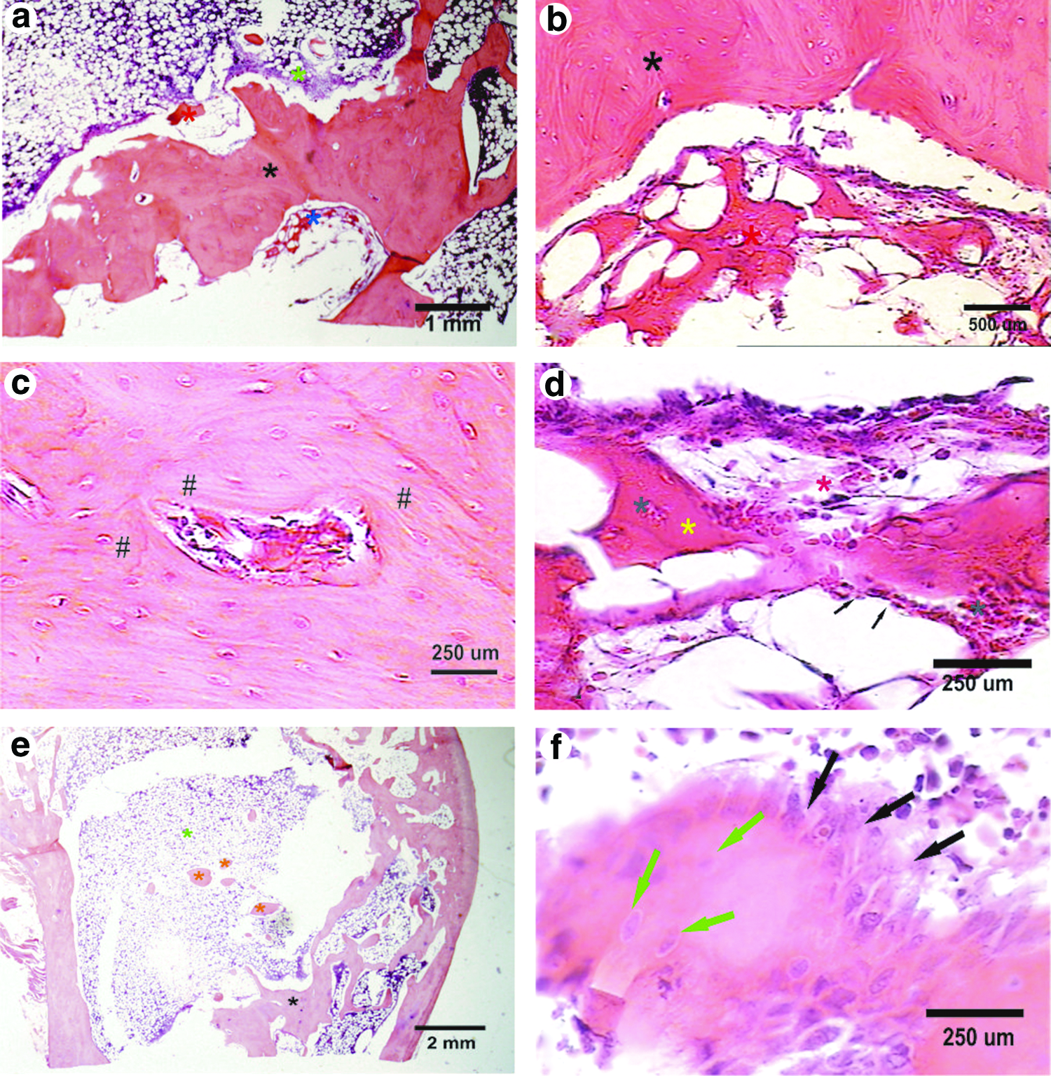
Microphotographs taken from decalcified femoral bone sections stained with hematoxylin and eosin.
Remnants of nonbiodegraded ELR hydrogels were observed inside the EFI center, showing a network or mesh shape and surrounded by microhemorrhages and congestive vessels. Rounded, triangular, rhomboid, or even amorphous structures were found inside the network, showing an eosinophil, granular, and mineralized pattern. These mineralized structures were surrounded by osteoblast-like cells, with osteocyte-like cells being found inside them, and angiogenesis could also be observed (Fig. 7d).
Hematopoietic bone marrow was observed surrounding the newly formed bone in the EFI, with scattered, rounded, nodule-like trabecular bone (Fig. 7e). This new trabecular bone was covered by two, three, or even more layers of prominent osteoblasts. Osteocyte cells were observed in the inner region (Fig. 7f). Furthermore, several congestive vessels were observed close to each trabecula.
Discussion
To address the main aim of this work, namely the regeneration of an FBI in 3-year-old female New Zealand white rabbits, two different bioactive ELRs have been developed and characterized to meet the requirements of novel biomaterials commonly used for that purpose. These novel ELRs were specifically designed to be osteoinductive, by fusing BMP-2 to one of them, and osteoconductive, by fusing RGD domains that promote cell adhesion, thus allowing surrounding cells to interact with the hydrogel and possibly promote bone formation even from inside the scaffold.
Initially, it was shown that the Tt is lower than body temperature, which may permit the formation of hydrogels once the ELR solution is injected into the body. In addition, this Tt is similar to that described previously for the nonbioactive ELR, which was found to be 13.0°C, 42 although an increase of 2.8°C and 2.3°C was observed for ELR-E-RGD and ELR-E-BMP-2, respectively. This can be explained by the lower hydrophobicity of the ELR molecule when other more hydrophilic peptides or proteins containing charged residues are fused to it.43,44
Regarding the rheological data, although this system is intended to be used for bone regeneration and the storage modulus is very low in comparison with bone tissue, this hydrogel was designed to be able to promote cell invasion and proliferation inside itself, acting as a temporary soft tissue that promotes optimal regeneration in a manner through which the implanted scaffold is substituted by host tissue. As such, although it may not be useful on its own for treating large bone defects, it has been shown to be very suitable in the FBI model used in this work since the hydrogel remains free from significant mechanical stress. 45
Biodegradation of the ELR molecules in solution has been confirmed in vitro, thus showing that this process can also be controlled by varying the quantity of elastase used. Although this fails to imitate in vivo conditions, it sheds light onto the biodegradation kinetics. The use of elastase-sensitive sequences should also allow the slow release of BMP-2 from the ELR molecule to exert its biological effect. Consequently, the ELR-based hydrogel acts as a drug-delivery system. Despite the fact that there are other examples in which rhBMP-2 and ELRs are combined as an encapsulation system, 46 this approach allows a more efficient production and application by taking advantage of recombinant DNA technology.
The excellent cell adhesion found on surfaces coated with ELR-E-RGD was similar to that obtained in other studies using RGD-containing ELRs. 47 As such, this work demonstrates that the inclusion of RGD sequences in the final ELR molecule by genetic-engineering methods promotes cell attachment and therefore provides a more ECM-mimetic environment that is also osteoconductive. ELR-E-BMP-2-coated substrates did not support cell adhesion due to the absence of cell adhesion motifs in the ELR itself and in BMP-2. With regard to cell viability, the lack of differences between the negative control (medium only) and the media supplemented with the recombinamers is in agreement with previous studies in which a cell culture medium was supplemented with ELRs. 48
Regarding the clinical and biochemical results of the implant process, although initially affected by the surgery per se, animal gait recovered rapidly to normal conditions. The lack of change in the biochemical parameters showed that neither the surgical procedure nor the subsequent possible matrix biodegradation had any effect on the animals, thus showing good biocompatibility.
The images obtained in the tomographic study with 3D reconstruction of the samples show promising results since the signal patterns processed in this work are correlated to bone tissue with similar characteristics to the surrounding tissue, with complete closure of the defect being achieved in six out of seven samples. Although a defect ∼1 mm in diameter was still visible in the remaining animal, this was only the case in three tomographic slices and may simply be a consequence of a lack of time for the regeneration process in this particular animal. However, the bone formed had the same characteristics as the other samples, and therefore, it can be concluded that these ELR-based matrices have a high osteogenic potential to restitute a bone defect of 6 mm diameter and 6 mm depth ad integrum in 90 days, most probably due to fusion of the BMP-2 protein to the ELR, which results in a BMP-2-loaded hydrogel.
The histological analyses showed that the FBI was replaced by dense, new lamellar bone. Although a few remnants of the ELR were observed at 90 days postimplantation, they were surrounded by congestive vessels and dense laminar bone. This supports the accepted knowledge through which new bone is only formed in the presence of blood irrigation. 49 This new bone is arranged randomly, with an irregular arrangement in various different directions, thus suggesting that the ELR-based hydrogels act as a carrier for BMP-2, with osteoprogenitor cells colonizing these hydrogels, depositing osteoid matrix, and mineralizing as pagetoid-like bone, probably driven by the network arrangement of the ELR-based hydrogels. 50 The new trabeculae obtained show a peculiar shape, as if they were obtained by the confluence of rounded isolated bone formations. The numerous layers of prominent osteoblasts and various shapes observed, which appear to simulate pseudostratification, could be a result of the activity of BMP-2.22,51,52
As observed in vivo from the microscopic results, the ELR-based hydrogel was found to be biodegraded as bone formation occurred since the cells involved in this phenomenon were stimulated by the BMP-2 released into the microenvironment, probably slowly enough to allow the differentiation of stem and progenitor cells. As such, in this situation, elastase (matrix metalloproteinase-12, MMP-12) secretion by osteoclasts might be increased as a consequence of matrix remodeling due to the formation of de novo bone tissue, as suggested before. 53 This could lead to degradation of the ELR-based hydrogel, which is sensitive to MMP-12 as a result of inclusion of the Val-Pro-Val-Ala-Pro-Gly (VPVAPG) sequence, as described previously. 54 On the contrary, ELRs without cleavable domains are not supposed to be biodegraded. In this regard, Sallach et al. reported a long-term stability (up to 1 year) of a physically crosslinked ELR-based hydrogel, similar to the one used in our work, when implanted in vivo. 55 In our case, biodegradation might happen simultaneously with bone regeneration, thus resulting in a resorbable matrix that maintains bone integrity until full regeneration. In addition, the peptides resulting from the degradation of VPVAPG have been reported to exhibit a strong cell proliferation activity that may promote tissue repair, as described previously. 56 Furthermore, RGD sequences provide anchoring points for cells that help them to migrate and proliferate inside the scaffold, thereby promoting self-regeneration of the damaged tissue.
Although several approaches have been developed in the field of tissue engineering, to the best of our knowledge this is the first work describing the use of ELR-based hydrogels for the successful regeneration of a bone defect in vivo. Previous examples make use of ELRs in combination with other materials,57,58 and most of them have only been tested in vitro, although with promising results. 59 Besides, the ELR-based hydrogel described in this study overcomes different issues regarding the use of biomaterials in bone tissue engineering. For instance, BMP-2 is not only loaded inside the hydrogel, but it is part of it. Hence, there is no need to add this osteogenic factor during the preparation of the scaffold, in contrast to other works, 60 reducing its cost. In addition, this acellular system has shown to be able to promote optimal bone regeneration, while other studies report good outcomes only in the presence of mesenchymal stromal cells.61,62 On the contrary, another acellular scaffold has been described, showing its usefulness in bone regeneration. 63 However, this system is not injectable and thus requires the use of invasive methods for its implantation. Moreover, the adaptation of this scaffold to the shape of the defect depends on the mold used in its development, reducing its versatility.
In conclusion, this work shows that a mixture of the originally designed ELRs is able to self-assemble into an appropriate BMP-2 carrier, namely an injectable and biodegradable hydrogel, which allows the slow release of this osteogenic factor, thereby stimulating progenitor and stem cell differentiation and osteoblast proliferation. Furthermore, the resulting ELR-based hydrogels also demonstrated an osteoconductive behavior since they provide an ECM-like environment as a result of the inclusion of RGD sequences. These two bioactivities (RGD and BMP-2), together with elastase sensitiveness, were easily included in the final ELR molecules in a controlled manner, due to their recombinant nature. Endogenous cells were able to migrate and proliferate into these hydrogels, thereby favoring bone neoformation at the femoral injury, as confirmed by CT, radiography, and histology.
Footnotes
Acknowledgments
The authors are grateful for funding from the European Commission (NMP-2014-646075, HEALTH-F4-2011-278557, PITN-GA-2012-317306, and MSCA-ITN-2014-642687), the MINECO of the Spanish Government (MAT2013-42473-R and MAT2013-41723-R), the Junta de Castilla y León (VA244U13 and VA313U14), and the Centro en Red de Medicina Regenerativa y Terapia Celular de Castilla y León. Dante J. Coletta has been funded by the Consejo Nacional de Investigaciones de Ciencia y Tecnología de la Nación (CONICET, Argentina). They also thank Dr. Pedro Esbrit from the Jiménez Díaz Foundation.
Disclosure Statement
No competing financial interests exist.
References
Supplementary Material
Please find the following supplemental material available below.
For Open Access articles published under a Creative Commons License, all supplemental material carries the same license as the article it is associated with.
For non-Open Access articles published, all supplemental material carries a non-exclusive license, and permission requests for re-use of supplemental material or any part of supplemental material shall be sent directly to the copyright owner as specified in the copyright notice associated with the article.