
Abstract
This review is focused on the use of membranes for the specific application of bone regeneration. The first section focuses on the relevance of membranes in this context and what are the specifications that they should possess to improve the regeneration of bone. Afterward, several techniques to engineer bone membranes by using “bulk”-like methods are discussed, where different parameters to induce bone formation are disclosed in a way to have desirable structural and functional properties. Subsequently, the production of nanostructured membranes using a bottom-up approach is discussed by highlighting the main advances in the field of bone regeneration. Primordial importance is given to the promotion of osteoconductive and osteoinductive capability during the membrane design. Whenever possible, the films prepared using different techniques are compared in terms of handability, bone guiding ability, osteoinductivity, adequate mechanical properties, or biodegradability. A last chapter contemplates membranes only composed by cells, disclosing their potential to regenerate bone.
Introduction
A
In the last 20 years, we have been observing an increasing interest in using membranes for the particular application in bone regeneration, but no attempt has been made to systematize a revision of such recent endeavors.
Critical bone defects created by trauma, infection, tumor resection, and skeletal abnormalities, or cases in which the regenerative process is compromised, including avascular necrosis, atrophic nonunions, and osteoporosis, are beyond the potential of self-healing.7–9 The gold standard strategies to address issues such as critical bone defects involve the use of autologous bone graft, allografts, and materials such as ceramics and metals.7,10–13 All of these therapeutic approaches have limitations such as scarce availability of tissues, donor-site morbidity issues, immunogenicity problems, and lack of integration in the host tissue that limit their application range and their overall performance.7,14,15
Tissue engineering (TE)-based strategies have been proposed to solve the above-referred problems.15,16 Many original works and review articles have highlighted the importance of such 3D devices in bone TE.15,17–22 However, we have been assisted to an increased interest in the use of quasi-2D structures (membranes) in the same field explored in this review.
The use of membranes in bone-related applications could be questioned taking into account that bone is a thick tissue and plays relevant structural functions. First of all, membranes can be used to mimic important particular structures of bone. Bone reconstruction occurs from two very vascularized membranes: the periosteum and endosteum.23,24 While the periosteum completely surrounds the bones except at sites of articulation, the endosteum is the thinner layer that lines internally the surface of the bony tissue. Both the periosteum and the endosteum are capable of producing osteoblasts, which are involved in bone production.25–28 In this case, membranes are used as biomimetic structures to copy anatomical compartments where they actively assist bone healing (Fig. 1II).
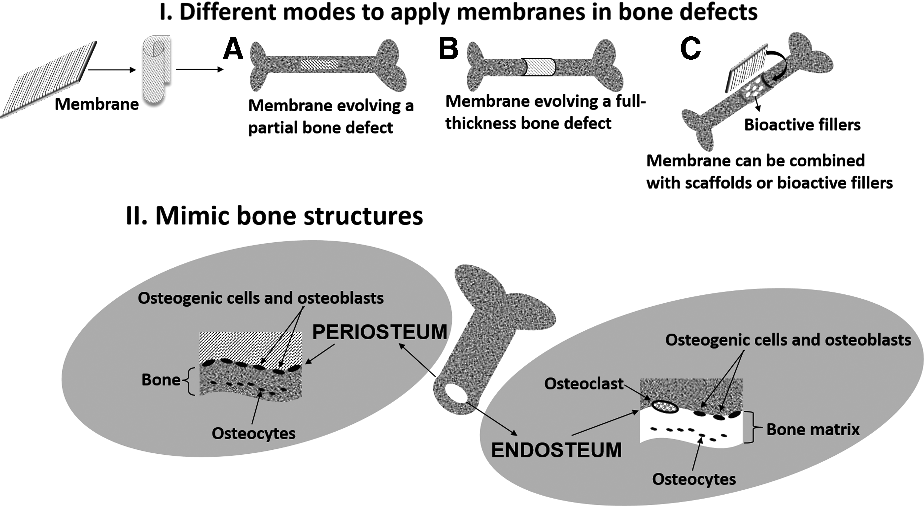
Schematic representation of the roles of membranes in bone regeneration.
Membranes may also have the function to cover bone defects, fully (Fig. 1IB) or partially (Fig. 1IA). In this context, they have been used for the restoration of bone defects, in which they can be used alone or in combination with other materials 29 (Fig. 1IC). These membranes can be used for guided tissue regeneration (GTR)/guided bone regeneration (GBR) and bone reconstruction. 30 GTR consists in the application of a membrane that acts as a physical barrier to protect the defect site and to prevent soft tissue (such as epithelial cells) reaching the injured area. GBR involves the GTR principles and “guides” the bone regeneration process.31–33 For such applications, membranes should be mainly used in sites subjected to limited mechanical loads. However, they have also been suggested for the regeneration of large bone defects, using, for example, the Masquelet technique,8,34–36 where the membrane is presented as a biological model; it was reported that such membranes possessed osteoinductive, osteogenic, and angiogenic properties.34,37
Membranes can be also used in combination with other scaffolds, such as hydrogels. 38 The examples described above clearly indicate that the use of membranes could find broad applications in the field of bone. However, when designing membranes for this purpose, several anatomic and physiological factors should be considered: (1) the time period for complete bone regeneration is not the same throughout the body, and thus, the choice of the membrane type depends largely on the required duration and function of the bone aimed to be reconstituted 39 ; (2) some bones are more vascularized than others—depending on the application envisaged, the membrane should possess adequate permeability, porosity, and perforation size33,40; and (3) the mechanical environments are different in distinct anatomic locations.
In this review, the design principles of membranes to be used in bone regeneration therapies are discussed. Incorporation of bioactive inorganic components, production of graded functionalized structures, surface functionalization, and incorporation of growth factors (GFs) are some of the modifications that will be addressed. Less conventional strategies will be also overviewed, such as the production of nanostratified membranes or the use of membranes composed by cells. The recent advances in their processing and characterization are also reviewed.
General Specifications
Membranes for bone regeneration should possess a series of properties to face the complex tissue environment and guide the formation of new bone (Fig. 2). Such devices should exhibit the following general specifications:
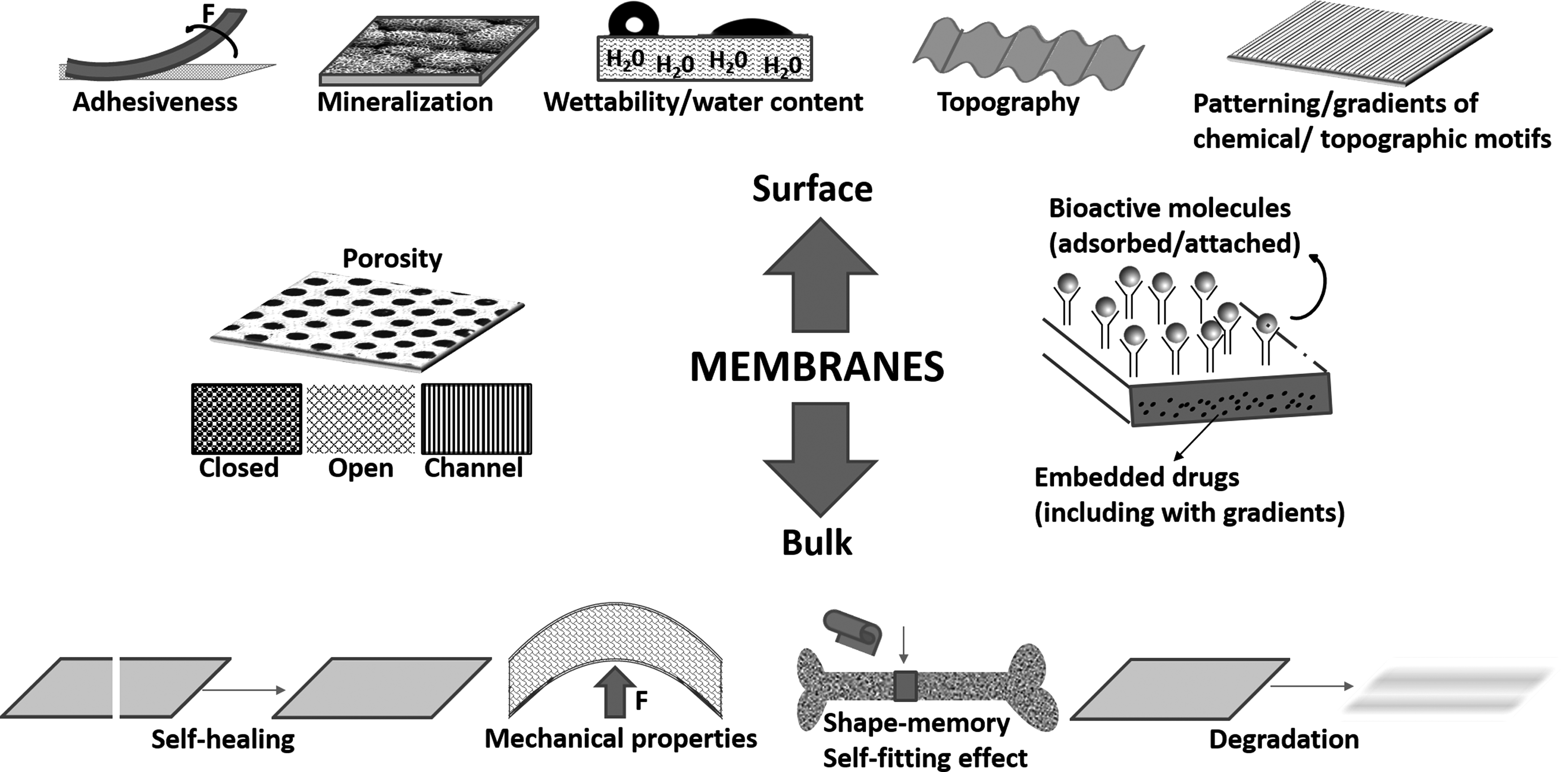
Relevant properties that should be considered in membranes to be used in bone regeneration applications.
(1) be biocompatible and nonimmunogenic: they should be able to prevent and not generate adverse inflammatory or immune response and to resist bacterial invasion and colonization;
(2) be strong enough to support the content or the emptiness of the defect and the exerted forces;
(3) the in vivo performance is also dependent on the interaction of the membrane with the extracellular matrix (ECM) components, enzymes, and cells 41 ;
(4) be easy to shape to fit the defect and regional anatomy;
(5) be easily fixable in situ and not prone to migration;
(6) be ideally bioactive to accelerate bone formation;
(7) be ideally bioactive to induce the bonding of the membrane with the contacting bone—in this case the term bioactivity is related to the ability of the formation of an apatite layer in vivo in the interface between the implant and the bone;
(8) be preferentially bioresorbable with minimal foreign body reaction to avoid a second surgical procedure to remove the membrane. The adsorption rate should be sufficient to maintain the physical barrier capability during the time of new bone formation;
(9) adequate porosity could be required to either enhance accessibility of bone forming cells to the bulk or prevent the invasion of undesired cells from the soft connective tissue;
(10) exhibit adequate permeability characteristics permitting controlled exchange of nutrients and oxygen avoiding necrosis;
(11) present adequate topography and surface chemistry to control cellular attachment, differentiation, and proliferation. The first response of the biological medium when in contact with the membrane is the coating of the biomaterial with a layer of proteins. The composition of this layer is strongly affected by the biomaterial surface chemistry and energy. This initial process is critical for the first steps of cell attachment and posterior cell behavior;
(12) exhibit, in some cases, adhesive properties, to prevent the use of sutures; and
(13) be able to submit to a sterilization process without losing their properties.
Different bioresorbable and nonresorbable materials have been used to produce membranes (Table 1). Nonresorbable membranes (e.g., expanded polytetrafuoroethylene) maintain their structural integrity on implantation and have superior space-maintaining properties and capacity for cell occlusion. However, a second surgical procedure is required for removal of the device. Such surgical procedures can hamper the development of the newly regenerated tissues 42 and the chance of secondary infection increases on exposure of the membrane.43–45 Infections in the treated locals reduce both the insertion gain and tissue regeneration. If infections occur, the deterioration of the membrane can be accelerate thus, a membrane with good occlusion capacity and with antimicrobial protection is necessary. 46 Bioresorbable membranes have been developed to avoid such a limitation once such kind of membranes do not need a second surgical procedure. However, attention must be paid to the following: (1) the degradation of the membrane should not interfere with bone healing (the products of degradation should be nontoxic and able to get metabolized and cleared from the body) and (2) the degradation should not be completed before the osseous regeneration takes place (sometimes, they tend to collapse depending on the size of the defect).47,48 As a main drawback, the bioresorbable ones lack control over the rate of membrane resorption, which is influenced by factors such as the local pH and the biochemical environment.49,50 Bioresorbable membranes are produced from synthetic or natural polymers. The natural ones have been produced using collagen (COL) 50 or chitosan (CHI) 51 among others (Table 1). Many other natural polymers that have been used in other biomedical applications52,53 could be explored in the context of membranes for bone repair. The natural-derived membranes are often characterized by their intrinsic biocompatibility, nonantigenicity, and bioresorption tuned by crosslinking treatment. 54 Synthetic bioresorbable membranes are typically made of aliphatic polyesters such as poly(lactic acid), poly(glycolic acid), poly(ɛ-caprolactone) (PCL), poly(hydroxyl valeric acid), poly(hydroxyl butyric acid), and their copolymers. 39 In general, they have been widely considered for orthopedic applications due to their manageability, processability, tuned biodegradation, and drug-encapsulating ability. 55
Segmental bone defects are related to maxillo-facial or critical mandibular defects.
Such polymeric membranes often possess relatively low stiffness, lack functional groups for cell adhesion, and are absent of osteoconductive/osteoinductive properties. It has been reported that mechanical properties of the membranes can be improved by crosslinking56–59 or by incorporating bioactive inorganic fillers.60,61 The biological performance of the membranes can be enhanced by the incorporation of GFs62–64 and therapeutic drugs 65 or through surface modifications (e.g., chemical/biochemical grafting or plasma treatment).66–70 These and other strategies are overviewed in this review.
Strategies for the Enhancement of Bone Regeneration
Bone membranes should be able to integrate osteoconduction, osteoinduction, and osteogenesis, which are inherently associated with the development of new bone. Thus, when engineering membranes for bone regeneration, key factors should be taken into consideration, such as topographical and biochemical cues as well as surface and bulk properties of the membranes. Due to the quasi-2D nature of membranes, it is easier to integrate such factors into the membrane design than into the design of 3D scaffolds. For instance, as the first events between the biomaterial and the cells take place at the surface, membranes can be a good choice for the inclusion of bioactive molecules. Besides, the modification of topography is easier to achieve in membranes. It is furthermore possible to modify both sides of the membranes independently.
Inorganic fillers/composites
Inorganic bioactive fillers have the ability to bond to bone by the formation of an apatite layer contrary to other materials, which after implantation form a fibrous capsule around the implant leading to clinically unacceptable results such as nonunion or encapsulation of the implants. The reaction mechanism of the apatite layer formation follows five stages that can be found elsewhere.116,117 The advantages, properties, and synthesis methods of these materials are not within the scope of this review but can be found elsewhere.118–124
Several components are being used to produce such inorganic bioactive components. Calcium is the major component of the skeleton and silicon is one of the most essential trace elements in human body playing an active role in mineralization processes in bone formation. Thus, their use to produce the inorganic part is being extensively used. Nowadays, such inorganic components are also doped with different ions 122 to confer different properties to them. For example, the inclusion of magnesium into the composition of inorganic bioactive fillers has been found to enhance the adhesion of bone cells 125 ; zinc has been also included once and is fundamental for bone cell growth, development, and differentiation. Zinc deficiency is associated with skeletal growth retardation and alterations in bone tissue calcification 126 ; strontium (Sr) possesses known therapeutic effects for postmenopausal osteoporosis treatment and had osteoconductive properties. Moreover, it has the dual role of promoting osteoblast differentiation and reduction of osteoclast activity thereby aiding bone formation127–129 ; the incorporation of copper, silver, or bismuth has also been studied to impart antibacterial properties once bacterial colonization or infections are a serious clinical issue, often leading to the failure of implants.130–132
The major issue related with this class of materials is that they lack toughness and elastic properties of the bone tissue and sometimes are difficult to process. One way to circumvent such limitations is the fabrication of composite materials. Polymeric materials are many times combined with such inorganic bioactive components, such as hydroxyapatite, β-tricalcium phosphate, bioactive glasses (e.g., Bioglass®), and glass ceramics.133–135 The obtained composites become a suitable option to fulfill the requirements of bioactivity, degradability, adequate biological response, and mechanical performance.60,136–139 Benefits of each class of material can be combined in the production of such composites. The polymer serves as the matrix to support cell growth and provides properties such as processability, biodegradability, biocompatibility, and flexibility. The incorporation of inorganic cargo has the dual aim to improve the mechanical properties and to provide bioactivity to the device. Thus, these materials have the ability to reproduce the structural and biological functions of the damaged hard tissues in a more efficient way.140,141 However, their combination is not always an easy task once the ratio between the polymer and inorganic bioactive component can hamper their efficacy resulting in nonhomogeneous composites with cracks and inadequate mechanical properties, and sometimes, the aggregation/low affinity of the ceramic with the polymeric matrix can result in nonhomogeneous composites.
Recently, studies have reported the use of nano-sized inorganic particles in the production of composites instead of the more conventional micron-sized ones.
142
The use of nanoparticles has proved to have the following advantages:
• A higher specific surface area forms a tighter interface with the polymer matrix in composites, leading to a high performance in both the mechanical properties and processability.
143
• The higher specific surface area allows for a faster release of ions (higher bioreactivity).
144
• The use of nano-sized inorganic particles, mimicking the dimension of mineral crystals present in the natural bone, has been shown to promote significantly an increase in protein absorption and cell activities, such as cell adhesion and proliferation, in comparison with their micro-sized counterparts.122,145 • The use of nanoparticles in the polymeric matrix mimics more closely the structure of natural bone, which contains nanoscale hydroxyapatite crystallites, being responsible for the high strength of bone, combined with the polymeric phase of collagen.
Although such strategy can be applied to any kind of scaffolds, the quasi-2D structures of the membranes allow a better dispersion of the inorganic particles along their thickness. Moreover, as the particles are located close to the surface, the physiological fluids can penetrate faster through the membrane and a faster mineralization can be obtained. The specific use of membrane composites containing both nano- and micron inorganic particles is described along this review.
Use of GFs
Transforming growth factor-β (TGF-β), bone morphogenetic protein (BMP), insulin-like growth factor (IGF-I), fibroblast growth factor-2 (FGF-2), and platelet-derived growth factor (PDGF) are the GFs commonly used in bone regeneration strategies, of which recombinant human BMP-2 and BMP-7 are already being used in clinical practice as osteoinductive agents. 146 Their release results in several benefits such as rapid healing around the implant and more complete remodeling over shorter time periods. Although their use seems promising, when introduced into the body, they are rapidly eliminated due to their short half-life. Because of that, in clinical practice, supraphysiological and expensive dosages in the tens of milligram range are being administered to obtain bone healing. However, their uncontrolled release leading to high initial concentrations followed by periods of rapid clearance limits possible therapeutic effects 147 and their administration in excess may lead to undesirable incidences. It is crucial that, in a tissue repair situation, the target cell population must be exposed to them throughout the whole healing process or at least for an extended period. Furthermore, their amount and kinetic release are essential to the biological response. Also, it is very important that the biomaterial carriers have affinity with the GFs to sustain their bioactivity in which they preserve their adequate conformational structure. Thus, adequate vehicles are required to obtain suitable controlled release systems and to present them in precise spatiotemporal patterns.
New bone formation should also be accompanied with the existence of a vasculature able to transport nutrients essential for cellular tissue development. In this perspective, angiogenesis is crucial for bone regeneration once the growth process and restructuring of bone tissue are greatly enhanced. 148 Angiogenesis can be stimulated through the inclusion of specific GFs, such as vascular endothelial growth factor, VEGF, and FGF.41,149 The combination of both osteo- and angiogenic GFs can have a synergistic effect to enhance bone regeneration.150,151 However, the timing and ratio of their application should be carefully administered to have desired effects. 152 In vivo introduction of more than one GF would initiate the different cellular cascades necessary for robust implant integration. Current research usually evaluates one or a combination of two GFs, which does not reflect the complex physiological process of bone formation. 29 Instead, the local controlled delivery of a broader selection of GFs (such as the content in platelet-rich plasma [PRP]) should be considered. Moreover, the sterilization of GFs is also a hot topic once it is fundamental that the GFs do not lose their bioactivity. Research is ongoing to develop novel membranes with adequate GF delivery capability to accelerate bone regeneration.38,153,154 Membranes incorporating molecules such as GFs should diffuse their content faster more than bulky 3D systems due to the high surface area. Release kinetics in membrane can be controlled by an adequate choice of the biomaterial. In particular, its degradation will have an active role in the release profile of the immobilized agents. In addition, due to the membrane geometry, GFs will be more accessible to cells once they are localized and maintained in the cell/biomaterial interface inducing higher intracellular signaling. More examples that combine membrane processing and inclusion of GFs are presented along this review.
Functionalizing surfaces with bioactive elements
On implantation, it is on the surface of a biomaterial that the first events take place, such as material cross talk with proteins, cells, and surrounding host tissues. Plasma modification 66 and blending155–157 are some of the approaches that are used to tailor surface functionality and proved to be effective in improving cellular behavior. It is well known that the ECM affords a number of biochemical (peptides, GFs, cytokines, etc.) and physical (stiffness, topography, etc.) cues to cells that will dictate their fate.
Peptides and other bioactive elements
Engineering surfaces by grafting bioactive macromolecules and peptides is another strategy to obtain bioinstructive biomaterials, 70 including membranes for bone repair. Such bioactive elements on the surface of the membrane accelerate ECM secretion and tissue regeneration158,159 once they support cell adhesion, proliferation, and osteoblastic differentiation.160–162 Peptides are signaling molecules that are crucial to regulate cell phenotype and contribute toward the regeneration of tissues, namely bone. Their incorporation can be done by simple adsorption, covalent attachment, or by enzymatic incorporation. However, care must be done to avoid desorption or undesired and time-dependent conformational changes, leading to loss of bioactivity. Several parameters such as peptide concentration, spacer arms, peptide type, immobilization technique, and spatial distribution of peptides should be taken in account when incorporating them into biomaterials to obtain an adequate cellular response.163–167 Small peptide fragments are being used to mimic specific functions of the ECM.165,168,169 They can be even designed to target an interaction with a specific cell population. 170 One of the limitations associated with short peptides is their lower biological activity, in comparison with complete protein found in the native environment, due to the partial absence of complementary domains. 171 The use of the entire molecule may provide important secondary actions regulating cell binding, signaling, cell proliferation, and differentiation. Larger molecule fragments can be produced that will not only increase the bioactivity but will also reduce the antigenicity constituting advantages over the whole molecule. 172
The most well-known peptides for bone regeneration are the proline-rich peptides and the RGD sequences.165,171,173 The proline-rich peptides are responsible for the deposition and growth of apatites into endoskeletal mineralized tissues. 174 During the formation of biological minerals such proteins may function as mineral-binding domains, protein/protein interaction domains, or internal molecular spacers. Several artificial peptides based on the common characteristics of the proline-rich region in hard tissue ECM proteins are being designed for the induction of bone formation and biomineralization.175–177
Although the real application of such materials is still limited, it seems that these peptides might be useful as bioactive agents for osteoblastic differentiation and as surface-modification ingredients in bone regenerative therapies. More examples about grafting of bioactive molecules will be given later.
Another very interesting approach for bone regeneration is the use of antibodies once they are able to act only in target cells due their high antigen specificity and affinity. 178 Thus, membranes with immobilized antibodies will guide cellular mobilization and function. Such an approach could be a very interesting strategy once it is possible to recruit both osteogenic and endothelial cells necessary for bone regeneration. In this context, it is also desirable to control spatially the exposure of proteins and antibodies over the surface, which can be done using adequate chemical routes. 179
Effect of topography
Another way to functionalize membranes' surface is to optimize surface micro- and nanotopography.180,181 Roughness and microtexture may have a strong influence on the enhancement of bone formation at the cellular and molecular level.182,183 Several techniques such as e-beam lithography, lithography, dip-pen nanolithography, soft lithography, and microfluidics, among others, have been applied to confer motifs at the material surface. The characteristics of these techniques are not within the scope of this review but they can be found elsewhere. 70 The creation of such motifs can be important on the recreation of important bone structures. In the specific case of the periosteum, it is known that its topographic surface consists of longitudinally oriented cells and collagen fibers. Such approach is much easier to perform in membranes than in 3D scaffolds due to their geometry. Examples that make use of this strategy are reported along this review.
Functionally graded and multilayered membranes
Mimicking structural characteristics of native tissue can be achieved by the fabrication of functionally graded membranes (FGMs). FGMs are materials that present gradual transition of their components (e.g., microstructure and/or composition) along their structure conferring to them different regional features and properties. 184 The research on FGMs is encouraged by the need for properties that are unavailable in any single material and the need for graded properties to offset adverse effects of discontinuities for layered materials. FGMs can be a good strategy to develop implantable nonhomogeneous devices able to promote bone regeneration. It is known that the microstructure of the bone tissue is characterized by the presence of spatial differences in the orientations as well as the concentrations of its mineral and organic constituents. 185 We can learn from the structural organization of bone to tailor and design membranes with different properties that will retain the structural, dimensional, and mechanical integrity as occurring in bone. Moreover, combined with spatially and temporally controlled delivery of exogenous GFs, the FGM approach may provide an effective scheme to engineer devices promoting the regeneration of tissues and organs, such as bone.5,30,166 The development of such nonhomogeneous structures along the thickness could also be interesting for the regeneration of bone interfaces. For instance, FGMs could be attractive for osteochondral TE as gradient-based signals can induce both osteogenic regeneration and chondrogenic regeneration in the interfacial area. 186
The development of membranes exhibiting continuous change along the thickness may be technically quite challenging. It is much easier to build multilayered constructs with discrete variations of compositions and properties. The main disadvantage of such an organization is that, in general, such constructs present distinctive interfaces between the layers and may cause delamination between the layers and weaken the structure. 187
Some FGMs are being proposed to promote bone regeneration.188–190 More examples of FGMs and multilayered membranes are given along this review.
Processing Methodologies: “Bulk”-Like Polymeric Membranes
Most of the methodologies for membrane development require the use of aqueous-based or organic solvents. Melt-based techniques are also used on the fabrication of membranes, avoiding the use of solvents. However, such methods require often high temperatures that could lead to the thermal degradation of sensitive biomaterials and molecules. This section explores different processing methods focusing on the development of membranes for bone regeneration.
Electrospinning
Electrospinning (ES) has attracted tremendous interest in the research community as a simple and versatile technique to produce polymeric ultrafine fibers 191 that have diameters within the range of nanometers to microns. This technique consists of the ejection of a polymer solution that is dispensed in a controlled manner by a syringe pump from a spinneret. Due to the application of an electric field, the solution undergoes extensional flow and fibers are formed and deposited over a collector. This happens because when a pendant droplet of the polymer solution in the tip of the spinneret is subjected to an electric field strong enough to overcome its surface tension, a tiny jet is ejected in the direction of the collector. Along the way from the spinneret to the collector, the solvent mostly evaporates leading to the deposition of long and very thin fibers, giving rise to nonwoven nanofibrilar membrane-like structures. 192
In the case of TE, this technique has received tremendous attention once very thin fibers can be produced by this method, closely resembling the structure and size scale of the native ECM.193–196 Due to their very small diameter, polymeric nanofibers exhibit properties such as high-specific surface area, flexibility in surface functionalities, and superior mechanical properties, which may improve the level of protein adsorption and subsequent cellular attachment, morphology, migration, and function.192,197 Ultrathin fibers from synthetic and natural origin polymers were successfully obtained by this technique.198,199 To date, the majority of electrospun fibers obtained are from synthetic polymers once their solubility in volatile solvents is better than for natural polymers. 192 Nevertheless, natural polymers are also being electrospun providing new opportunities and enhanced possibilities for their use in TE applications. Such remarkable properties make ES a suitable technique to process membranes for bone regeneration.200,201 Some reviews portray the use of several polymers in the production of electrospun polymeric biodegradable membranes for bone regeneration,202,203 and other works even reported the use of ES for the reconstruction of bone interfaces.129,204 ES membranes proved to be effective in in vivo experiments by implanting them,205–208 or combinations with differentiated bone cells, 209 into bone defects. In addition, the ES fibers can be also functionalized by encapsulation or attachment of bioactive molecules such as GFs to control the differentiation and proliferation of seeded cells and they can also be hierarchically assembled by manipulating their alignment, stacking, or folding as disclosed later.
Shape memory
Although not always feasible in clinical settings, the use of minimally invasive surgery is highly appreciated for well-known reasons, such as patient compliance and costs. In this context, polymers with shape memory could be a very interesting approach for the production of ES membranes.210–212 This principle lies in the fact that a compacted temporary shape can be implanted into the human body and then, under a certain stimulus, can revert back to its original bulky shape in vivo. Thus, self-fitting that provides structural support for the stabilization of defects can be obtained by using such smart membranes. Some authors even envisaged such kind of ES membranes for the development of screw-like membranes 210 as in bone fracture healing proceedings the removal of hardware is sometimes necessary. The removal of the hardware, generally, leads to localized microtrauma and residual screw holes may act as stress risers, exposing the patient to a risk of refracture. Thus, the development of screw-like membranes must (1) permit osseointegration where the material developed must tightly fit into a bone screw hole—an ill-fitting interface caused by poor contact between the material and surrounding bone tissue increases the risk of bone nonunion; (2) have adequate bioactivity to recruit the cells from the adjacent tissues—thus, the developed membrane must recreate relevant aspects of the natural ECM to allow cell attachment, migration, proliferation, and differentiation; and (3) offer appropriate stress to the surrounding bone tissue—mechanotransduction in bone regeneration is crucial in dynamically modulating and maintaining the balance between osteogenesis and bone resorption. Based on these concepts, the abovementioned authors produced membranes with shape memory capability for the development of a biologically mimicking and mechanically self-actuated nanofibrous screw-like scaffold/implant for in situ bone regeneration. 212
Surface properties
The surface properties are also a pivotal issue when developing fibers by ES, which contribute to the cellular performance of the processed membranes. Thus, the influence of hydrophilicity and roughness should be analyzed and optimized. To enhance the biological performance of ES membranes, some authors make use, for instance, of plasma treatment. This treatment allows changes on the roughness and the hydrophilicity/hydrophobicity of the membrane surface, which will further impact the behavior of cells. 197 This kind of treatment could be very interesting in the case of GBR membranes. In GBR, it is crucial that the membrane has a surface that allows the growth of bone cells but, in contrast, it is crucial that the membrane is able to prohibit soft tissue invasion into the localized bone defect. To achieve this end, one possible strategy is to render the surface of an ES membrane hydrophobic through a hydrophobization process by using plasma treatment. 213
Porosity
Although it is important to prohibit the invasion of undesired cells into the defect, it is also important to improve cell penetration of the ones that we want to colonize into the defect and nutrient diffusion for their survival; such selectivity capability can be achieved by controlling the porosity.214,215 Larger pores may be required for ingrowth of cells to nanofibrous networks. However, making the diameters of the fibers smaller might not be the way to improve cell proliferation on such substrates. 216 ES membranes that possess high specific surface area and porosity might aid attachment and migration of the cells inside the membrane.
In ES, the pores are always interconnected because the fibers are essentially deposited around the pores, 215 contrary to other techniques such as solvent casting/particulate leaching. In solvent casting/particulate leaching, the pores are obtained by washing out a sacrificial phase to form an interconnected pore structure within a continuous polymer phase. However, depending on the relative amounts of the particulate phase to the polymer, the pore structure is not necessarily interconnected.
Although in ES the pores are interconnected, it is difficult to control the distribution and size of pores. To circumvent this issue, an ES membrane with controlled microarchitecture was proposed: ES membranes were prepared and afterward, by using a computer-assisted design approach, they were subjected to a laser ablation process. 214
Another approach to obtain porous membranes with controllable pore size is the use of blends in which one of the polymers [e.g., poly(ethylene oxide)] can be used as the porogen agent. As expected, as the percentage of the porogen increases, more pores are formed on the membrane after leaching. One aspect that could be very interesting is that, instead of leaching it out with water, to use, for example, simulated body fluid (SBF) solution. This SBF solution would enhance synergistically both pore formation and mineral deposition once it allowed the simultaneous removal of the porogen agent and deposition of calcium phosphate. Like this, the pore structure formed can enhance the anchoring points for cells and the mineral deposits can promote osteoconductivity and induce direct bonding with native bone, which consequently affects cell proliferation and differentiation. 217
Blends and composite membranes
The majority of the ES membranes produced for bone regeneration are composed of polymer blends and/or composites. The most difficult when designing membranes by ES is the adjustment of the adequate parameters to facilitate the ES process and obtain structures and ES membranes with the intended properties.218,219 Thus, depending on the materials and solvents used for the conception of the membrane, several experimental ES parameters should be optimized, such as applied voltage, tip-to-collector distance, and flow rate.60,200,218,220 In addition, the adequate concentration of polymer/blends and/or inorganic bioactive part should be found to achieve better cellular adhesion, growth, and mineral formation suitable for the regeneration of bone. 200 Several strategies are being used to confer bioactivity on an ES system. The most common one is the use of traditional inorganic bioactive components. There are several ways to introduce the inorganic bioactive part: they can be mixed with the polymeric solution,200,220–222 or the bioactive inorganic suspension and the polymeric solution can be prepared separately, loaded into different syringes, and electrospun on the same region. 223 Other works reported the production of inorganic nanofibers that are further mixed with the polymeric solution where the membrane is obtained by subsequent solvent evaporation.224–226 Recently, ES composite membranes were obtained by combining the ES technique in which a protein was electrospun and simultaneously the inorganic bioactive component was added by electrospraying. 227
Conductive polymers can be used to engineer ES composite membranes for bone regeneration once it was hypothesized a mechanism in which the piezoelectric signals can regulate bone growth. With this purpose, an electrically conductive and composite ES membrane was prepared. 228 For that, a polymer blend composed by a nonconductive polymer and a conductive one was prepared by ES and the inorganic bioactive component was added by electrodeposition. 228
One of the major issues in the fabrication of blends and/or composite membranes by ES is the presence of nonuniform fibers. New approaches are being developed in this sense. For instance, the use of surfactants showed to be a good choice for a homogeneous dispersion of the inorganic bioactive component within the polymeric matrix.60,229
Another strategy to produce an ES composite membrane is the emulsion ES, also called as “Green Electrospinning.” In that case, the oil-in-water emulsion of both polymer and inorganic bioactive part is formed in the presence of a template polymer [e.g., poly(vinyl alcohol)] where the emulsions are made using minimal quantity of an organic solvent and are then electrospun. 230
Another very interesting approach to produce bone-like composite membranes lies in the fact that (1) substantial changes in both biochemical and biomechanical properties take place during the early development of vertebrate bone, and (2) the increase in numbers of intra- and interfibrillar crosslinking of collagen network is followed by mineralization during the maturation, thus achieving maximum stiffness and toughness. To mimic this process, a versatile and easy-to-perform method was proposed. 231 It consists of the preparation of an ES membrane composed by collagen (COL) doped with catecholamines and calcium chloride (CaCl2), where the presence of divalent cations during ES triggered oxidative polymerization of catecholamines, thus promoting the crosslinking of COL in situ. The membranes were then dried to remove any residual solvent and subjected to subsequent exposure to ammonium carbonate, (NH4)2CO3. The mineralization of the membrane was promoted by exposure to (NH4)2CO3 that leads to complete oxidative polymerization of catecholamines and in situ precipitation of CaCO3. 231
To mimic the hierarchical structure and biological functions of ECM, another strategy to prepare composite membranes was proposed. 232 For that, CaCO3/casein microspheres were encapsulated in PCL using a cosolvent-based ES method. The casein that is a proline-rich phosphoprotein was used to prepare and to stabilize the crystallization of the microspheres; and as the PCL is hydrophobic and lacks functional groups for cell adhesion, the membranes were first treated with NaOH solution to partially hydrolyze the polymer and then gelatin was grafted. 232
The use of blends can also impart bioactivity. 233 It was reported that blends were produced by a free liquid surface ES method and the produced membrane was afterward submitted to a post-treatment with CaCl2. The chelating ability of one of the polymers coordinates bonding between COO− and Ca2+, eliciting a homogeneous Ca/P crystal nucleation that is a crucial osteogenic property an ideal membrane should possess for bone regeneration. 233 An uncontrolled mineralization could be detrimental when the mineralization is too low, thus not providing adequate bioactivity to the system or when the mineralization is too high. In that case, mineralization could reduce the pore size and therefore may prevent cell penetration within the membrane. 234 Moreover, ES membranes can be engineered with microbial protection. This can be especially useful in GTR/GBR to prevent infection. To achieve this end, anti-infective drugs can be incorporated in the membrane 235 or the inorganic component can be doped with specific ions, such as bismuth, to promote microbial protection. 132
Although different types of composite membranes can be engineered through ES, the abovementioned examples reported that the produced membranes were bioactive, with improved mechanical properties, and were able to enhance cell adhesion, proliferation, and differentiation of different types of bone cells thus demonstrating potential to be used in bone TE. In addition, some in vivo experiments demonstrated that ES composite membranes possessed good biocompatibility and bone-forming ability with the ES membranes only236–238 or with ES membranes precultured with bone cells.239,240 It was also reported that ES composite membranes promote bone regeneration by supporting adhesion, spreading, and proliferation through upregulating integrin-mediated signaling cascades that control cytoskeletal organization and through activating BMP/Smad signaling pathways of osteogenic cells both in vitro and in vivo. 241 As described in vitro, when implanted, such membranes promoted osteogenesis. In addition, when the produced membranes precultured with bone cells were also implanted, an even higher osteogenic behavior was observed because the transplanted cells may directly differentiate into osteocytes and they may modify the healing environment by secreting a number of trophic molecules such as soluble ECM glycoproteins, cytokines, and GFs. 241
GFs and peptides in ES membranes
ES membranes combined with GFs have emerged as a new alternative treatment in bone repair and regeneration. Care must be taken to maintain the GF integrity and its natural conformations after undergoing the process of ES if we intend to incorporate the GF at the same time that the biomaterial is processed. Some studies reported the production of ES membranes where the GF was just mixed in the polymeric solution; and their efficacy was proved both in vitro and in vivo.63,242,243
The use of organic solvents for the preparation of membranes through ES constitutes a major issue on the design of membrane with GFs. In particular, GFs can lose their activity when in contact with organic solvents. One way to circumvent this issue is the preparation of membranes by coaxial ES. In coaxial ES, two concentrically aligned capillaries are used to enforce the formation of the fibers with a core-shell structure, allowing different materials to diffuse out sequentially. Such method has the dual role to protect the GF from direct exposure to organic solvents and control its release. 244
Additional positive effects could be obtained by combining composites with GFs. 245 It is crucial that the developed system is able to allow sustained release of GF once the amount of inorganic phase can influence the release profile due to its hydrophilic nature.
The presence of both osteoconductive and osteoinductive factors could enhance the potential of the membranes as a better environment for osteogenic cells can be provided by the osteoconductive property, and the recruitment of stem cells and osteoprogenitor cells to the defect site can be provided by the osteoinductive property thus facilitating osteogenesis. In this context, even more interesting properties could be attained when performing membranes by coaxial ES. 246 It was reported that the shell provided stiffness and bioactivity and acted as a transport barrier to the GF while the core conferred the osteoinductive properties to the construct. Another important characteristic is that by increasing the thickness of the shell, different release profiles could be obtained. 246
The combination of GFs offers advantages over the use of only one GF. 247 In this sense, it was reported that ES composite membranes possessed significant improvement of properties in terms of biocompatibility, mechanical strength, cell viability, and proliferation. At the same time, the incorporation of dual GFs on the composite membrane promoted a better behavior at supporting cell adhesion, proliferation, ECM formation, and bone-associated gene expression. 247
Instead of incorporating the GF inside the biomaterial, it is also possible to coat it at the surface of the membrane just by immersing the membrane in a GF solution.63,248 Although presenting different release profiles, both strategies proved to be effective in bone regeneration where neovascularization was also observed. 62
Membranes made from the self-assembly of peptides were also prepared by ES showing ability to guide cells through regenerative processes. 249 Moreover, the use of peptides associated with other features such as fiber alignment can be used to direct cellular behavior on nanofiber mesh membranes, enhancing the migration, proliferation, and osteogenic differentiation. 250
Other bioactive cues, such as cytokines, can also be loaded via physical adsorption on ES blend membranes, where positive effects in bone formation in vivo were observed. 251
FGM or layered ES membrane
ES has been used as well in the production of FGMs or layered membranes to tune/mimic specific properties and structural features of bone. For instance, a sequential layer-by-layer (LbL) ES method was adopted in which one layer was composed only of polymer and the other layer was composed of polymer, inorganic component doped with Sr and PRP providing a combination of osteoconduction and induction. 129 Such approach protects and sequestrates the GF within the membrane, where different release profiles could be obtained and could better mimic the in vivo environment. Its use facilitates osteogenic differentiation and aids in bone formation. This approach could also improve the mechanical properties of the produced membrane due to in situ stacking. 129
Bone matrix is intimately associated with different types of soft connective tissues, such as muscles and nerves. Thus, a membrane exhibiting composition gradients could help guiding tissue regeneration so that it proceeds more effectively. A good example for that is the periondontal membrane used in dental surgery. Such membrane is placed in the defect region between the soft tissue (periodontium) and the hard tissue (alveolar bone). Initially, it acts as a defect coverage by preserving the stability of autologous or synthetic bone grafts and then guides specific cell migration and promotes the regenerative potential of the bone tissue. Other examples in which FGMs or layered membranes could have an important role include the interface of tissues such as bone/cartilage, bone/ligament, and bone/tendon 252 due to the gradual transition of both physical and chemical properties. To date, the majority of the ES FGMs developed for bone regeneration include the use of at least two different layers, in which one is constituted by a composite and the other one is composed only by the polymer component.143,253,254 The composite segment promotes the attachment and proliferation of bone cells, conferring good osteoconductivity to the membranes, and the polymer segment offers mechanical stability to the construct. However, more complex structures interfacing with several tissues such as bone or epithelial tissues can be produced in which anti-infective drugs can even be incorporated. 255
Fabrication developments have combined ES with other techniques such as electrospraying, bioprinting, particulate leaching method, and solvent casting.256–259 In this context, it is possible to engineer multilayered membranes depending on the tissue or interface that is intended to recreate where it is possible to conjugate blends, composites, and even GFs. For example, layered membranes were designed by combining solvent casting and particulate leaching for the production of a porous composite membrane. 259
As discussed above, the design of membranes that are able to regenerate tissue interfaces, such as the bone-to-ligament one, is primordial and very challenging. To mimic such structural environment, a membrane with a linear gradient of materials and mechanical and structural properties was fabricated by combining ES and bioprinting. 258 A membrane with three different regions was obtained in which each zone presented different porosities and also possessed a gradient of physical and mechanical properties that elicited different biological responses. Through the combination of different biomaterials at different scales, the construct better mimicked the structural biological environment of bone-to-ligament interface. 258
Remarks
Although ES shows possibilities for the creation of biologically active membranes, some drawbacks are also associated with this technology. The porosity of the meshes is difficult to control, and typically, pore sizes prevent the infiltration and colonization of cells into the structure.193,216,260,261 Consequently, this can limit tissue ingrowth and vascularization in vivo. However, this could be used as an advantage in membranes for periodontal regeneration, as they could prevent the invasion of cells from the connective soft tissue.
In the last years, modifications to the conventional ES technique have been proposed. One such method is called “cell electrospinning,” where the polymeric material and living cells are directly electrospun. Basically, a coaxial ES is used in which the cell suspension flows through the inner needle and the polymeric solution flows through the outer layer. Several types of cells have been found to be viable and no cellular damage was observed during the fabrication of such structures.262,263
More recently, efforts have been made to produce ES membranes in a highly controlled manner to comply with good manufacturing practices (GMP) for the production of advanced healthcare materials for regenerative medicine, and to test cellular behavior on these. 264 Such strategy constitutes a breakthrough in this field once it is important to produce membranes under GMP if the intent is to commercialize the final product. 264
Solvent casting
In this method, the polymer is dissolved into a suitable volatile solvent and the resulting solution is casted into an appropriate mold, such as a Petri dish. Subsequently, the solvent evaporates giving rise to a membrane exhibiting the shape of the mold.265,266 If desirable, a leachable porogen (e.g., sodium chloride or sugar crystals), usually in the form of particles with controlled sizes, can be combined with the polymer solution. In this case, subsequent to solvent evaporation, these crystals are dissolved in water, producing a porous footprint in the polymer structure. 267 The pore size and porosity level of the membrane scaffold can be tailored by adjusting the particle size and relative content. Moreover, composites can be created by adding to the polymer solution inorganic particles. Solvent casting is an easy technique that does not require specialized equipment. The limitations of this technique are as follows: (1) typically just flat sheets are obtained; (2) common use of organic solvents that can be retained within the polymer affecting the quality and viability of the materials—if not completely removed this may negatively affect cell and tissue response on implantation 268 ; (3) the use of organic solvents can also lead to the denaturation of proteins and other molecules incorporated into the polymer, thus decreasing the activity of these bioactive molecules; and (4) for low volatile solvents, the preparation of the membranes may be time-consuming.
Membranes have been prepared by solvent casting to be used specifically in bone regeneration.269–273 Some particular aspects related to such devices prepared using this technology will be particularized.
It was demonstrated that stiffness influences cell behavior, 274 and thus, optimization of mechanical properties of membranes is essential. Crosslinking is probably the most used strategy to control the mechanical, permeability, and other bulk properties in polymeric membranes. 58 For instance, the dynamic mechanical properties of chitosan membranes were measured in physiological-simulated conditions. 58 With increasing crosslinking degree, less swelling could be observed, together with an increase in the elastic modulus. For uncrosslinked membranes, the storage modulus, E′, presented a reduction of a factor of 50 when the membranes were hydrated. 275 It has been also reported that the swelling process takes place in few minutes during which the glass transition of the biopolymer occurs. Such glass transition process was investigated in membranes immersed in mixtures of water/ethanol with different compositions by using dynamic mechanical analysis (DMA). 276 Such kind of studies strengthened the importance of performing mechanical tests in biomaterials with water uptake capability under physiological-like conditions. Moreover, such glass transition also proved to influence the transport properties of small molecules. 276 It is crucial that membranes possess permeation properties that could be useful for the controlled delivery of bioactive agents. 277 Such findings demonstrate that the successful development of polymeric drug delivery systems and biomedical devices requires a comprehensive understanding of the mechanical/viscoelastic properties of polymers. 278
Membranes should possess an adequate porosity in which they allow the diffusion of nutrients and oxygen, but, at the same time, they should have adequate mechanical properties and degradation profiles. Moreover, in bone regeneration, the membranes could be designed in a way that they possess closed smooth face on one side to prevent inner migration of conjunctive and epithelial cells, and on the other side the membrane presents open porosity allowing ingrowth of osseous neotissue. In addition, when conceiving membranes for bone regeneration, the introduction of drugs could be very beneficial to prevent infection. As aforementioned, the porosity of membranes produced by solvent casting is generated by the introduction of porogen agents. Thus, it is vital that they do not provoke deleterious alterations of the drug. 271
The ability of membranes to induce the formation of an apatite layer in the interface to bone enhances their integration in vivo. 49 The simplest strategy to obtain bioactive membranes is to include bioactive elements in the polymeric matrix.124,279–281 When engineering composite membranes, several aspects must be taken into account in which the introduction of the inorganic bioactive filler could lead to several different properties. 282 For example, the mechanical properties of the composite membranes depend on the affinity that the polymer matrix has with the inorganic component, as well as the ratio between them. Also, the introduction of the inorganic bioactive component in the polymeric matrix can modify the glass transition temperature that influences molecular mobility 283 and also influences the degradation of the polymeric matrix and the release of loaded drugs.49,284 Moreover, the introduction of the inorganic bioactive part should also be well dispersed throughout the polymeric matrix; the introduction of a polymer [e.g., poly (ethylene glycol)] could be one option to enhance dispersion. 285
The examples presented have been important to highlight the relevance of developing bioactive membranes in the orthopedic field, and have also pushed the interest of understanding in more detail the calcification process occurring in these devices. Several solutions (e.g., SBF) and protocols (static, dynamic,..) have been tested to comprehend the mechanism of mineralization in vitro. The majority of the studies characterize the calcified layer formed at predetermined time points after immersion in aqueous physiological-like fluids. However, useful information could be obtained following the biomineralization process in real time. Only a few studies addressed this possibility.286–288 In this context, the mineralization process was followed in situ by using DMA.289,290 On immersion in SBF, chitosan/bioactive glass particle composite membranes induced the formation of an apatite layer. Offline DMA experiments revealed an increase in the storage modulus, E′, with the increase of soaking time in SBF that could depend on the displacement amplitude of the same. Such stiffening effect induced by the immersion of the composite membranes in SBF was attributed to the development of an apatite layer over the two surfaces of the samples. Moreover, the mineralization process was followed using online DMA experiments where the changes of the viscoelastic properties of the samples and the calcification process were monitored with time. The variations observed on the mechanical properties could be explained by the combined effect of the dissolution process of the inorganic particles and the apatite precipitation.
Several works suggested the addition of nano-sized inorganic particles to polymeric membranes to produce composite membranes for bone regeneration. Such nanocomposite materials appear to be relevant for use in bone regeneration approaches since they exhibit enhanced performance in terms of mechanical properties and bioactivity and promote significantly an increase in protein absorption and cell activity, such as cell adhesion and proliferation, in comparison with their micro-sized counterparts.122,145,291 For instance, it was reported that several effects were verified by the addition of nano particles: (1) a nanostructured topography was induced on the surface of the composites; (2) a significant stiffening effect; (3) an improvement of the total protein adsorption; (4) high level of bioactivity; and (5) higher water absorption. 270 Online DMA experiments also demonstrated that a stiffer and faster kinetic of apatite layer formation occurred by using nanoinorganic components in the conception of composite membranes. 144 Composite membranes composed of different types of nanoinorganic particles have been proposed for bone regeneration269,292–294 where degradation, superior bioactivity, improved mechanical properties, and greater promotion of cell matrix mineralization by bone cells can be attained making them a promising material for orthopedic applications. Moreover, bone cells can even be cultivated on the composite membranes and frozen repeatedly without losing their differentiation potential. 295 In vivo experiments were conducted on such osteoblast-conditioned composite membranes with cells and an enhanced biocompatibility and osteoinductivity were observed. 295 Such approach could be a very good option for personalized therapies. Recently, the fabrication of membranes with spatial control of biomineralization was proposed, which are able to stimulate specific cellular responses in a way that cells can adhere, proliferate, and differentiate just by patterning regular motifs. 296 To achieve this end, solvent casting can be conjugated with microcontact printing where the inorganic component is stamped in the polymeric membrane. Therefore, such bioactive patterns will provide a versatile tool for biological studies, since they are more resistant to temperature, storing, and sterilization procedures than the molecular components often used in biology research. Although the use of composites has been most used in the design of solvent casting membranes for bone regeneration, the use of blends also demonstrated to be beneficial for bone regeneration. Moreover, applying plasma treatment to the blends by activating functional groups of polymers can even enhance their biological performance. 297
The sterilization process is crucial for bone regeneration. Thus, this process must maintain the pristine properties of the developed membrane. It was reported that a GBR membrane was submitted to β-radiation sterilization and no substantial changes were detected on the studied properties, with the exception of the surface energy that was found to be slightly increased for higher applied doses. 298
FGMs or layered membranes were also engineered by solvent casting in conjugation with other techniques such as ES, wet spinning, thermally induced phase separation, and particulate leaching65,257,299–301 where their performance was even demonstrated in vivo. 302 Several methodologies can be used to design such types of membranes. For instance, authors envisaged the development of an asymmetric membrane to enhance bone regeneration, in which the contact with the bone region takes place in just one of the sides of the membrane. In that way, the membrane will be in contact with distinct biological environments in which osteointegration should be promoted just on one side of the membrane. Asymmetric membranes can be obtained in a one-step methodology where the asymmetry of the composite was obtained by the slow evaporation rate of the solvent combined by the gradual deposition of the inorganic particles due to gravity.61,303 As abovementioned, there are advantages if membranes for bone regeneration are porous. At the same time, a membrane should also be sufficiently cell occlusive to prevent the invasion of undesired cells into the bone defect. The majority of the traditional scaffolds that exist nowadays have larger pores and cannot function as a barrier membrane. One way to circumvent such issue is to design a bilayered membrane with a thin-compact layer to stop the growth of fast-growing fibrous tissues and with a thick and porous layer to support the osteoblast activities. To produce such kind of membranes, a modified-solvent casting and evaporation technique was used. Remarkably, the produced membrane allowed good biological performance of bone cells and the upper surface of the membrane even prevented the down-growth of the fibroblasts. 301
Another method to produce FGMs that possess one porous side to allow cell growth and a smooth opposite side to inhibit cell adhesion is using an LbL casting method to develop a three-layered membrane. 189 The organization of such membrane allowed adequate in vitro degradation. 139 For bone regeneration, bioactivity is also required allowing the membrane bond to bone on implantation. Thus, the inorganic bioactive component of a double-layered membrane can be doped with, for example, magnesium to confer superior bioactivity owing to the more similarity to inorganic component of natural bone. Such double-layered membrane was developed by combining solvent casting with ES. 257
A triple-layered membrane was designed in which an antimicrobial agent was dissolved in the polymeric matrix. Such membrane was produced by solvent casting and thermally induced phase separation/solvent leaching techniques. 299 The membranes for bone regeneration must allow good cell adhesion, proliferation, and differentiation. While the use of synthetic polymers can be advantageous in a way that they are much easier to process and their degradation rates are predictable, they do not have any cell recognition functional chemical groups. Thus, grafting proteins such as collagen would provide the necessary active sites for cellular recognition once it enhances cell attachment and proliferation through interactions between the Arg-Gly-Asp (RGD) domains in collagen molecules and integrin receptors in the cell membrane. 304
Phase inversion
Phase inversion is another method to produce membranes. In this method, a homogeneous polymer solution undergoes a demixing process where this initial solution is transformed in a controlled manner from a liquid to a solid state. In other words, the polymer solution is cast over a suitable substrate to form a thin film. After that, the polymer film is immersed in the nonsolvent (coagulation bath, often volatile) where the solvent diffuses into the coagulation bath, and the nonsolvent diffuses into the cast film. The exchange of solvent and the coagulation bath continues until the solution becomes thermodynamically unstable, and demixing takes place leading to the formation of an asymmetric porous membrane.1,305 Depending on the formation of mechanisms that are dependent on the thermodynamic properties of the polymer, the solvent and nonsolvent, sponge-like pores (closed cells), open cells, and finger-like pores can be obtained by this technique. Comparing it with the other methods already reported some advantages and disadvantages that can be disclosed. For example, when producing membranes by ES, it is possible to obtain small-diameter fibers, but it is difficult to make them with adequate pore sizes. Solvent casting has the benefit to control pore sizes just by manipulating the size of the salt particulate. However, such membranes can have limited interconnectivity. By phase inversion, membranes with a great number of interconnected pores can be obtained, but precise control is limited and pore size varies greatly.
The use of membranes made by phase inversion could be very interesting for bone regeneration because their asymmetric nature allows the formation of a porous structure that is essential for cellular adaptation and for nutrient permeation. In this sense, several works were developed to improve bone regeneration. Membranes for bone regeneration were produced by blending polymers where their ratios and the rate and mechanism of precipitation determine membrane morphology, hydrophobic/hydrophilic domains thus influencing bone cell attachment, proliferation, and function. 306 Although satisfactory results can be obtained, it is necessary to look at other properties such as degradation and mechanical and mass transfer characteristics. Attempts were also made to incorporate drugs or even GFs in the membranes making them adequate drug delivery systems.307,308 It is necessary that the molecules are able to be gradually released and at adequate doses from the membrane to the defect side. Also, when incorporating drugs in membranes made by phase inversion, several factors must be considered. First, the drug must have affinity with the solvent to be dissolved in the polymeric solution. Then, the morphology of the pores must be checked since the dissolved drug can affect the structure of the membrane.
As aforesaid, phase inversion is ideal to obtain asymmetric membranes. As the solvent on the exposed surface evaporates faster than that on the interior, asymmetric membranes can be obtained across their thickness, in which the pores become bigger from the micropore layer to the spongy layer. The gradient in porosity generated a porous surface that may be beneficial to promote cell immobility and differentiation into a mature phenotype producing mineralized matrix and the less porous surface can forbid the invasion of undesired cells. 309
By phase inversion, generally, a dense layer (i.e., denser as the solvent is faster in evaporating) and a porous layer are obtained. However, some authors defend that the structure must be totally porous once the dense layer may hinder nutrient flux and cellular adaptation. FGMs or layered membranes are being used to obtain structures completely porous where one strategy could be to cast the phase inversion membranes on knitted mesh membranes. 308 In this case, the polymer that evaporated will adhere to the surface of the membrane mesh that will in turn influence the orientation of the polymer avoiding the formation of the dense layer. On the contrary, other authors defend that the presence of a dense layer is needed. In this sense, such methods already present positive effects where the synthetic polymer is sandwiched by a natural polymer. Such membranes presented an outer layer with a rough and more porous surface that was suitable for cell attachment and growth in vitro and the inner layer with less porosity provided mechanical strength to the membrane. Moreover, such asymmetric structure demonstrated bone formation in vivo where the membrane also prevented soft tissue migration into the bone defect and enabled the repopulation of pluripotent bone cells migrated from the bony defect. 310
Other methods
Other methods have been proposed for the preparation of membranes intended for bone regeneration. Such methods include precipitation methods,311–313 phase separation,314,315 liquid-induced phase separation,305,316,317 evaporation, 308 and compression molding. 318 All these methods had clearly contributed toward the advance in the engineering of membranes for bone regeneration with distinct characteristics and final properties.
Nanostructured Membranes Using LbL
The aforesaid membranes were produced using “bulk”-like methods. Hereafter, the production of nanostructured membranes using a bottom-up approach is discussed.
Bone is organized in very complex structures with different degrees of arrangements, ranging from a nano- to a micro-organization, being a combination of small units assembled over several scales. A good understanding of tissue organization from the molecular level up to the macroscopic scale and understanding how the different components interact with one another will serve as a good guide for the rational design of the artificial bone substitute. Thus, building artificial bone structures in a bottom-up assembly seems a good strategy. Moreover, as bone is already complex, advantages could be taken if control of the properties of each piece can be achieved in a simple manner. In particular, LbL is a technique that allows the production of thin film (commonly called polyelectrolyte multilayer films [PEMs]), where the films are usually built by the nanostructured assembly of polyelectrolytes. 319 Basically, such technique consists of the deposition of oppositely charged polyelectrolytes with rinsing steps between the deposition of the polycation and the polyanion. The popularity of such method is due to its user-friendly preparation, possibility to incorporate various biomolecules, fine control over the film architecture, and robustness of the production under ambient and physiological conditions.319,320 Its versatility has allowed a broad range of materials such as polymers,321–323 nanoparticles, 324 lipids, proteins, dye molecules,325–327 or peptides 328 to be assembled on various substrates (either planar, colloidal particles or in the form of porous solids). 329 Not only electrostatic interactions but also hydrogen bonding,319,330–334 hydrophobic interactions, 335 covalent bonding, 336 even by “click chemistry,” 337 and complementary base pairing 338 can be used for the buildup of PEMs. In addition, a simple control can be obtained by varying the deposit conditions such as the pH of the polyelectrolyte solution,339–346 the ionic strength,341,346,347 the polymer functionality and charge density, 348 the number of layers,349,350 the order in which the layers are deposited,351,352 and through postassembly modifications.343,353–355 An extensive discussion of how these parameters can tune the buildup of the films is not within the scope of this review and can be found elsewhere.147,320,331,356–360 Such technique also allows the buildup of more than two species. 361 Owing to its versatility, supported PEMs have been investigated for a wide range of applications, including microelectronics, nanofluidics, drug delivery, coating of biomaterials, and TE.331,360,362–364
In this review, applications of PEMs in bone regeneration are discussed in two main parts. The first part is related to supported membranes in which multilayered films are deposited over membranes produced by the “bulk”-like methods (see the Processing Methodologies: “Bulk”-Like Polymeric Membranes section). The second part gives examples of free-standing (FS) membranes entirely produced by using LbL.
Supported LbL membranes
LbL as coatings for bone regeneration
One of the major limitations of implants proposed for bone regeneration is their lack of ability to bond to bone, forming a fibrous capsule around the implants resulting in nonunion or encapsulation of the implant. LbL membranes provide the possibility to build biologically active coatings, and this could be especially important for the implants already approved by regulatory agencies. Such bioactive LbL coatings are of major importance once the surface properties are the ones that are responsible for guiding cell response in contact with the biomaterial. In this sense, several works reported the use of PEMs as coatings on other polymeric membranes365,366 that could be conjugated with bioactive fillers 367 at the surface of the PEM. Other interesting approach is that the multilayer films could be made by incorporating bioactive fillers.368–372 The incorporation of the inorganic bioactive fillers into PEMs can render them osteoconductive and can even provide a more porous and interconnected structure through which cells can migrate and form new bone tissue. All these strategies demonstrated the use of LbL films as coatings that improved adhesion, growth, and differentiation of bone cells. Moreover, the presence of bioactive fillers could mimic the structure, composition, and biological function of bone ECM. This approach opens new perspectives for the design of nanostructured bioactive implant devices. The properties of the coatings will depend on several parameters such as the type of polyelectrolyte used, the total number of layer pairs, the outer layer, pH, and ionic strength. All these variables will have a strong influence on the surface roughness and porosity, and thus, they should be carefully considered depending on the type of application envisaged. For example, for bone regeneration, some studies already reported that the polyelectrolytes selected as terminal layers have a crucial role in cellular adhesion, phenotypic properties, and inflammatory response of bone cells. 373
The cell microenvironment is composed of a complex and anisotropic matrix where the cellular processes are dictated by several physical and biochemical cues. Together with a microfluidic device, LbL has proven to be a very interesting way to investigate cell/material interactions by creating a surface gradient on the PEM. For example, it has been reported that preosteoblastic cells are sensitive to stiffness gradient. 374 Such surface gradients allow the screening of a large range of conditions such as biochemical or physical cues, and reduce the need to consume large quantities of expensive or sensitive materials in a single experiment. These gradients could be very valuable for tissues that face interfacial areas once PEMs with anisotropic biochemical and mechanical properties could be created. In this sense, such technique provides an efficient platform for finding optimal culture parameters for bone cells.
LbL films functionalized with GFs and peptides for bone regeneration
Until now, a myriad of possible assemblies were demonstrated by using PEM. Many of such films demonstrated possessing good properties to promote bone regeneration. An interesting strategy is to incorporate adequate signals in the membranes that are usually associated with the maintenance of the biological activity in native tissues. Such biomimetic systems should be able to support the growth and proliferation of target cells. 147
LbL films functionalized with peptides
The LbL architectures enable the design of biomaterials with bioinstructive cues once it is possible to introduce different molecules in both the surface and among the nanolayers. Thus, a wide range of responses, promoting cell adhesion and migration, can be modulated by tethering peptide sequences to PEM.375,376 To date, most of the peptides used in LbL for bone regeneration are immobilized at the surface of the LbL membrane. The immobilization of peptides could provide positive effects on even synthetic biomaterials that lack the natural mechanisms that mediate cell attachment. Although positive effects could be obtained, their efficacy is dependent on the pH of the film buildup once this affects the internal and surface structure of the film. 377 Incorporating peptides on LbL films made of natural polymers that already possess similarities with the ECM also promotes adhesion, proliferation, and phenotype maintenance of bone cells. The peptide density also affects the cellular behavior of bone cells, but when the peptide density reaches a certain level, this effect is leveled off reaching a plateau that is due to the limited number of active sites available for peptide binding. 378 One of the major drawbacks in using natural polymers is their softness and degradation rate. One way to circumvent these issues is to crosslink them. Moreover, by crosslinking LbL films, better cell adhesion could be obtained. Thus, the combination of crosslinking with the incorporation of peptides in LbL membranes can have an important role for the modulation of bone cell behavior. 375
LbL films as reservoirs for GFs
The incorporation and controlled release of GFs from PEMs could be an interesting strategy to enhance the biological efficacy of coatings and films. One main reason is that the PEMs could protect them from degradation by enzymes that are present in tissue fluids, allow their spatiotemporal accessibility, and permit their sustained delivery. In fact, LbL is a strategy that has advantages over other techniques to incorporate GFs: (1) the films are produced by a mild aqueous process that does not require the use of organic solvents or high temperatures thus preserving the fragile GF; (2) due to its stratified structure, LbL can be seen as an adequate reservoir of such bioactive molecules; (3) different release profiles could be obtained by incorporating the GF in different locations. In fact, LbL allows the incorporation of GFs in several locations, but the bioactivity and efficacy are dependent on their location. The location of the GF in the LbL influences its release profile and affects the behavior of bone cells 379 ; and (4) LbL offers the possibility to deliver precise doses of multiple GFs where control over the spatial and temporal release profile of the biologic is desired being a promising strategy to maximize efficacy during the healing process. The delivery of multiple GFs is an important issue for bone TE once the action of several GFs is required at distinct stages during the differentiation of many stem cell types. 152
Together with microfluidic device, gradients of several GFs can be created allowing their spatially controlled diffusion along the LbL film. In addition to the above benefits (see the LbL as Coatings for Bone Regeneration section) in using such combination, very interesting outcomes can be obtained by creating gradients of GFs either parallel or opposite through LbL coatings. Thus, this technology could be used to quickly test a large range of GF concentrations for optimization purposes and also to investigate the effects of several GFs. Such combination is really versatile once it is possible to select any polyelectrolyte pair and the deposition of several GFs. In this context, it is possible to recreate specific interactions to deliver GFs locally to cells. Moreover, several cellular events can be studied, such as adhesion, proliferation, migration, and differentiation on an ECM mimetic biomaterial. Once gradients of GFs are naturally generated during morphogenesis in which tissue patterns are guided by their presence, such approach constitutes a step forward in the recreation of the natural cellular microenvironment once it is possible combine both biochemical and topographical properties.380,381
The incorporation of GFs in the PEM can be made by (1) using the GF as an LbL ingredient or (2) postloading the GF after the LbL assembly by adsorption in a “matrix-bound” manner. In the later, after LbL film production, the film is incubated in the GF solution at determined conditions, such as pH and temperature. The advocates of this approach defend that the presentation of the GF to cells in a “matrix-bound” manner is the more efficient way to release it once the GF is localized and maintained at the cell/biomaterial interface inducing higher intracellular signaling.382,383 The amount of GF adsorbed within the PEM can be tuned by varying the initial solution concentration of GF, by varying the film thickness, or even by controlling the degree of crosslinking. The crosslinking degree can influence the mechanical and degradation properties of the LbL films, the response to macrophages, as well as the release of the GF. Of course that, in such cases, the choice of the polyelectrolyte has a crucial role once it is necessary that the GF is able to bind to it and get trapped in the LbL film. Indeed, if the GF has a strong affinity with the LbL film, it is able to remain trapped in it even after extensive washing. 382 In addition, the nature of the LbL films, such as their density, their capacity to be more or less crosslinked, and their capability to be more or less hydrated will influence the diffusion of the GF, thus influencing the amount of GF that they could incorporate. 383 Some studies have reported that the loading is made at adequate pH conditions and its release can be manipulated by playing with the solubility of the GF by changing the pH of the rinsing solution. 382 Note that the GF must be trapped in the film but its release is also required and will be dependent on the interactions (ionic and hydrogen bonds and hydrophobic interactions) established between GF and the polyelectrolyte. It is hypothesized that these interactions probably occur in vivo participating in their delivery regulation and protection against proteolysis. 383 However, in such strategy, the GF cannot be delivered sequentially once it is surface bound and not sequestered along the LbL and some GF desorption may occur.
In the former, several strategies could be adopted: (1) the GF can be used as a film component being used as a layer of the assembly.152,369,379 In that case, the loading of the GF will be dependent on the specific binding interactions with the polyanion (considering that the GF is used as the polycation), which will allow better film packaging and increased GF density. Moreover, their loading and release will be dependent on the type of polycation used, such as when they are assembled with a hydrolyzable polymer. In this context, some authors play with hydrophobic units of the polymers that are adjacent to the hydrolytically cleavable bond, thus decreasing water access to the bond that results in longer degradation times of the polymer leading to longer release periods384,385; or (2) by incorporating them covalently to a polymer before the generation of the multilayered films. Some authors argue that this method seems to be the preferable one in terms of controlling the loaded amount of GFs. However, their release will be only possible through the degradation of the PEM, and the choice of the polyelectrolytes should be chosen carefully. Moreover, such method could change their native structure compromising the biological activity by impairing the availability of functional epitopes for cell surface receptors. Thus, the success of such approach will be dependent on the polyelectrolyte biodegradation by the location of the GF that should be located in a position available for cells or by the combination of both. 386
For a proper regeneration of bone, it is crucial that the substrate possesses adequate signals that can support the attachment and direct the growth, proliferation, and differentiation of bone cells. In this context, LbL also offers the possibility to conjugate, in the same coating, both osteoconductive and osteoinductive properties. By using such approach, synergistic effects could be obtained in which the simultaneous presentation of an osteoconductive (inorganic component) and an osteoinductive (GF) agent may be most beneficial to active bone deposition and remodeling, thus accelerating the regeneration of stable bone tissue. In such conditions, the inorganic component does not need high-temperature processes protecting the GF to denature. Moreover, the inorganic component will offer a platform on which the mesenchymal stem cells can proliferate and differentiate and the GF will mediate interactions between the host tissue and the implant. 369
Although all strategies aforementioned reported the GF release in quite a good way and even with quite good in vivo outcomes,152,385,387–389 no consensus is still established for the ideal GF delivery and even no consensus has been established for the adequate doses to be used. The main reason for that is the myriad of different parameters that are used by the different research groups (formulation conditions, GF doses used, loading conditions, among others), being difficult to directly correlate the different studies. Notwithstanding, LbL is indeed an ideal drug delivery system where the loading and release are affected by both polyanion and polycation used; the delivered dose and the time span of release of the GF from these LbL films can be easily tuned simply by changing the number of layers used. Once the GF bioactivity is directly related to its secondary and tertiary structure, LbL coatings seem to be an adequate carrier to preserve it. It was reported that the structure of GF when confined in the LbL film remained close to the one it had in solution. 390 All the presented examples suggest that the use of such PEM could be a very promising strategy, compared, for example, with the collagen sponges used nowadays in clinical practice. However, to reach such stage, further studies must be accomplished such as the degradation profile, soft tissue ingrowth, and packaging and storage conditions of the PEM coatings. Indeed, one of the factors to reach clinical practice is the sterilization of medical devices after packaging while maintaining their functional characteristics. Several complications arise in this topic once the presence of GF makes these devices more difficult to store and sterilize than other traditional biomaterials hampering their clinical use. One of the main reasons is the structural modifications that the GF suffers on dehydration. Such process removes part of the hydration layer, which may disrupt its native state and cause its aggregation once GF conformation is very often lost on storage in dry conditions. 391 In this context, LbL could be a promising strategy once the structural elements of the GF in solution was comparable to the one found when the GF was trapped in the multilayers in hydrated state and, to a lesser extent, in dry state. 390 Bioactivity of GFs could be maintained in the PEM coatings and depends on the crosslinking degree.366,387
As aforementioned, bone is a highly vascularized tissue where a multistep process involving migration, proliferation, differentiation, and activation of several cell types is necessary. 392 Several studies investigated the use of PEM specifically in strategies to enhance and promote new blood vessel formation.393–399 Beneficial effects could be obtained by the combination of GFs that enhance the regeneration of bone and the ones used to promote angiogenesis. Also, such coatings can be conjugated with scaffolds,152,378,387–389 permitting the development of hierarchical structures with improved multifunctionality.
FS membranes
Recently, the LbL technique has been extended to the fabrication of FS membranes. While such FS membranes are able to keep the relevant properties already commented in the last section, they also allow the direct measurements of several physicochemical and mechanical parameters, including ion or biomolecule permeation and mechanical properties, usually not accessible when the PEM is attached to a substrate. Such parameters are particularly important if the intent is to design new membranes to be tuned for real-world applications. In this context, valuable information can be achieved not only about the surface but also about the bulk composition of the FS. As commented in the Introduction section, permeation is crucial when designing membranes. Through the use of different model molecules, it is possible to determine such property400,401 and detect which factors could affect it. It has been demonstrated that the permeation of such molecules through the FS membrane is dependent on the molecular weight of the drug, porosity of the membrane, swelling of the FS, and potential to retain such molecules. The diffusion is also dependent on the molecular mobility of the components of the FS membrane that can be attained by the determination of the glass transition (Tg).402,403 Moreover, it was demonstrated that the pH of the environment leads to film modification that could influence the release of molecules.334,404 Also, FS membranes are generally porous, which could be beneficial to enhance nutrient transport to cells or to increase the surface area of the constructs allowing some tissue ingrowth.400,405 The mechanical/viscoelastic properties can be also obtained.402,403,405–409 Such properties can be determined for native FS and if they do not possess the requirements for a specific application, it is possible to tailor them to obtain particular features. For instance, depending on the assembly conditions, wet FS membranes presented a Young's modulus of 100 MPa. 408 Generally, the crosslinking of FS membranes is one of the most popular methods for the improvement of their mechanical properties. Furthermore, the crosslinking reaction can be even followed in real time giving valuable information about the mechanical/viscoelastic properties along the reaction time. For instance, the crosslinking reaction was monitored in real time by DMA and an E′ higher than 100 MPa was obtained after 20 h. 409 Moreover, the FS can be even covalent and ionically crosslinked giving new features to the membrane. For example, it was demonstrated that the former could have an effect on the stiffness of the FS and the latter could induce an increase in the maximum extension. 405 The potential for self-repair can also be evaluated by performing simple mechanical tests in which two FS are just overlapped.405,408 The mechanical/viscoelastic properties can also be assessed at the same time that the degradation of the FS occurs. 410 Transparent FS membranes can be obtained,407,408 which can be very advantageous for cellular behavior studies. Thus, by fully understanding the aforesaid properties, it will be possible to establish fundamental concepts for the design of new LbL FS membranes. Moreover, such FS membranes possess two accessible surfaces that can be independently tailored. 411 Such concept can be a way to create entirely new materials after removing them from the solid substrate onto which they were deposited. With these properties, FS membranes may be used in TE as drug delivery systems or as wound dressings.400,407,411–416 The production of FS is quite simple: like in the supported LbL films, the deposition is made onto a substrate. The difference lies that the assembly does not remain attached to the substrate. One of the major concerns in producing FS is the detachment conditions. The detachment must be done very carefully without damaging the construction of the assembled film.403,414,415,417 Several methods have been proposed to detach FS membranes, including dissolution of the substrates,414,415 presence of a sacrificial layer,418,419 and neutralization of charged layers.340,420,421 All these methods require the application of a postprocessing step that could be a disadvantage if it alters the original structure of the film. The direct peeling of the film from a low-surface energy substrate may be an alternative way to obtain defect-free FS membranes exhibiting the same structural features as the multilayer coating.403,407,422 Besides the obvious role of supporting the deposition of the PEM, the substrate can have an active role. This could be very interesting in the conception of bioactive patches carrying specific drugs where, depending on the substrate used and the detachment conditions, FS with micrometric holes or nonporous and very smooth could be obtained. 416 Also, the conception of FS is so versatile that, with adequate substrate, patterned FS can be created. 181 Such patterning can be very useful for cell study behavior or even for the creation of patches with cells. 423
FS membranes can allow the production of composite membranes combining organic and inorganic components. 424 That combination could result in an ordered brick-and-mortar arrangement that resembles the structure of nacre and bone. Moreover, it has been proven that such assembly possesses some mechanical properties that match with the ones of nacre and bone (at a relative humidity of 32% and at a T = 25°C, the Young modulus was ∼10 GPa). 414 This achievement is possible by the production of FS membranes in which we can have an individual structure so that they can be easily handled with tweezers, rolled, folded, and/or curved without damage. 400 FS can be also used to recreate membrane-like native tissues such as the periosteum that have important physiological roles. FS mimicking periosteum was already produced where the hierarchical topographic surface of the periosteum was recreated. In this sense, the substrate has an active role to allow the patterning of the FS that regulates cell and ECM arrangement in a similar manner to natural periosteum. Moreover, the film itself can have a sacrificial layer in which its role is to facilitate the FS to be placed on the desirable site. 425 Depending on the parameters used for the design of the FS such as the crosslinking degree or the dose of GF introduced, FS mimicking periosteum can also act as reservoir for GF turning it osteoinductive. 406 The design of a periosteum-like membrane ideally implies the elaboration of a thick membrane suitable for both muscle and bone formations. Mechanically resistant FS membranes were already produced being both myogenic and osteogenic in vitro. 406
Until now, the FS obtained was produced by the sequential deposition of polyelectrolytes. Recently, instead of playing with the multistep deposition process, thick FS membranes were produced using a one-step procedure by self-assembling two oppositely charged polymers. 426 Moreover, GF can also be added just by adding it to one of the polyelectrolyte solutions. Interestingly, by using such methodology, the initial burst release of the GF that generally occurs was suppressed, but the osteogenic potential was maintained being confirmed in vivo. 426 Such approach can be very useful in cases where the burst release of the GF should be avoided.
Recently, saloplastic membranes were proposed for biomedical applications,427,428 including for cartilage regeneration. Such kind of membranes could be interesting for the regeneration of bone.
Cell Sheet Engineering
Until now, the development of polymeric membranes for bone regeneration has been discussed. However, recent research has also considered the use of membranes consisting essentially of cells that would permit the reconstruction of tissue without the use of scaffolds. The advocates of this strategy argue that the 3D biodegradable scaffolds used in TE often failed to produce the desired results due to cell necrosis at their bulk related to deficient gas and nutrient diffusion. Moreover, some scaffolds possess poor biocompatibility and immunogenicity, they may have donor-site morbidity issues, lack integration in the host tissue, and, in the case of autografts, scarce availability of tissues is also considered a problem. In this sense, cell sheets (CS) have been proposed for the regeneration of tissues and already proved to succeed on recreating in vitro tissues with high therapeutic potential. 429 The CS can be recovered by using a cell scraper or by using thermoresponsive methods. The most successful one is the so-called cell sheet engineering that has been notably established by the group led by Okano. 430 Essentially, this technology makes use of thermoresponsive materials such as poly(N-isopropylacrylamide) (PNIPAAm), grafted on culture dishes. PNIPAAm undergoes a transition at the lower critical solution temperature of 37°C from hydrophobic (above that value) to hydrophilic. The success of such technology is that when the cells reach confluence in such grafted surfaces, they spontaneously detach by merely reducing the temperature. For that reason, such approach is minimally invasive to the ECM and cell-to-cell and cell-to-ECM interactions, in opposition to the conventional enzymatic methods, which are deleterious to important biological structures such as ECM and cell membrane proteins. Such method is even more advantageous than the one that makes use of a cell scraper once it has been demonstrated that the mechanical recovery of cells by scraping depletes ECM proteins from the resulting sheets. 431 Like this, the thermoresponsive method preserves ECM proteins that will act as a natural glue avoiding the use of sutures after transplantation. The major drawback of this approach is the detachment and the manipulation of the CS. It has been reported that the hydration of the grafted surfaces is essential for cell detachment, thus some polymeric substrates were suggested to improve the detachment of the CS.432–435 On confluence, the CS are harvested from the thermoresponsive dishes and the cellular contractile forces exerted by the cytoskeleton are no longer supported by the forces of adhesion to the solid surface. Consequently, the CS start to shrink 436 and to aggregate. 437 This impairs both their therapeutic application and their correct characterization. To overcome this problem, the use of membranes on top of the CS has been proposed. It has been reported that when the temperature is decreased, the CS are released but still trapped to the supporting membranes. Such membranes provide a physical support for the manipulation of the fragile CS avoiding the shrinkage of the CS and enabling their transfer from the thermoresponsive surface to the desired substrate.436,438–440 Other studies even reported that the use of polymeric membranes can increase the mass transfer area for nutrients and metabolic wastes and that, when fully hydrated, they are easily adaptable to several anatomical shapes, owing to their flexible mechanical properties, facilitating the transfer of either single CS or layered CS constructs directly to the host site with minimal manipulation. 441 So far, this concept of “cell sheet engineering” has been successfully applied to the regeneration of a variety of tissues.442–444 Indeed, CS engineering is already been used in clinical treatment of the cornea, heart, and esophagus. Moreover, the development of this approach already allowed to build automated fabrication systems to produce large quantities of CS and thick tissues or organs in aseptic conditions. This approach is probably one with more valuable outcomes of TE in which the intent is the industrialization and supply of high-quality and reliable CS. In addition, to overcome problems faced in medical transplants due to a shortage of organs, the creation of vascular network has also been envisaged—CS-based tissue and organ factory.
This concept was extended for bone regeneration.445–447 For the regeneration of bone, mechanical support is required, which together with its peculiar biological properties renders this task rather complicated. Also, CS are often fragile and difficult to manipulate. To overcome such issue, CS were used conjugated with membranes448–451 or with scaffolds.445–447 The use of membranes are mostly used in periodontal regeneration where the membrane will prevent the movement of the CS and facilitate manipulation. CS wrapped around scaffolds can enhance the mechanical properties but it was verified that the new bone formed was limited at the periphery of the scaffolds and poorly vascularized and again, as aforementioned, necrosis could occur.
More recently, CS were used for bone regeneration but without any kind of scaffold and where the lack of vascularization was addressed.452–454 It has been reported that such CS when cultivated in osteogenic medium possess the expected features for osteogenic phenotype such as collagen-rich and thick ECM with abounding mineralized areas. 454 Moreover, on implantation, it was verified that new bone tissue was formed through the presence of osteocytes, vascularization, and bone marrow formation from a single osteogenic CS. 454 Although this is a promising finding, the lack of mechanical properties or the time span needed for the formation of an adequate bone volume and the lack of vascularization could be considered limitative. Moreover, such single osteogenic CS was difficult to manipulate once several mineral nodules were attached to the dish on the recovery of the CS. In addition, bone is a thick tissue, and thus, a 3D CS possessing vascularization should be envisaged to avoid necrosis. It has been reported that necrosis affects nonvascularized transplanted tissues thicker than 100 μm. 455 Based on these cues, workable CS with sufficient ECM to allow CS detachment and with robustness, and with a mineralization degree that cannot hinder detachment were produced through supplementation of the osteogenic medium with ascorbic acid at a concentration three times higher than the standard conditions. 452 Also, CS composed of osteogenic, endothelial, and perivascular cells 452 or composed of osteogenic and endothelial cells 453 were produced. Such conjugation takes advantage of the mutual beneficial effects of the cross talk between the different cell types and of the intimate relationships between blood vessels and bone formation that allowed to accelerate the vascularization of the engineered construct in vivo and thus contributed to survival. For that, coculture systems were created where angiogenic cells were seeded over the osteogenic cells few days before the end of the osteogenic differentiation period. Also, as a 3D construct should be envisaged, stacking of osteogenic CS with coculture CS in between was performed. As the recovery of the CS preserves ECM proteins, the adhesion of the successive CS occurs naturally allowing the stacking of the different CS.452,453 All these findings indicate that these 3D CS constructs have the capacity to form vascularized osteogenic tissue in vivo with improved maturation and vessel stability reinforced constituting a suitable starting strategy to further develop thicker cell dense constructs for bone regeneration. Such technology has been also proposed for minimally invasive surgeries. 456 Together with their attachment, potential new ways to deliver CS are being developed.
Conclusions
We reviewed the different aspects related with the development of membranes to be used in bone regeneration. We overviewed the processing methodologies that are more suitable to fabricate membranes for such specific application. Several physicochemical, mechanical, and biological requisites are necessary so that such devices could perform adequately for such purpose. For example, it is now widely accepted that for establishing bond with bone, a biologically active apatite surface layer must be formed at the material/bone interface. Several bioactive composite membranes were presented exhibiting adequate ability to strongly interact with living tissue. Many such composite membranes made by the combination of polymers with bioactive inorganic components have also been proven to promote osteogenesis at the implant site. Their ionic dissolution products have a direct effect in stimulating bone cell genes toward the path of regeneration and self-repair. Such composites can incorporate specific molecules or ions and their release can elicit appropriate therapeutic effects. In this sense, composite membranes can be a good alternative for bone regeneration. The mechanical properties, the degradation rate, and the bioactivity of the membrane can be tailored by choosing bioactive inorganic particles with different solubilities and sizes and polymers with adequate biodegradability profile, crosslinking degree, and chemical nature.
Some native tissues present gradient properties in their interfaces as in the case of musculosketal or osteochondral tissues. An alternative to improve the in vivo performance of membrane systems is the insertion of distinct transition zones to improve load transfer between the device and the tissue. Moreover, the change in composition along the thickness of the membrane may adequate its properties to the different nature of the tissues in contact with the two faces of the membrane.
The initial acceptance and integration with the surrounded tissue of a membrane are dictated by the cross talk of its surface with the bioentities present in the physiological environment. The biological performance of the membranes can be enhanced if bioactive elements are exposed on their surface or within their structure. In this review, several methods were presented to provide biocompatible surfaces that control cellular interactions, and to confer osteoconductive and osteoinductive properties. The grafting of biomolecules and peptides, patterning/topography adjustment, and other chemical modifications allow not only a better understanding of cell/membrane interactions but also provide relevant indications toward the enhancement of bone formation. In particular, the incorporation of GFs offers the possibility to spatially control cell migration and proliferation to improve bone formation. However, for clinical efficacy, it is necessary that they are delivered in a controlled and sustained manner with prolonged delivery of adequate concentrations to the desired site of bone repair. To date, the majority of the systems used present several limitations. The main issue is that it is difficult to retain and locally concentrate the GF in a tunable amount and to avoid initial burst release. Consequently, such systems are not efficient to generate long-term bone repair. Moreover, the conventional systems used require the initial encapsulation of large doses of GF, which raises concerns regarding the safety, cost, and effective delivery of these supraphysiological amounts of proteins. Although in this review some recent and improved solutions were presented (e.g., the use of polyelectrolyte multilayers as reservoirs for GF), the design of a safe and effective delivery system that immobilizes GF still remains a challenge: more efforts will be needed to ensure the sustained release of active compounds at therapeutic levels over proper periods of time for bone induction, immobilize them near the surface in a way that cells could recognize them through their membrane receptors, and to degrade without eliciting unexpected side effects. Moreover, important outcomes can be expected by the integration of distinct abovementioned signals, permitting to approach the natural processes involved in bone repair, including the promotion of vascularization. Most of the membranes that have been proposed have a polymeric or a composite nature—experimental evidences have shown that membranes combining cells and biomaterials, or even membranes made of cells alone, could also be very interesting in the context of bone regeneration. In this case, regulatory issues could be more restrictive in introducing such systems into the market, but the results obtained so far clearly indicate that efforts in such developments should be pursued.
To date, despite the limitations of autograft materials, such as donor-site morbidity and limitations on the amount of tissue that can be collected, surgeons still consider them as the “gold standard” for bone repair. Thus, although in vivo experiments were performed, more animal and preclinical tests are necessary to determine the real performance of the membranes. These tests should also be accompanied by other important issues, such as the development of membranes with adequate soft tissue ingrowth and with mechanical stability over time as well. Such characteristics are fundamental for bone repair and are some of the limitations associated with membranes thus limiting their clinical use. In addition, the establishment of the most adequate sterilization protocols, the usability of the devices by surgeons, and the commercialization potential should also be improved. To accomplish all these requirements, continuous cooperation among researchers, materials scientists, biologists, chemists, and surgeons will be required.
Footnotes
Acknowledgment
This work was financially supported by the Foundation for Science and Technology (FCT) through the scholarship SFRH/BPD/96797/2013 granted to S.G.C.
Disclosure Statement
No competing financial interests exist.