
Abstract
Adipose-derived stem cells (ADSCs) have the capacity to differentiate into neural precursor cells which can be used for nerve regeneration. However, their inherently low neurogenic differentiation efficiency limits further clinical applications. This study was designed to promote neurogenic differentiation efficacy of ADSCs by integrating conductive hydrogel-based microwells with electrical stimulation (ES). We hypothesize that ADSCs will differentiate more efficiently into neural precursor cells when electrically stimulated in conductive hydrogel microwells. To make the conductive hydrogel-based microwell, polyethylene glycol (PEG) diacrylate aqueous solution mixed with poly(3,4-ethylenedioxythiophene):polystyrene sulfonate (PEDOT:PSS) was patterned with the polydimethylsiloxane mold and exposed to UV light to induce photo-cross-linking of the conductive hydrogel. After seeding the ADSCs in the microwells, the cells formed distinct cell spheres in PEG microwells and wide disks in the PEG/PEDOT:PSS microwells. Although the microwells yielded varying three-dimensional (3D) cell aggregate structure, cell viability was not affected. After neurogenic differentiation with ES, the ADSC aggregates in PEG/PEDOT:PSS microwells with ES expressed greater positive neuronal differentiation markers compared to nonstimulated PEG/PEDOT:PSS microwells. Although all neuronal gene expression levels were greater in PEG microwells with ES, the increased rates of gene expression levels between treated and untreated PEG/PEDOT:PSS microwells were much higher compared to PEG microwells. This would mean that electrically stimulating ADSC aggregates in conductive microwells is an effective method in increasing neurogenic differentiation. Therefore, we propose a most effective strategy taking advantage of a 3D conductive culture system which can be useful in a wide variety of electrical application.
Introduction
S
To fabricate 3D cellular aggregates, conventional 3D cell culture techniques such as suspension culture in hanging drops or nonadherent plates have been widely used. However, it may be too difficult to produce in large quantities using conventional techniques. Because of these, techniques require more time and inconvenient processes. 9 In addition, the resultant cell aggregates obtained from this method exhibit heterogeneous size and shape leading to uncontrolled and inefficient differentiation rates. 10 To address this challenge of generating cellular aggregates of uniform size and shape, hydrogel-based microwell systems have been developed using nonbiofouling materials such as hyaluronic acid, polymethyl methacrylate, and polyethylene glycol (PEG). PEG-based hydrogels are the most widely used microwell culture systems which produce microwell-mediated control of cell aggregate size.
In this study, we applied electrical stimulation (ES), which can improve many fundamental biological processes such as stem cell differentiation, neural tissue regeneration, and neural signaling. 11 ES is widely used in tissue engineering application such as cardiac tissue regeneration and neural tissue engineering. 12 To allow the ES transference, we used a conductive polymer in this study. Among the various conductive polymers, a poly(3,4-ethylenedioxythiophene):polystyrene sulfonate (PEDOT:PSS) has been known for better chemical stability, electrical properties, and biocompatibility and allows for more direct interaction between the PEDOT:PSS and cell/tissue in comparison to other conductive polymers. 13 Therefore, the PEDOT:PSS conductive polymer was used to enhance the electrochemical properties of hydrogel-based microwells in this study. We thoroughly evaluated neuronal differentiation of ADSC aggregates cultured in the hydrogel-based microwells with ES. Therefore, this strategy taking advantage of a 3D conductive culture system may be an applicable platform for regulating stem cell differentiation and providing neural differentiation culture system.
Experiment Section
Materials
PEDOT:PSS aqueous solution (Clevios P) was purchased from Heraeus (Hanau, Germany). PEG diacrylate (Mn = 700), ethylene glycol, and 3-(trimethoxysilyl) propyl methacrylate (TMSPMA) were purchased from Sigma-Aldrich (St. Louis, MO). The photoinitiator, bis(2,4,6-trimethylbenzoyl)-phenylphosphine oxide (BAPO), was purchased from BASF Corporation (Florham Park, NJ). Silicone Elastomer Kit (Sylgard®) was purchased from Dow Corning Corporation (Auburn, MI). All chemical solvents were used as received without further purification.
Fabrication of photocurable conductive hydrogel
The photocurable conductive hydrogel was prepared based on previously reported method. For incorporation of PEDOT:PSS inside of the PEG hydrogel, PEDOT:PSS solid will be obtained from PEDOT:PSS aqueous solution using freeze-drying method. Seventy milligrams of the fully dried PEDOT:PSS solid was dissolved in 7 mL of an aqueous solution mixed with distilled water (DW) and ethylene glycol (8:1). Subsequently, diacrylated PEG (Mn = 700) containing 0.5 wt% photoinitiator (BAPO) was added to the PEDOT:PSS solution at 30 wt%. After that, mixed solution was poured into a glass petri dish and cross-linked by exposed UV light for 60 s. To remove the extra ions and impurities, the resulting hydrogels were washed in DW overnight before using them to characterize the surface morphologies and electrochemical properties. This sample was named as “PEG/PEDOT:PSS.”
To fabricate the conductive hydrogel-based microwell (PEG/PEDOT:PSS), the polydimethylsiloxane (PDMS) mold with 500 μm in diameter was generated by pouring a mixture of silicone elastomer solution and curing agent (Sylgard) on the micropatterned silicon water. Micropattern with 500 μm in diameter was generated on a silicon wafer using SU-8 photoresist (MicroChem Corp.).
The PDMS mold was used to fabricate the hydrogel microwells by micromolding of diacrylated PEG mixed with PEDOT:PSS and photoinitiator. To enhance the adhesive strength between cover glass and hydrogel, glass substrates were treated with TMSPMA at 70°C overnight. The photocurable mixture was placed on the TMSPMA-treated glass surface, covered with the micropatterned PDMS mold, and cross-linked by exposed UV light for 60 s. After removing the PDMS mold from the cover glass, the hydrogel microwells were washed with DW and Dulbecco's phosphate-buffered saline (DPBS) overnight before using them to culture the ADSCs.
Characterization of the fabricated conductive hydrogel-based microwell
After fabrication of the hydrogel-based microwells (PEG and PEG/PEDOT:PSS microwells), their optical images were acquired by optical microscopy and used to calculate the average size of microwells by ImageJ (NIH Freeware). To examine the microwell morphologies, all of the hydrogel microwells were sputter-coated with platinum for 5 min and confirmed their surface morphologies through scanning electron microscope (SEM; FEI Teneo). The electrochemical properties were investigated at room temperature by CV measurement using a multichannel potentiostat (DY2013; Digi-Ivy, Inc., TX). CV was measured with a standard three-electrode system, which consists of a working electrode, a platinum counter electrode, and an Ag/AgCl reference electrode. The voltammogram curves were obtained after five repetitive potential scans in the range of −0.8 to 0.8 V at a scan rate of 100 mV/s.
ADSC culture
ADSCs (StemPro™ ADSCs isolated from human lipoaspirate; Thermo Fisher Scientific) were used to study neurogenic differentiation. The cells were cultured in MesenPRO RS™ medium composed of MesenPRO RS growth supplement, 2 mM glutamine, 1% penicillin/streptomycin and passaged with TrypLE™ Express Enzyme (Thermo Fisher Scientific) when cells were covered with 75–85% of cell culture flask. The cells were maintained in standard cell culture conditions and used between three and five passages. For biological experiments, the hydrogel-based microwells were placed in the six-well plates. Then, ADSCs were harvested and seeded on the hydrogel-based microwells at a cell density of 2 × 10⁶ cells per each well plate in 200 μL medium. After allowing the cells sink to the microwell bottom for 4 h, 1 mL of DPBS was added to detach the cells on the surface of the hydrogel by DPBS flow. The detached cells in DPBS were removed, and a fresh medium was added. After allowing the cell aggregation in the microwells for 24 h, ADSCs were cultured in Neurogenic differentiation medium (C-28015; PromoCell) for neurogenic differentiation studies. To evaluate the effect of the ES on the neural differentiation, ES was applied with two parallel stainless steel electrodes that were placed on two ends of each custom-made well plate cover. ADSC-seeded microwells were placed at the center of the two electrodes, and the ES was applied with 1000 mV/sample of the steady-state direct current electric field for 10 days.
Cell viability assay
The cell viability was evaluated using a calcein-AM/ethidium homodimer-1 (EthD-1) LIVE/DEAD Assay Kit (Invitrogen). After 10 days culture later on the formation of the cell aggregation in the microwells, the cells were rinsed with DPBS and treated with the test solution of 2 mM calcein AM and 4 mM EthD-1. After that, the stained cells were observed and imaged using a Zeiss 710 confocal laser scanning microscope.
Immunofluorescence staining analysis
To confirm that ADSC aggregates induce neural differentiation by ES, immunofluorescent staining was performed with specific antibodies, neurofilament (Abcam) and Tuj1 (BioLegend), to visualize the neurogenic gene expression. ADSC-seeded microwells were rinsed thrice with DPBS, fixed in 10% formalin for 60 min, permeabilized in 0.1% Triton X-100 for 30 min, and blocked with 2.5% bovine serum albumin for 60 min at room temperature. Primary antibodies diluted in blocking solution were treated to the samples overnight at 4°C. After three washings with DPBS, the samples were treated with secondary antibodies diluted in blocking solution for 1 h at room temperature, washed with DPBS, and stained with DAPI. All stained samples were imaged using confocal laser scanning microscope.
Quantitative real-time polymerase chain reaction
To evaluate the neurogenic gene expression levels of neurogenin 2 (Ngn2), neurogenic differentiation 1 (NeuroD1), neuron specific enolase (NSE), and TAU, real-time polymerase chain reaction (PCR) was performed after 10 days of neurogenic differentiation culture. The total RNA of ADSCs cultured on the PEG and PEG/PEDOT:PSS microwells with and without ES was isolated using an RNeasy Plus Mini Kit (QIAGEN) according to manufacturer's instructions. Quantitative real-time PCR (qPCR) was performed using the following primers: for Ngn2: 5′-CCT GGA AAC CAT CTC ACT TCA-3′ (sense); 5′-TAC CCA AAG CCA AGA AAT GC-3′ (anti-sense); for NeuroD1: 5′-CCA CGG ATC AAT CTT CTC AG-3′ (sense); 5′-CAT GAT GTG AAT GGC TAT CG-3′ (anti-sense); for NSE: 5′-TAA CTT CCG TAA TCC CAG TGT-3′ (sense); 5′-AAG AGG TCA GGT AAG CCA A-3′ (anti-sense); and for TAU: 5′-TAC AGA CCT GCG GCT TCA TAA-3′ (sense); 5′-CCA GAA ATA GTC CTG CTC AAC A-3′ (anti-sense).
Statistical analysis
All values are presented as the mean ± standard deviation (SD). Multiple comparisons were analyzed using one-way analysis of variance (ANOVA) with Dunnett's T3 post hoc comparison test, and paired comparisons were analyzed using student's t-test with Bonferroni correction. The differences with p-values (*p < 0.05) were considered statistically significant.
Results and Discussion
Fabrication and characterization of the conductive hydrogel-based microwell
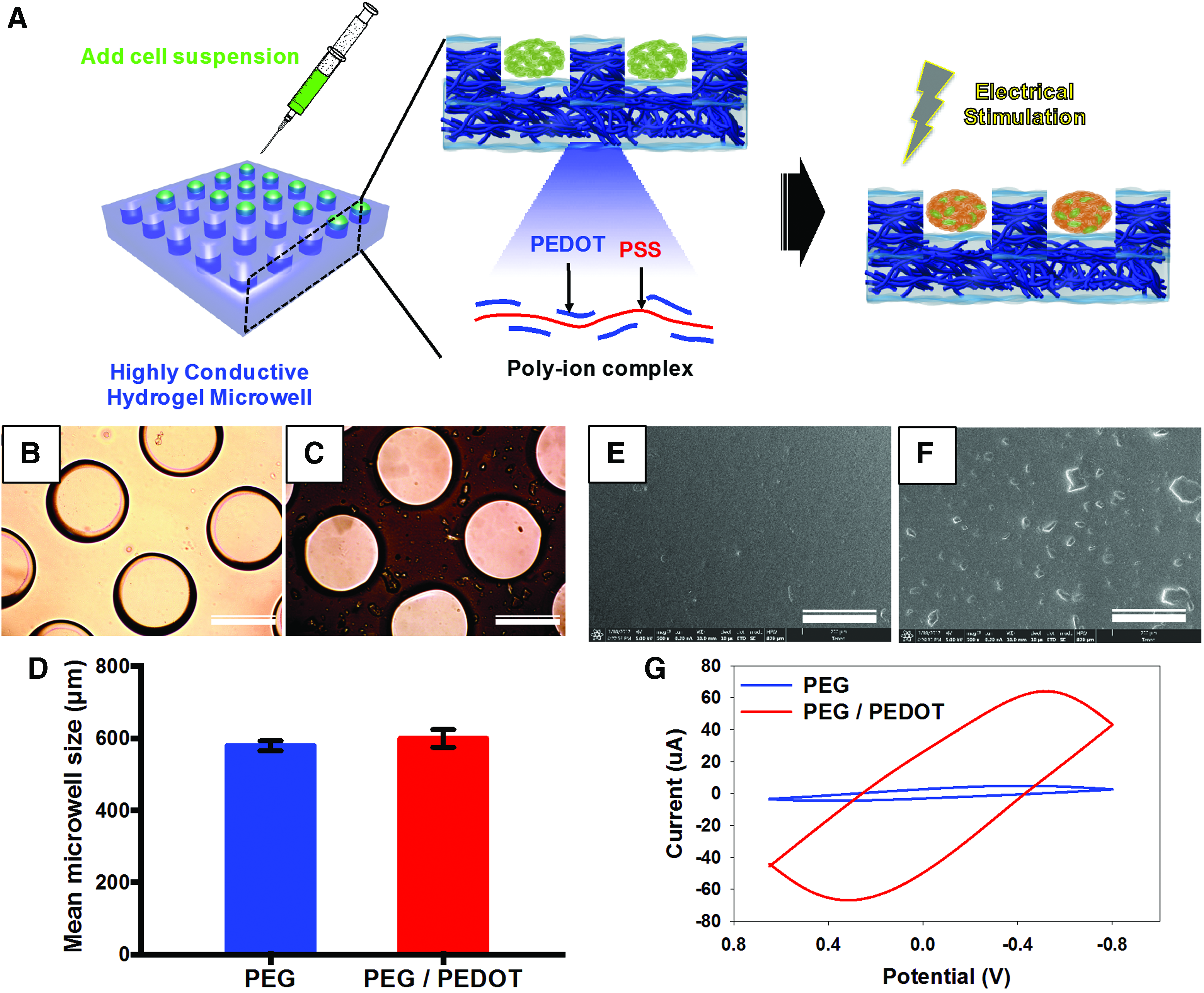
The objective of this research was to increase neurogenic differentiation efficacy of ADSCs through 3D microwell culture and ES transfer. Figure 1A shows a schematic diagram of the overall process used in this study. First, micro-contact printing technique was used to fabricate the hydrogel-based microwells due to its quick, simple, and cost-effective manufacturing capacity. Through this technique a micropatterned elastomeric stamp produced a predesigned pattern upon the glass surface which served as a negative mold for the photocurable hydrogels yielding well-formed microwells. To enhance the adhesive stability between glass and hydrogel, a chemical treatment with TMSPMA was performed to create the methacrylate monolayer on the glass surface. As shown in the optical images (Fig. 1B, C), the PEG and PEG/PEDOT:PSS hydrogel were well coated on the glass surface with well-formed microwells which had average diameters of 579.4 ± 14 μm and 599.7 ± 25 μm, respectively (Fig. 1D). After their surface morphologies were characterized, PEG hydrogel showed a smooth surface; however, PEG/PEDOT:PSS hydrogel had an increased surface roughness and uneven morphology (Fig. 1E, F). It is well known that PEDOT:PSS exhibits a bicontinuous structure and can aggregate with each other producing well-crystallized nanofibrils.14,15 Therefore, PEG/PEDOT:PSS hydrogel surface showed aggregations of a nanosized granular structure. In addition, cyclic voltammetry (CV) measurement was performed to confirm the improved electrochemical properties due to the addition of the conductive component (Fig. 1G). The CV curve area of PEG/PEDOT:PSS microwell significantly increased with all scanning range of −0.65 to 0.8 V compared to PEG microwells. The PEG/PEDOT:PSS microwell with high CV value induces greater electrical conduction through the incorporated PEDOT:PSS. In addition, we used the freeze-dried PEDOT:PSS for enhancing the electrochemical properties. After freeze-drying process, the PEDOT:PSS solid changed to well aggregated and closely packed structure compared with the as-received PEDOT:PSS solution. Hence, their electrochemical properties were improved by the organizational change. 16 However, low or no electrochemical reaction occurred at the PEG hydrogel. 17
ADSC aggregate formation and cell viability on the hydrogel-based microwells
After microwell characterization, the hydrogel-based microwells were used to form ADSC aggregates. The microwells consist of a side well and bottom layer to provide a structural barrier protecting direct fusion of individual cell aggregates. Figure 2A shows that ADSCs seeded on the hydrogel surface readily aggregate. After rinsing with DPBS to remove the cells from the top layer, the PEG microwells showed well-formed cell aggregates with uniform size and round morphology (Fig. 2B) primarily attributable to PEG's inhibitory binding of nonspecific biomolecules. 18 Therefore, the microwell surface composed of PEG led to the formation of cell aggregates due to its nonbiofouling property. However, ADSCs cultured in PEG/PEDOT:PSS microwells attached and spread on the surface exhibiting less aggregation compared to PEG. The addition of PEDOT:PSS to the PEG hydrogel induced cell adhesion and spreading through the binding of media proteins to the PEDOT:PSS surface. In addition, the cell viability of PEG microwell was observed by live and dead assay using confocal laser scanning microscope compared to that of PEG/PEDOT:PSS microwell (Fig. 2C). Confocal microscopy images show that all of cell aggregates displayed higher cell viability after 10 days of proliferation. The cell aggregate in the PEG microwell had a near-spherical shape with viable cells, whereas the morphologies of the cell aggregate culture in the PEG/PEDOT:PSS microwell were well spread and branched. This is due to the presence of PEDOT:PSS in the PEG hydrogel which was agglomerated in a form of well-crystallized nanofibril. Similar results have been reported showing that the addition of nanoparticles or crystalline structures into a PEG hydrogel increased cell adhesion and proliferation.18–21 For example, Castro et al. presented that the adhesion, proliferation, and osteochondral differentiation of human mesenchymal stem cell were significantly improved in a PEG hydrogel with addition of nanocrystalline hydroxyapatite. 21 In a similar approach, Lee et al. showed that the addition of core-shell nanoparticles to a PEG hydrogel results in increased surface roughness which enhanced cellular behavior. 19 Moreover, other work has reported that the nonspecific binding property of PEG hydrogels can be decreased through the addition of many types of biomaterials.18,20
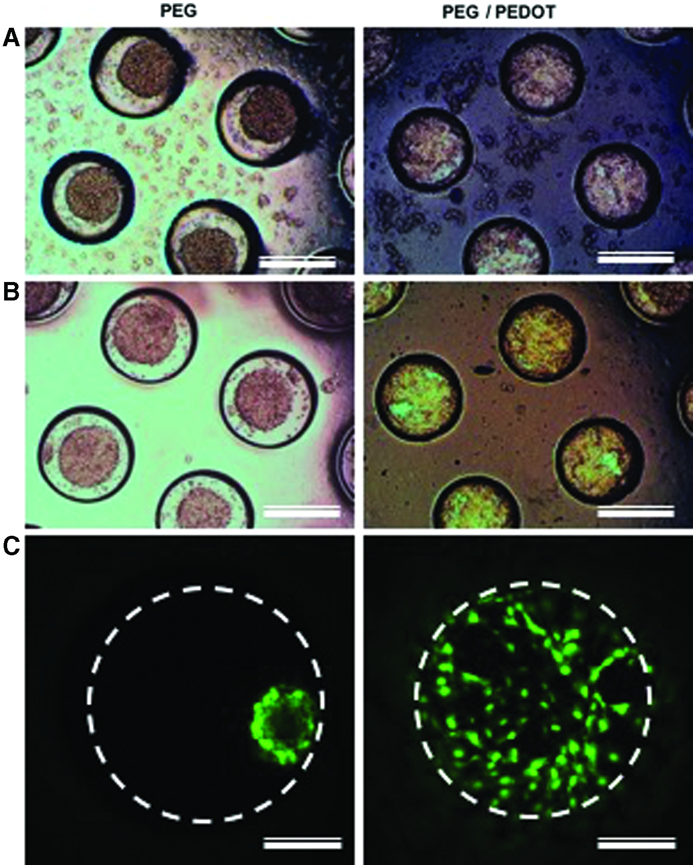
ADSCs seeding on hydrogel microwell and their morphology
Effects of ES on the neuronal differentiation capability of ADSC aggregate culture on the hydrogel-based microwells
To investigate whether ES treatment induced neural differentiation, ADSC aggregates in PEG and PEG/PEDOT:PSS microwells were cultured with and without ES for 10 days and examined by immunofluorescence. The confocal microscopy images showed ADSC aggregates derived neuronal differentiation in four different biological environments (Fig. 3). After 10 days of differentiation, all ADSC aggregates expressed positive neuronal markers, including Tuj1 and neurofilament. But no definite differences were observed between ES treated and untreated PEG microwells. In contrast, neuronal gene expression showed a significant difference between treated and untreated PEG/PEDOT:PSS microwell groups. PEG/PEDOT:PSS microwells without ES showed weak gene expression compared to other groups; however, gene expression greatly increased when treated with ES. In addition, ADSC aggregates in PEG/PEDOT:PSS microwells with ES were more interconnected with neurites to form neuronal networks than untreated groups. In addition, the morphologies of the ADSC aggregates exhibited a hydrogel-dependent difference. ADSC aggregates seeded in PEG microwells showed orbicular shape, whereas those in the PEG/PEDOT:PSS microwells were well spread and branched.
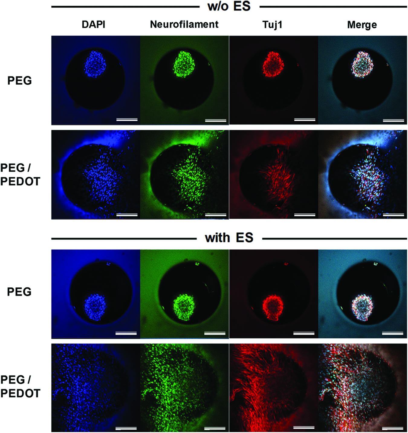
Immunofluorescence images of PEG and PEG/PEDOT:PSS microwells with ADSC aggregates that were cultured in neurogenic differentiation media, with or without ES for 10 days. Color indicates presence of each neurogenic differentiation marker: green for Neurofilament, red for Tuj1, and blue for DAPI. Scale bars = 200 μm. Color images available online at www.liebertpub.com/tea
Furthermore, qPCR was performed to quantitatively examine differences among the experimental groups using specific primers for Ngn2, NeuroD1, NSE, and TAU to quantify gene expression of neural differentiation (Fig. 4a). All neuronal gene expressions in the PEG microwells were significantly higher than those in the PEG/PEDOT:PSS microwells, regardless of whether they were treated with ES or not. In particular, the gene expression of Ngn2, NeuroD1, and Tau in the PEG microwells without ES was several fold higher compared with the PEG/PEDOT:PSS microwell without ES. It is because ADSC aggregates seeded in the PEG microwells have more 3D-like shape than those in the PEG/PEDOT microwells. The 3D tissue-like structure with a microenvironment can allow for direct cell–cell signaling and cell-ECM interaction. Hence, ADSC aggregates in the PEG microwells can have improved in vitro biological functionality over those in the PEG/PEDOT:PSS microwells. In the case of ES treated groups, differences between PEG and PEG/PEDOT:PSS microwells decreased with the PEG microwell exhibiting higher gene expressions compared to the PEG/PEDOT:PSS microwell. To confirm the differences with and without ES treatment, each group was used as control and all of analyzed genes were recalculated as shown in Figure 4b. Ngn2, which is a proneural determination marker, is present in neural progenitor cells and expressed in the neuronal differentiation. 22 Corresponding mRNA expression levels in PEG microwells with ES increased more than twofold, whereas Ngn2 expression in the PEG/PEDOT:PSS microwells with ES showed a greater than 10-fold increase compared to each of untreated groups. mRNA expression of the immature neuronal marker, NeuroD1, significantly increased in the PEG/PEDOT:PSS microwells with ES (7.4 ± 1.5). In comparison, NeuroD1 expression in the PEG/PEDOT:PSS microwells with ES increased by a factor of 2.1 ± 0.3. In addition, NSE and TAU which are the most important mature neuronal markers involved in the process of neurogenesis 22 exhibited similar expression patterns with remarkable increase rate in the PEG/PEDOT:PSS microwells with ES than that without ES. In comparison to PEG microwells, the PEG/PEDOT:PSS microwells showed highly increased expression rates of all neuronal differentiation markers after ES treatment. These positive effects are attributed to systematic transfer of ES to the ADSC aggregates by the PEG/PEDOT:PSS microwells leading to enhanced neuronal differentiation. Similar results have been reported by other groups showing that ES treatment increases protein synthesis in neurons and highly induces neuronal differentiation of stem cells.23–25 Thrivikraman et al. showed that ADSCs internalized with gold nanoparticles could highly induce neurogenic differentiation under ES treated culture condition. 24 In a similar approach, Pires et al. described that the cross-linked PEDOT:PSS substrate could enhance neural stem cell differentiation into neurons and astrocytes by ES. 25 Recently, many researchers have demonstrated that ADSCs can be capable of undergoing transdifferentiation into mature cells not related with their original lineage by specific induction media, special growth factor, and biological stimulation. 26 When appropriate culture condition and stimulation are applied, ADSC can transdifferentiate into desired cell types.22,24,27 As previously discussed, it is important to prevent the differentiation of stem cells into unwanted cell types, as well as control the differentiation toward desired phenotype before clinical application. 6 Interestingly, ADSCs showed a higher potential of neuronal differentiation compared to mesenchymal stem cell from other sources.28,29 For these reasons, in this study ADSCs were cultured on the hydrogel-based microwells in aggregate form for giving them microenvironment-like native tissue and applied ES for biological stimulation. In consequence, ADSCs cultured on 3D conductive culture system effectively induced neuronal differentiation. Based on these findings, our conductive hydrogel-based microwell with ES would provide a highly effective neural culture system for neural tissue engineering.
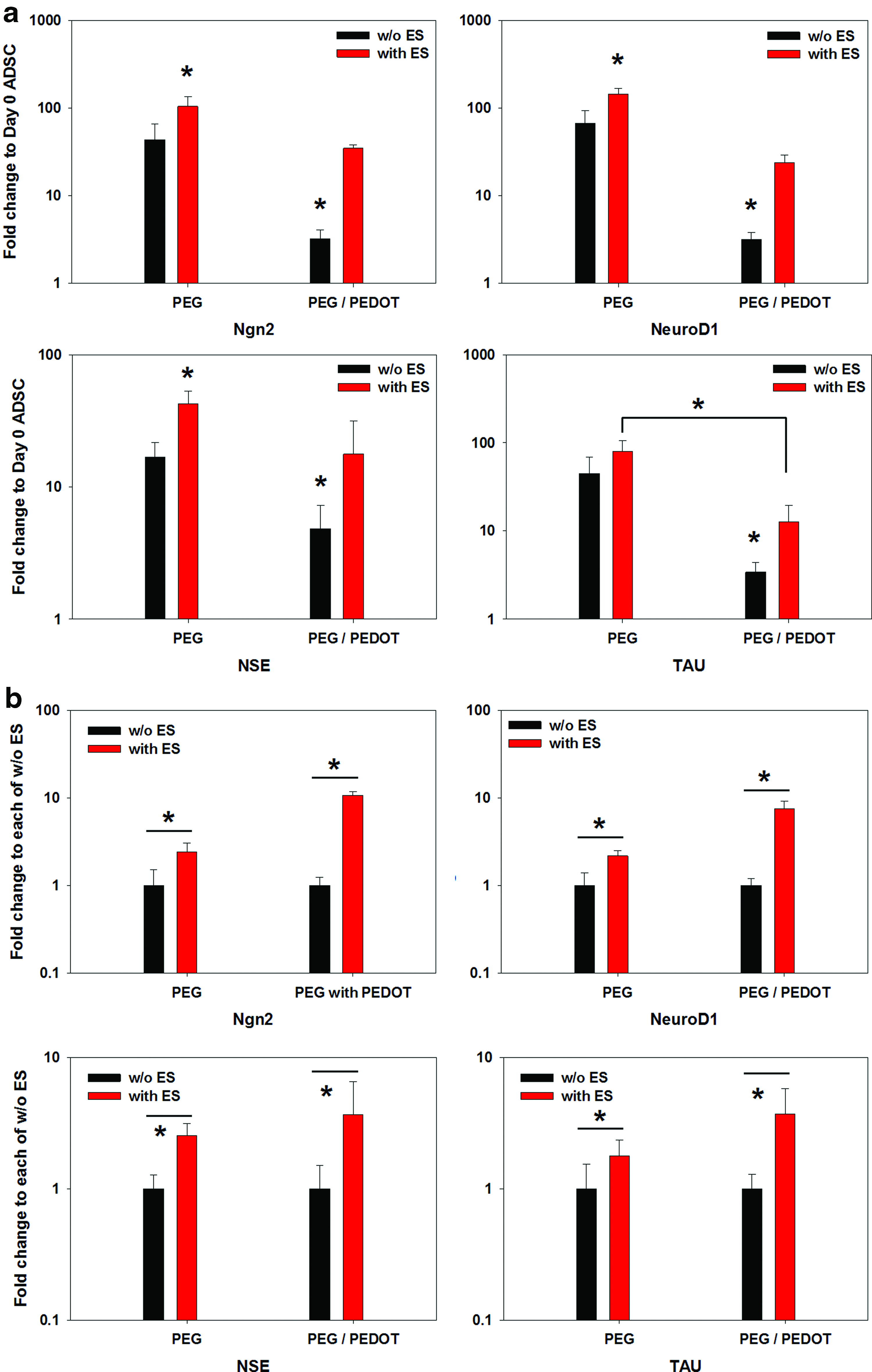
Gene expression levels of ADSC culture at day 10 for neurogenic differentiation markers: Ngn2, NeuroD1, NSE, and TAU. Relative gene expression of each gene (mean ± SD), normalized to the expression of the housekeeping gene GAPDH and compared with
Conclusion
In conclusion, our results corroborating previous reports show conclusively that (1) the presence of PEDOT:PSS within a microwell provides a conductive substrate, (2) the conductive hydrogel-based microwell significantly improved electrical potential with electrochemically driven current on the surface compared to nonconductive hydrogel-based microwell, and (3) microwell transmitted ES to ADSC aggregates effectively induces neuronal differentiation. In addition, we confirmed that ADSC aggregates in PEG/PEDOT:PSS microwells with ES expressed more positive neuronal differentiation markers than PEG/PEDOT:PSS microwells without ES. Although all neuronal gene expression levels were greater in PEG microwells with ES, the increased rates of gene expression levels between treated and untreated PEG/PEDOT:PSS microwells were higher compared to PEG microwells. Therefore, we propose a most effective strategy taking advantage of a 3D conductive culture system which can be useful in a wide variety of electrical application, including biological signal recording devices, stimulation electrodes, neural differentiation culture system, and neural tissue regeneration. However, our 3D conductive culture system requires further improvement with regards to low adhesion affinity to form a round shape and uniform cell aggregate for stem cell differentiation in near future.
Footnotes
Acknowledgment
This work was supported by March of Dimes Foundation's Gene Discovery and Translational Research Grant.
Disclosure Statement
No competing financial interests exist.