
Abstract
Previously, we synthesized an articular cartilage extracellular matrix (ECM)-derived oriented scaffold for cartilage tissue engineering, which was biomimetic in terms of structure and biochemical composition. However, the limit resource of the cartilage-derived ECM is a hindrance for its application. In this study, we developed a new material for cartilage tissue engineering—human umbilical cord Wharton's jelly-derived ECM (hWJECM). The hWJECM has an abundant resource and similar biochemistry with cartilage ECM, and the use of it is not associated with ethical controversy. We adopted the method previously used in cartilage ECM-derived oriented scaffold preparation to generate the oriented hWJECM-derived scaffold, and the scaffold properties were tested in vitro and in vivo. The three-dimensional scaffold has a porous and well-oriented structure, with a mean pore diameter of ∼104 μm. Scanning electron microscopy and cell viability staining results demonstrated that the oriented scaffold has good biocompatibility and cell alignment. In addition, we used functional autologous chondrocytes to seed the hWJECM-derived oriented scaffold and tested the efficacy of the cell-scaffold constructs to repair the full-thickness articular cartilage defect in a rabbit model. Defects of 4 mm diameter were generated in the patellar grooves of the femurs of both knees and were implanted with chondrocyte–scaffold constructs (group A) or scaffolds alone (group B); rabbits with untreated defects were used as a control (group C). Six months after surgery, all defects in group A were filled completely with repaired tissue, and most of which were hyaline cartilage. In contrast, the defects in group B were filled partially with repaired tissue, and approximately half of these repaired tissues were hyaline cartilage. The defects in group C were only filled with fibrotic tissue. Histological grading score of group A was lower than those of groups B and C. Quantification of glycosaminoglycan indicated that newly formed cartilage in group A rabbits was comparable with normal cartilage. In conclusion, hWJECM-derived oriented scaffolds loaded with autologous chondrocytes induced cartilage repair in rabbit knees, which was comparable with native cartilage in terms of macroscopic view, microstructure, and biochemical composition.
Introduction
A
Scaffold is a critical factor for tissue engineering, which should be biocompatible and with sufficient mechanical property, good cell affinity, and suitable pore size.7,8 Scaffolds fabricated with various biodegradable materials have been developed and used in tissue engineering. Synthetic materials [e.g., poly (lactic-co-glycolic acid), PLGA] commonly are biodegradable and have good mechanical properties and low immunogenicity, however, its surface is hydrophobic and not fit for cell adhesion. Another category of scaffold material includes collagen, chitosan, alginate, fibrin, and hyaluronan (HA). These natural materials have good cell affinity and compatibility, and are more conducive to the adhesion and proliferation of cells.
ECM became prospective material for tissue engineering scaffold preparation in recent years because of its ideal composition, which was most biomimic to natural chondrocyte microenvironment, and was favorable for cell adhesion, proliferation, and matrix synthesis. In a previous study, we have developed a three-dimensional (3D) porous scaffold derived from articular cartilage ECM, which was closely biomimic in biochemistry with cartilage ECM and conducive to cell adhesion, proliferation, and differentiation because of the informational signals (e.g., the Arg-Gly-Asp sequence).9–11 Furthermore, the articular cartilage ECM-derived scaffold, combined with autologous adipose-derived mesenchymal stem cell (MSC), induced cartilage repair tissue comparable to native cartilage. 12 However, the limited human cartilage resources restrict its application, and many difficulties are also associated with the use of xenogenous cartilage-derived ECM, so we attempted to find a new material that might be suitable for cartilage tissue engineering scaffold.
Human umbilical cord Wharton's jelly (hWJ) is a gelatinous substance that surrounds the umbilical cord vessels. It contains very few cells, but is rich in collagen, HA, and sulfated glycosaminoglycan,13–19 which is similar with cartilage ECM. In addition, the functions of WJ are to protect the umbilical vessels against external forces and to guarantee venous and arterial umbilical blood flow, which is also in some extent similar to that of cartilage. Moreover, as in cartilage, small blood vessels, nerves, and lymph are absent from WJ. Therefore, WJ is similar to cartilage in structure, biochemistry, and biofunction. What's more, human umbilical cord is an appendix of delivery with a wide range of sources and no ethically controversial. Based on all the properties mentioned above, we propose that WJ is a prospective biomaterial for scaffold preparation in cartilage tissue engineering.
An ideal scaffold for cartilage tissue engineering should be biomimetic in not only biochemical composition but also in the morphological structure. The structure of scaffolds is critical for directing cell alignment and matrix synthesis as well as mechanical performance.20,21 Therefore, a scaffold with a vertical microtubule structure would be most likely to mimic the physiological and biomechanical functions of native cartilage. The cell source is another important consideration in tissue engineering strategies, and autologous chondrocyte implantation has been used in cartilage repair for many years. 22 So in this study, we produced an oriented hWJECM-derived scaffold, characterized its properties, and populated it with chondrocytes to explore its potential for the repair of cartilage defects in vivo.
Materials and Methods
Decellularization of hWJ
Human umbilical cords were collected from the Department of Obstetrics and Gynecology at the Chinese PLA General Hospital, with the approval of the General Hospital Ethics Committee. They were kept on ice and transported to the laboratory in sterile phosphate-buffered saline (PBS) solution (pH 7.6). Umbilical arteries and vein were removed, and the WJ was dissected from the umbilical cords using toothed forceps under aseptic conditions. The decellularization method combined physical and chemical procedures as follows: WJ tissue was resuspended and rinsed in sterile PBS solution for three times. It was then homogenized using a tissue disintegrator to form a suspension slurry and centrifuged (Beckman Allegra X-22R) for 20 min at 2000 rpm in an F0850 rotor. The microparticles in the supernatant were separated from the precipitated macroparticles and centrifuged for another 20 min at 3000 rpm, then for 20 min at 6000 rpm, and finally for 35 min at 9500 rpm. The precipitate from the final centrifugation step was retained and rinsed twice with sterile PBS solution. It was then digested successively using 0.25% trypsin in 1% TritonX-100 (containing 0.1% ethylenediaminetetraacetic acid [EDTA], 0.1% sodium azide) under gentle agitation for 24 h at 4°C and then using 50 U/mL deoxyribonuclease I and 1 U/mL ribonuclease A (both from Sigma-Aldrich Ltd., St. Louis, MO) with gentle agitation for 12 h at 37°C to remove nuclear material. After decellularization, the hWJECM precipitation was centrifuged at 9500 rpm again. All nanofibrous precipitations were retained in sterile glassware for scaffold preparation. For scanning electron microscope view, the nanofibrous precipitation was fixed with 2.5% glutaraldehyde for 24 h at 4°C. The samples were dehydrated by a graded series of ethanol washes and dried at room temperature before being sputter coated with gold.
Fabrication of hWJECM-derived oriented scaffold
The decellularized hWJECM microfilaments (approximate diameter = 500 nm–5 μm) were washed intensively with sterile PBS and made into a 3% (w/v) suspension in PBS. hWJECM-derived oriented scaffolds were generated using a unidirectional solidification freeze-drying method, as we previously described. In brief, the hWJECM was poured into a cylindrical mold and the mold was placed vertically on a metal plate at −20°C for 30 min and then at −80°C for 1 h. The mold was then transferred into a freeze dryer and lyophilized for 48 h under vacuum. After lyophilization, the hWJECM-derived scaffolds were removed from the mold and cross-linked with ultraviolet light at 258 nm for 6 h. They were then immersed in 95% (v/v) ethanol solution [containing 50 mM 1-ethyl-3-(3-dimeth-ylaminopropyl) carbodiimide hydrochloride (EDAC) and 20 mM N-hydroxysucinimide; Sigma-Aldrich Ltd.] for 24 h at 4°C. Excess EDAC was rinsed from the scaffolds with PBS, and the oriented scaffolds (∼4 mm in diameter and 2 mm thick) were sterilized with 60Coγ-irradiation at 5 mRad.
Histology and immunohistochemical staining
The samples were mounted in Tissue-Tek O.C.T. compound (Sakura Finetek, Torrance, CA), cryosectioned at 5-μm thick, and fixed in acetone for 10 min at room temperature. Sections were stained with toluidine, safranin-O, and type II collagen antibody to highlight the presence of glycosaminoglycan (GAGs) and type II collagen. Vertical and cross sections were blocked using peroxidase blocking solution for 10 min, followed by 10% (v/v) goat serum solution for 30 min. The sections for immunohistochemistry staining were then incubated with anti-human type II collagen antibody at 1:100 dilution overnight at 4°C, and the biotinylated goat anti-mouse secondary antibody (Maxim Corporation) was applied for 20 min, followed by incubation with horseradish peroxide–conjugated streptavidin for 10 min. Primary antibodies were omitted for control. Finally, the diaminobenzine (DAB) coloration was carried out and the coverslips were mounted in resin and viewed by optical microscopy.
Isolation and cultivation of articular cartilage chondrocytes
With the approval of the Animal Care Committee of the Chinese PLA General Hospital, mature New Zealand white rabbits (weighing 2.5–3.0 kg) were anesthetized. Chondrocyte isolation and culture were performed as follows: cartilage samples were obtained from rabbit shoulder joints and digested with type II collagenase on a magnetic stirring apparatus for 1.5 h. After centrifugation at 1500 rpm for 5 min, cells were separated and washed twice with Hank's solution. Then, they were resuspended in regular growth medium containing chondrocyte culture medium supplemented with 20% fetal bovine serum and seeded in a 25-cm2 culture flask. Cells were cultured in a humidified atmosphere with 5% CO2 at 37°C. The medium was changed every 2 days until cells became 90% confluent. The cells were digested with 0.2% trypsin solution and passaged for regrowth. P2 cells were used in the following study.
Preparation of cell–scaffold constructs
The chondrocytes were resuspended in culture medium. Oriented scaffolds (4 mm diameter, 1.5 mm thick) were sterilized using ethylene oxide and placed in a 24-well culture plate before 20 μL cell suspension containing ∼1 × 10⁶ cells was seeded onto each scaffold to completely saturate it. The scaffolds were incubated in a humidified atmosphere containing 5% CO2 at 37°C for 4 h to allow cell attachment before being immersed completely in media. Every subsequent hour, 5 μL medium was added to the cell–scaffold construct and 2 mL culture medium was added to each well. After the cell–scaffold constructs had been cultured in chondrogenic medium at 37°C in a humidified atmosphere of 5% CO2 for 3 days, the constructs were washed with PBS and implanted into the cartilage defects.
In vivo implantation of chondrocyte-loaded hWJECM-derived oriented scaffolds
This study was performed under a protocol approved by the Institutional Animal Care and Use Committee at PLA General Hospital. After general anesthesia, rabbit knee joints were opened using a medial parapatellar approach. A full-thickness cylindrical defect (4 mm in diameter, 1.5 mm deep) was created on the patellar groove of the femur in both legs using a corneal trephine. The rabbit defects were classified randomly into three groups: (A) the chondrocyte–scaffold group; (B) the scaffold-alone group; and (C) the untreated group. The cell–scaffold constructs were cultured in chondrocyte medium for 3 days before implantation and were then inserted into the cartilage defects with no additional fixation. Ten repaired defects were assigned for each group at one time point, six for histological analysis and four for biochemical assays (Table 1).
Gross morphology evaluation
Specimens were harvested 3 and 6 months postoperatively, and the macroscopic evaluations were conducted by three blinded observers, based on gross visual examination of synovial fluid/membrane changes, the opposing articular surface, defect repair quality, and repair surface geometry. International Cartilage Repair Society (ICRS) gross scoring system 23 was used for evaluation, with scores assigned for degree of repair, integration, surface regularity, and total judgment.24,25 Scores for each category were combined and the maximum possible score was 12, with a lower relative score indicating “better” healing.
Histological evaluation
After gross examination, the specimens were fixed in 4% paraformaldehyde for 1 week and then were decalcified with 10% EDTA for 4 weeks. Subsequently, they were embedded in paraffin, cut into 5-μm sections, and then deparaffinized and rehydrated for histological staining, including hematoxylin and eosin, toluidine blue, safranin-O, and type II collagen immunohistochemical staining. Immunohistochemical staining for type II collagen was performed as previously described. 10 In brief, the sections were treated with 3% v/v hydrogen peroxide to block endogenous peroxidase activity and incubated with mouse anti-human type II collagen monoclonal antibody (Santa Cruz Biotechnology) at 1:100 dilution at 4°C overnight. Then, they were incubated with biotinylated goat anti-mouse immunoglobulin secondary antibody (Maxim Corporation) for 20 min at room temperature, followed by diaminobenzidine coloration and viewing through microscope. Sections incubated with PBS without primary antibodies were used as a negative control. The histological and immunohistochemical staining results were evaluated using the Wakitani histological grading system. This scoring system consisted of five categories (cell morphology, matrix staining, surface regularity, thickness of cartilage, and integration of donor with host adjacent cartilage), with total scores ranging from 0 to 14. 26
GAG quantification
Repaired tissues were harvested with a core bit (4 mm internal diameter) and the cartilage slices were used for GAG quantification. Cartilage of the same thickness from the surrounding area was used as controls. Samples were freeze-dried and digested in 1 mL papainase (1.25 mg/mL papain, 100 mM PBS, 10 mM EDTA, and 10 mM cysteine; pH 6.5) for 24 h at 65°C. GAG content was evaluated by dimethylmethylene blue (Sigma-Aldrich Ltd.) staining of chondroitin sulfate, as in the method developed by Farndale et al. 27
Statistical analysis
All quantitative data are analyzed and expressed as means ± standard deviations. Water uptake and macroscopic and histological results were examined with one-way analysis of variance using SPSS statistical software (ver. 17.0; SPSS, Inc., Chicago, IL). Significance was set at p < 0.05.
Results
Preparation of hWJECM
Histological staining of hWJ was positive for safranin-O and toluidine blue and immunological staining was positive for collagen II, demonstrating the existence of GAG and collagen II in WJ (Fig. 1). Because the biochemical constitution of WJ is similar to that of cartilage, hWJECM would likely be a good material for cartilage scaffold engineering. Toluidine blue and collagen II staining demonstrated that GAG and collagen II were retained, and scanning electron microscopy (SEM) view showed the nanofibers in the hWJECM suspension (Fig. 2).
Histological and immunological staining of human umbilical cord.
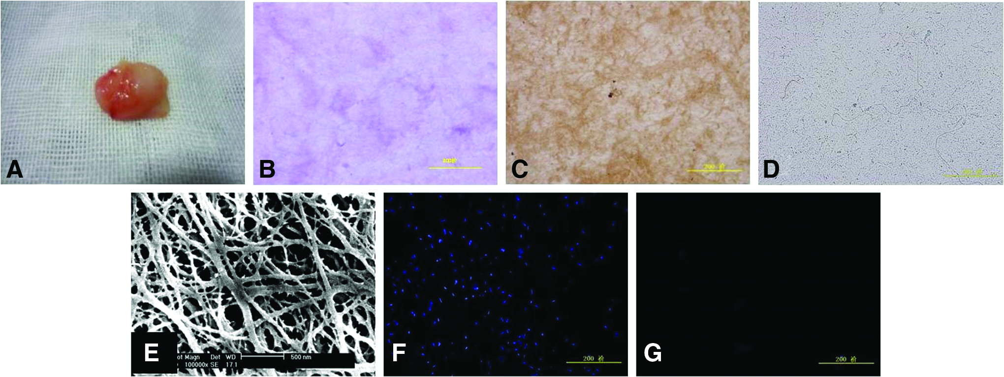
Observation of hWJECM.
Preparation of oriented scaffolds
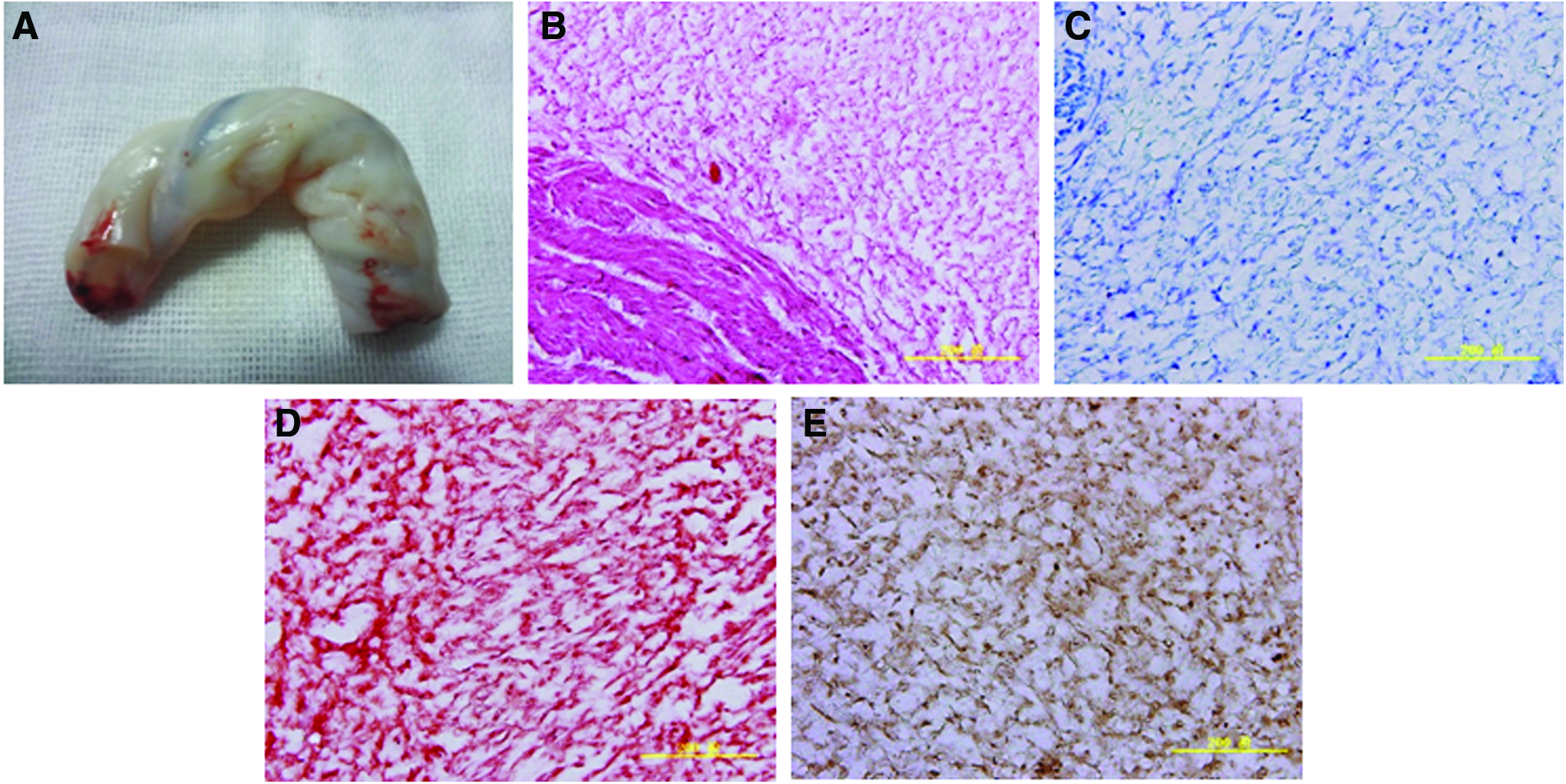
Macro- and microview of the hWJECM-derived oriented scaffold showed 3D structure of it, with interconnected and vertically distributed pores and some nanofibers connected across pore spaces. Figure 3B and C shows the cross and longitudinal section of the oriented scaffold. Histochemistry and immunohistochemistry staining were carried out to evaluate the biochemical composition of the hWJECM oriented scaffold. The positive results revealed the presence of collagen II and GAGs in the oriented scaffold (Fig. 3D, E).

Observation of hWJECM-derived oriented scaffold.
Macroscopic observation of rabbit cartilage repair
Three months postoperatively, defects in the chondrocyte–scaffold group consisted of a single smooth fully repaired surface with obvious boundaries with host adjacent cartilage. In the scaffold-alone group, the defects were depressed and irregular with poor integration with the host cartilage (Fig. 4A–C). Six months after implantation, the regenerated tissue had filled the defects completely with a smooth surface in chondrocyte–scaffold group, with hardly visible boundaries and slightly different color from that of the surrounding normal cartilage. In scaffold-alone group, the defects were filled with white tissue, with rough surfaces, and obvious boundaries. In untreated group, the defects were only partially filled with concave tissue (Fig. 4D–F). Statistical analysis demonstrated that the gross grading scores of repaired tissues in chondrocyte–scaffold groups and scaffold-alone group were consistently higher than those of repaired tissues in untreated group at both time points, and that the scores from chondrocyte–scaffold group were significantly higher than those from scaffold-alone group 3 months postoperation (Fig. 4G).

Macroscopic appearance and gross scores of cartilage defect repair at 3 and 6 months postoperation.
Histological observation
After 3 months, the restoration areas in group A were relatively flat and continuous, consisting of new cartilage that was thicker than the surrounding cartilage. More hyaline cartilage cells were present, and the repair junction was less well defined. In some deep areas of repaired tissue, the cells were organized in a columnar array. The subchondral bone ossification area was not obvious. In group B, the restoration areas had rugged border zones, demonstrating poor continuity/integrity. A very small number of border-zone hyaline cartilage cells were observed. The stroma was lightly stained, disordered, and uneven. Deep tissue repair still consisted mostly of fibrocartilage (Figs. 5A–F and 6). In group C, the cartilage defects still showed significant depressions. A small amount of fibrous tissue repair occurred in this area, but no integration at the repair junction, no cartilage, and little subchondral bone tissue formation was observed.

Histological staining and score of repaired tissue at 3 and 6 months.

High-magnification view of repaired tissue in group A at 6 months.
After 6 months, the cartilage repair areas were smooth, flat, and continuous in group A. A larger number of hyaline cartilage cells were present on the surface, but some gaps remained between the repair zone and the surrounding cartilage. In parts of the repair areas with large numbers of deep cartilage chondrocytes oriented in a columnar structure, repair junction integration was good, but some border-zone chondrocytes were more lightly stained and subchondral bone ossification was visible. In group B, the cartilage repair areas of defects were relatively flat and partially integrated at the border zones. Cartilage in the repaired area was thinner than in the surrounding matrix and only lightly stained. The majority of cartilage cells were fibrous, although a small number of hyaline cartilage cells were present (Figs. 5G–L and 6). In group C, central depressions remained evident in the cartilage defects; little tissue repair had occurred, and any new cartilage present was relatively thin. Few cartilaginous cells were present, and the majority of new cells were fibrous and poorly integrated.
The Wakitani histological grading scores demonstrated that the quality of repaired tissue was significantly better in group A than in groups B and C at 3 and 6 months postoperation, with a lower relative score indicating “better” healing. In addition, repair quality in group B was significantly better than that in group C at 6 months (Fig. 5M).
GAG content determination
Biochemical analysis revealed that the GAG content of repaired tissue increased over time. The GAG content in group A was 0.070 ± 0.007 mg/mg at 3 months and 0.099 ± 0.008 mg/mg at 6 months, reaching 57.04% and 80.93% of that of normal cartilage (0.122 ± 0.008 mg/mg), respectively. In group B, it was 0.036 ± 0.000 mg/mg and 0.055 ± 0.003 mg/mg, respectively.
Discussion
ECM has attracted a great deal of attention in tissue engineering and regenerative medicine because of their good biocompatibility and bioactivity, which can directly or indirectly regulate cell proliferation, adhesion, migration, and differentiation, as well as play key roles in homeostasis and regeneration of tissues and organs. We have previously developed a natural, acellular, 3D interconnected porous scaffold derived from cartilage ECM with use of hypotonic buffer, Triton X-100, a nuclease solution, and simple freeze-drying and cross-linking techniques. Histological results showed that most of the ECM components remained after cell fragments removal and SEM revealed a 3D interconnected porous structure of the scaffolds. The novel scaffold could provide a suitable 3D environment to support the cell adhesion, proliferation, and differentiation with no cytotoxic effects. But it faces a hindrance, lack of safe and abundant material resources, limiting its application in cartilage engineering.
The umbilical cord is a tissue which connects the fetus to the placenta in mammals. It contains a single vein and two arteries, which are surrounded by WJ and covered by a simple amniotic epithelium. The WJECM is rich in HA, collagen, and sulfated GAGs, which are also main components of articular cartilage ECM. The high HA content means that the tissue is well hydrated, and the high collagen and GAG content means that it can withstand the forces of extension and compression produced by the movement of the fetus and by uterine contraction. In addition, Sobolewski et al. indentified the presence of several growth factors, viz. acidic FGF, basic FGF, EGF, IGF-I, PDGF, and TGF-β in Wharton's jelly, which is to say, Wharton's jelly is a reservoir of peptide growth factors.19,28–30 Combining the good biocompatibility and presence of abundant peptide growth factors, researchers investigated the feasibility to use WJECM in tissue engineering and regenerative medicine.
Hao et al. found that Wharton's jelly extracts (WJEs)-derived substrate could clearly reduce a dynamic growth of human MSCs in late-phase culture, suggesting that the replicative senescence of MSCs was efficiently slowed by WJE. The related mechanism is that WJE-based culture efficiently suppressed the enhancement of intracellular reactive oxygen species (ROS), p53, and p16INK4a/pRb in MSCs. In addition, the decreased differentiation ability of MSCs after long-term culture was largely ameliorated by WJE. 31 Beiki et al. found that WJ-derived scaffold was able to improve attachment, penetration, and growth of the fibroblast cells and speed up the wound healing processes, not only promoting complete wound closing and disappearance of the scab but also promoting complete reepithelialization, newly generated epidermal layers, and appendages. 32
Dan et al. 33 used WJECM as a standardized and homogeneous coating substrate for vascular engineering and found that this coating surface was appropriate for both the adhesion and proliferation of human MSCs and mature endothelial cells, via the promotion of integrins α2 and β1 expression in the cells, even under the shear stress. A human angiogenesis array result confirmed presence of VEGF, FGF-2, TGF-b1, and endothelin-1 in the trypsin-digested WJECM solution. Chan et al. 34 have investigated the possibility of using decellularized human umbilical vein (HUV) with WJ as an allogeneic, acellular ECM scaffold for the engineering of vocal fold lamina propria in vitro. HUV specimens with WJ on the abluminal surface were dissected using an automated procedure and subjected to a novel, saline-based decellularization treatment to remove potentially antigenic epitopes. Human vocal fold fibroblasts from a primary culture were seeded onto the resulting acellular constructs and cultured. They found that the elastic modulus and dynamic viscosity of the resulting fibroblast-repopulated scaffolds were comparable to those of the human vocal fold lamina propria. In this study, WJ was regarded as important in the construction and tissue engineering process, due to the presence of a variety of proteins, including collagen (types I–VII), laminin, peptide growth factors, GAGs, and proteoglycans, in its ECM.
Recently, it was reported that Stocco et al. investigated the feasibility of polyvinyl alcohol (PVA)/ECM scaffolds for cartilage regeneration in vitro. They aimed to combine the biomechanical properties of the synthetic hydrogel PVA with the biomimetic microenvironment of Wharton's jelly ECM in cartilage regeneration. The results proved that the Wharton's jelly ECM, although nonspecific, promoted chondrocyte colonization of the scaffold, compared with PVA itself and more specific decellularized cartilage matrix. 35
Based on the researches mentioned above and the similarities in structure, biochemistry, and function between WJECM and cartilage ECM, we propose that WJECM might be an ideal kind of material for cartilage tissue engineering scaffold preparation. In our study, an acellular suspension of hWJ was produced by grinding, centrifugation, and repeated freezing and thawing. The oriented hWJECM-derived scaffold was then generated by unidirectional solidification freeze-drying and cross-linking method. The micrograph view verified the oriented structure and the histochemical and immunohistochemical staining results demonstrated the presence of GAGs and type II collagen in the prepared acellular scaffold. Specific features of the scaffold include the following: (1) the suspension of umbilical cord WJ microfilaments forms nanoscale structures. Research has demonstrated that the nanoscale structure of cartilage can promote cell adhesion, proliferation, and differentiation36,37; (2) The oriented structure of the scaffold facilitated biomimic of morphology.38–45
To evaluate its repairing effect, scaffolds loaded with autologous chondrocytes were implanted in vivo, and the gross/histological aspects of the repaired tissue were comprehensively evaluated, as well as its biochemical composition, and biomechanical properties. The gross and histological grading scores both showed the quality of repaired tissue and was significantly better in chondrocyte–scaffold group than in scaffold-alone group and untreated group, especially with the orientated alignment of the chondrocytes in newly formed tissues in chondrocyte–scaffold group. These results demonstrated that the biomimic composition and structure of the oriented hWJECM-derived scaffold promoted the chondrocytes adhesion, alignment, and proliferation in the scaffold, as well as the secretion of matrix during the repair of damaged cartilage. From a quantitative respect, the GAG content of specimens from chondrocyte–scaffold group was higher than those of scaffold-alone group and untreated group specimens.
From these results, we can find the bioactivity of the Wharton's jelly-derived ECM, and it could be a prospective material for cartilage engineering scaffold preparation. In addition, human WJECM is a kind of material with abundant resources and no ethics controversy, so it is prospective to be applied in clinic for cartilage and other tissue engineering. In the future study, we will investigate the effect of hWJECM on MSCs and chondrocytes proliferation and phenotype, as well as its mechanism, and furthermore, the repair effect of its derived scaffold on cartilage defects compared with other techniques already available to treat cartilage defects, such as microfracture and articular cartilage ECM-derived scaffold, will be explored.
Conclusions
A biomimetic scaffold derived from natural nanofibrous hWJECM was synthesized by an improved thermally induced phase separation (TIPS) technique. This novel hWJECM-derived oriented scaffold promotes cell repopulation and supports the formation of new cartilage tissue. The scaffold is hydrophilic and has good cell affinity, promoting cell adhesion and proliferation. Implantation of chondrocyte–scaffold constructs in rabbits with cartilage defects resulted in good-quality repair of cartilage that was comparable with native cartilage in terms of its mechanical properties and structure. The hWJECM-derived oriented scaffold is a good candidate material of cartilage tissue engineering scaffold, but further studies should utilize larger animal models to explore the feasibility of its clinical application.
Footnotes
Acknowledgments
This study was funded by the National Science Foundation of China (grant nos. 81071458, 81472092, 21134004) and the National High-Tech Research and Development Program (grant no. 2015AA020303).
Disclosure Statement
No competing financial interests exist.