
Abstract
Biomaterials with excellent osteogenic and angiogenic activities are desirable to repair massive bone defects. Decellularized matrix from porcine small intestinal submucosa (SIS) has attracted particular attention for tissue regeneration because it has strong angiogenic effects and retains plentiful bioactive components. However, it has inferior osteoinductivity and osteoconductivity. In this study, we developed porous composite of SIS combined with mesoporous bioactive glass (SIS/MBG) with the goal of improving the mechanical and biological properties. SIS/MBG scaffolds showed uniform interconnected macropores (∼150 μm), high porosity (∼76%), and enhanced compressive strength (∼0.87 MPa). The proliferation and osteogenic gene expression (Runx2, ALP, Ocn, and Col-Iα) of rat bone marrow stromal cells (rBMSCs) as well as the proliferation, angiogenic gene expression (VEGF, bFGF, and KDR), and tube formation capacity of human umbilical vein endothelial cells (HUVECs) in SIS/MBG scaffolds were significantly upregulated compared with nonmesoporous bioactive glass (BG)-modified SIS (SIS/BG) and SIS-only scaffolds. Western blot analysis revealed that SIS/MBG induced rBMSCs to osteogenic differentiation through the activation of Wnt/β-Catenin signaling pathway, and SIS/MBG enhanced angiogenic activity of HUVEC through the activation of PI3k/Akt pathways. The in vivo results demonstrated that SIS/MBG scaffolds significantly enhanced new bone formation and neovascularization simultaneously in critical-sized rat calvarial defects as compared with SIS/BG and SIS. Collectively, the osteostimulative and angiostimulative biomimetic composite scaffold SIS/MBG represents an exciting biomaterial option for bone regeneration.
Introduction
L
Recently, ECM biomaterials prepared from decellularized tissues or organs have gained enormous attention, as they contain complex ECM components and native tissue microstructure, and have exhibited bioactivity in tissue remodeling and regeneration.3,4 ECM derived from small intestinal submucosa (SIS) is an excellent matrix to induce bone regeneration because of its abundant bioactive components and angiogenesis-stimulatory effects, which are crucial in tissue regeneration.2,5 SIS consists of ∼90% collagen (mainly Type I), with growth factors such as vascular endothelial growth factor (VEGF) and basic fibroblast growth factor-2 (bFGF-2).6,7 SIS has been approved by the Food and Drug Administration, and can be preserved and used “off the shelf.”3,8 However, pure SIS has some disadvantages that limit its bone regeneration capacity: it lacks sufficient osteoinductivity (inducing cells for osteogenic differentiation) and its inferior mechanical strength can hardly maintain a three-dimensional pore structure and osteoconduction (promoting tissue ingrowth and nutrient transport into interconnected macropores).
To improve the bone regeneration capacity of SIS-based bone grafts, it may be a desirable approach to combine composite bioactive ceramic with SIS. Bioactive glasses (BGs) are a subset of oxide-based biocompatible ceramics that can uniquely integrate with hard and soft tissues and stimulate new tissue growth while dissolving over time, making them highly attractive materials for healthcare and regenerative medicine. 9 Substances such as Ca, P, and Si ions released from BG promote the osteogenic differentiation of bone marrow stromal stem cells (BMSC).10–12 In particular, Si plays a key role in vascular development. 13 Mesoporous bioactive glass (MBG) is a new class of BG with a well-ordered mesoporous structure, 2 to 50 nm in size. 9 Compared with non-MBG, porous MBG has a much higher surface area to volume ratio, a key property for enhanced drug delivery capability, in vitro apatite mineralization, and degradation.14,15 However, the material's inherent brittleness, high degradation rate, and surface instability are major disadvantages. 16 To address these challenges, a number of studies focused on the composites of polymers and MBG, such as collagen-MBG, 10 silk-MBG, 16 polycaprolactone-MBG, 17 and poly(lactic-co-glycolic acid)-MBG. 18 However, to the best of our knowledge, incorporating MBG into natural ECM material to induce bone regeneration has not been reported.
Considering these intrinsic properties of MBG scaffolds, it is of great interest to apply MBG to modify SIS. The aim of this study was to investigate whether the incorporation of MBG into SIS would improve the mechanical strength and physical property of SIS, and synergistically stimulate osteogenesis and angiogenesis in vivo and in vitro. Moreover, to elucidate the underlying mechanism of MBG/SIS on osteogenesis and angiogenesis, the alteration of intracellular molecular signals was tested in rat BMSCs (rBMSCs) and human umbilical vein endothelial cells (HUVECs).
Materials and Methods
Fabrication of BG, MBG, and MBG/SIS scaffolds
Nonmesoporous BG and MBG were prepared following the procedures described previously. 16 Twelve grams of Pluronic P123 (Mw = 5800; Sigma-Aldrich) was dissolved in 180 g of ethanol and stirred at room temperature for 1 h. Then, 20.1 g of tetraethyl orthosilicate (TEOS98%; Sigma-Aldrich), 4.2 g of Ca(NO3)2·4H2O, 2.19 g of triethyl phosphate (TEP, 98%; Sigma-Aldrich), and 3 g of 0.5 M HCl were added to the ethanol solution (Si/Ca/P = 80:15:5, molar ratio) and stirred for 24 h. The resulting solution was transferred to a Petri dish for evaporation at room temperature for 12 h. Then, the dry gel was calcined at 700°C for 5 h to obtain the MBG powder. BG powders were synthesized under the same procedures, but without additive Pluronic P123. The MBG and BG powders were ground and sieved through a 300 mesh. The inner microstructure of MBG powder was characterized by transmission electron microscopy (Tecnai G2 20; FEI, Inc., The Netherlands).
SIS solution was prepared by subjecting porcine jejunum to mechanical dissociation, degreasing, enzyme digestion, detergent treatment, lyophilization, sterilization, and pulverization in sequence. 19 A SIS solution (5% w/v) was prepared by dissolving an appropriate amount of the obtained SIS powder in an aqueous solution consisting of 3% acetic acid and 0.1% pepsin. The solution was stirred at room temperature for 48 h. To prepare SIS/MBG scaffolds, MBG was added to 5% w/v SIS solution to have a final concentration of 0.2% or 0.5% w/v and stirred for 24 h. The solution was transferred to a cylindrical polymethyl methacrylate mold, frozen at −70°C overnight, and freeze dried at −4°C in a freeze dryer (SCIENTZ-10N) for 24 h. The scaffolds were crosslinked with 1% 1-ethyl-3-(3-dimethylaminopropyl) carbodiimide in 95% ethanol for 20 min, followed by freeze drying and sterilization with ethylene oxide. Cylinder-shaped scaffolds of various sizes (Φ 5 × 2 mm, Φ 8 × 2 mm, and Φ 8 × 10 mm) were prepared. The SIS/# MBG (# is 0.2 or 0.5 depending on the MBG concentration used in scaffold fabrication) composite scaffolds were obtained. In the later section of the text, SIS/0.2 MBG is simply referred to as SIS/MBG as it was the primary composite scaffold under investigation. For comparison, scaffolds composed of SIS only (referred to as SIS hereafter) and composite scaffolds of SIS combined with regular BG (nonmesoporous, 0.2%) (referred to as SIS/BG hereafter) were prepared under the same conditions.
Structural characterization
The pore structure and surface microstructure of the pore wall of SIS, SIS/0.2 MBG, and SIS/0.5 MBG scaffolds were characterized by scanning electron microscopy (Sirion 200; FEI, Inc.). Captured images were further processed with NIH ImageJ software, and 10 pores per sample (n = 3) were randomly selected for pore size determination. 1 Energy-dispersive spectrometer (EDS) was used to confirm the surface compositions of three scaffolds.
The porosity of SIS, SIS/0.2 MBG, and SIS/0.5 MBG scaffolds was measured according to Archimedes' principle.
20
Five samples (Φ 8 × 10 mm) for each group and water as liquid medium were used for the measurement. The porosity (P) was calculated according to the following formula:
To evaluate the mechanical properties of SIS, SIS/0.2 MBG, and SIS/0.5 MBG scaffolds (Φ 8 × 10 mm), five samples per group were used for compressive mechanical test by using a computer-controlled universal testing machine (ElectroPuls™ E1000; Instron, Inc.) at a crosshead speed of 0.5 mm/min. 21
The components of SIS, MBG, and SIS/MBG composite were characterized by using Fourier transform infrared (FTIR) spectroscopy. FTIR spectra were recorded on a VERTEX 70 spectrometer (Bruker Daltonics, Inc., Germany) at 4.0 cm−1 resolution.
Cell culture
rBMSCs were purchased from the China Center for Type Culture Collection (Wuhan University, Hubei, China) and maintained in a growth medium composed of DMEM plus 10% fetal bovine serum. The osteogenic medium for rBMSC consisted of the growth medium plus 100 nM dexamethasone, 10 mM beta-glycerophosphate, and 0.05 mM ascorbic acid. 22 HUVECs (Cat. No. 8000; ScienCell, San Diego) were maintained in ECM (ScienCell). 11 In general, cells were cultured in a humidified incubator maintained at 37°C and 5% CO2. The medium was replenished every 2 days. After culturing in growth medium to 80–90% confluence, cells were detached and passaged. Passage 4 cells were used for this study.
In vitro osteogenic ability of rBMSCs on SIS/MBG scaffolds
Assessment of cell proliferation of rBMSCs on SIS/MBG scaffolds
The proliferation of rBMSC on scaffolds was analyzed using the BrdU Cell Proliferation Assay Kit (Roche) as previously described. 2 rBMSCs were seeded on SIS, SIS/BG, and SIS/MBG scaffolds (Φ 5 × 2 mm) at a density of 5 × 10⁴ cells/scaffold in 96-well tissue plates and cultured in a growth medium for 1, 3, 5, and 7 days. At prescribed time points, cells were labeled with the BrdU solution for 4 h. Afterward, the cells were fixed for 30 min at 37°C and incubated with the anti-BrdU-peroxidase solution for 2 h. The substrate solution was added to observe the color reaction after several rinses of phosphate-buffered saline (PBS). After stopping the reaction by adding 1 M H2SO4, the absorbance was measured with a microplate reader at a wavelength of 450 nm.
Osteogenic gene expression of rBMSCs on SIS/MBG scaffolds
rBMSCs were seeded on SIS, SIS/BG, and SIS/MBG scaffolds (Φ 8 × 2 mm) at a density of 2 × 10⁴ cells/scaffold, and cultured in an osteogenic medium for 7 and 14 days. The total RNA was isolated with the TRIzol reagent (Invitrogen), and the complementary DNA (cDNA) was reverse transcribed from 1 mg of total RNA using Superscript III (Invitrogen). The forward and reverse primers for the osteogenic genes are listed in Table 1. The relative mRNA expression level of each gene was measured using real-time polymerase chain reaction (PCR) (ABI prism 7900HT sequence detection system; Applied Biosystems) with SYBR Green PCR Master Mix and normalized to the housekeeping gene, glyceraldehyde-3-phosphate dehydrogenase (GAPDH).
Osteogenic pathway involved in rBMSCs on SIS/MBG scaffolds
rBMSCs with a density of 2 × 10⁴ cells/scaffold were seeded on SIS, SIS/BG, and SIS/MBG scaffolds (Φ 8 × 2 mm), and cultured in an osteogenic medium. The inhibitor ICG-001 (10 μM; Selleck) for Wnt/β-catenin pathway was added to culture medium when medium was replenished. 2 On day 7 after seeding, the total proteins were extracted from the cells/scaffolds as previously described. 2 Then, cell lysates were centrifuged at 14,000 g for 15 min at 4°C, supernatant protein samples were harvested, and the total protein concentration was measured using the BCA Protein Assay Kit (Beyotime Biotechnology, China). Western blot analysis was carried out with antibodies against β-catenin, Runx2 (1:1000; Abcam), and β-actin (1:5000; Sigma-Aldrich). Primary antibodies in 1× TBST containing 1% BSA were added onto the polyvinylidene difluoride (PVDF) membranes and then incubated overnight at 4°C. The membranes were rinsed three times in TBST, and the secondary anti-rabbit IgG-HRP conjugate (1:2500; Santa Cruz Biotechnologies) was added, and the membrane was incubated for 1 h at room temperature. The protein bands on the PVDF membrane were visualized using an enhanced chemiluminescence detection system (Beyotime Biotechnology).
In vitro angiogenic ability of HUVECs on SIS/MBG scaffolds
Assessment of cell proliferation of HUVECs on SIS/MBG scaffolds
HUVECs were seeded on SIS, SIS/BG, and SIS/MBG scaffolds (Φ 5 × 2 mm) at a density of 5 × 10⁴ cells/scaffold, placed in 96-well tissue plates and cultured in ECM for 1, 3, 5, and 7 days. The BrdU cell proliferation assay was performed as described in the “In vitro osteogenic ability of rBMSCs on SIS/MBG scaffolds” section.
Angiogenic gene expression of HUVECs on SIS/MBG scaffolds
HUVECs with a density of 2 × 10⁴ cells/scaffold were seeded on SIS, SIS/BG, and SIS/MBG scaffolds and cultured in ECM for 7 and 14 days. The total RNA was harvested and the angiogenic genes (VEGF, bFGF, and KDR) were analyzed by real-time PCR. The forward and reverse primers for the angiogenic genes are listed in Table 1.
In vitro tube formation assay
The in vitro angiogenesis assay was conducted on a Matrigel (BD Bioscience). The Matrigel was coated onto 24-well plates at 37°C for 30 min to allow for gelation according to the manufacturer's instructions. HUVECs (1 × 10⁴) were seeded onto each Matrigel-coated well, and a transwell insert loaded with scaffolds (Φ 8 × 2 mm) was placed in the upper part. After 4 h, the cells were photographed under an inverted light microscope (IX-71; Olympus, Japan). Triplicate samples were tested for each condition, and random microscopic images were collected for the measurement of the numbers of branch points (nodes) and total tubule length.23,24
Angiogenic pathway involved in HUVECs on scaffolds
HUVECs were seeded on SIS, SIS/BG, and SIS/MBG scaffolds (Φ 8 × 2 mm) at a density of 2 × 10⁴ cells/scaffold, and cultured in ECM. The inhibitor LY-294002 (30 mM; Selleck) for PI3k/Akt pathway was added to culture medium when medium was replenished. 2 The whole cell lysates were collected on day 7 and protein expression of angiogenic pathway PI3k/Akt (p-Akt, Akt, p-eNOS, and eNOS) was analyzed by western blot with primary antibodies against p-Akt, Akt, p-eNOS, and eNOS (1:1000; Cell Signaling Technology) and β-actin (1:5000; Sigma-Aldrich).
Animal experiments
The animal experiments were approved by the Animal Ethics Committee of Huazhong University of Science and Technology. Critical-size calvarial bone defects were created in male Sprague Dawley rats (250–280 g, acquired from Tongji Experimental Animals Center, Tongji Medical College of HUST) and used to test the bone regeneration of scaffolds in vivo. The rats were randomly divided into four groups (five rats per group) and subjected to the following treatments: (1) control group: defects left untreated; (2) SIS group: defects implanted with SIS; (3) SIS/BG group: defects implanted with SIS/BG; and (4) SIS/MBG group: defects implanted with SIS/MBG. Before the surgery, the rats were anesthetized with chloraldurat (1 mg/kg, i.p.). A 1.5-cm incision in the sagittal direction was made on the dorsal surface of the cranium. A circular bone defect of 8 mm in diameter was created using a trephine. The defects were constantly irrigated with sterile saline during the drilling procedure. Scaffolds (Φ 8 × 2 mm) were implanted into the defects. Animals were sacrificed to explant the cranium at 8 weeks postsurgery.
Micro-Computed Tomography analysis of bone defects
The cranium was explanted and fixed in 4% paraformaldehyde. The regeneration efficacy of cranium defect was evaluated by using a micro-computed tomography(micro-CT) scanner (Skyscan 1176 X-ray microtomography; Bruker, Belgium). Scanning was performed at a resolution of 18 μm. After three-dimensional reconstruction, the bone mineral density (BMD) and bone volume fraction (bone volume/total volume, BV/TV) in the defect regions from each group (n = 5) were used to calculate new bone formation using the CT analysis software.25,26
Histological and immunohistochemical staining assessment
After micro-CT scanning, specimens were decalcified and sectioned for histological analysis with Hematoxylin and Eosin (H&E) and Masson's Trichrome. Semiquantitative image analysis of H&E-stained sections from each group (n = 5) was performed with Image-Pro Plus software to assess the new bone fraction in defects. One section from the central part of the implant for each rat was analyzed. The fraction of new bone was calculated as the new bone area divided by the bone defect area. The new vessel density was determined by the number of new blood vessels in the defect area divided by the entire defect area.
Immunohistochemical (IHC) staining was performed to detect the expression of the osteogenic marker osteocalcin (Ocn) and angiogenic marker CD31. 27 Deparaffinized sections were first incubated in hydrogen peroxide for blocking endogenous peroxidase activity, followed by incubation with primary mouse monoclonal antibodies: anti-Ocn (1:200 dilution, ab13418; Abcam) or anti-CD31 (1:100 dilution, ab119339; Abcam) at 4°C overnight. After washing thrice with PBS, sections were incubated with secondary antibody (Yiqing Biotechnology Co., Ltd., China) for 20 min, followed by buffered 3,3-diaminobenzidine tetrahydrochloride (DAB) as chromogen.
Statistical analysis
All experiments were performed in triplicate datasets unless specially stated. All data are presented as mean ± standard deviation. Statistical significance was tested using analysis of variance (ANOVA) followed by a post hoc Tukey's test. p < 0.05 (confidence level of 95%) was considered statistically significant.
Results
Scaffolds characterization
Mesoporous MBG has a well-ordered channel structure with a pore size of ∼5 nm (Fig. 1a). The SIS-based scaffolds, that is, SIS, SIS/0.2 MBG, and SIS/0.5 MBG, were highly porous (Fig. 1d–f), with nearly identical porosities (78% ± 3%, 76% ± 4%, and 76% ± 2%, respectively). The SIS scaffolds had interconnected pores and a smooth pore surface (Fig. 1d). The pore size ranged between 20 and 150 μm in all groups. A wide-range pore size may be a superior structure 20 : macropores with a pore diameter in the range of 50–300 μm are required for cell penetration and neovascularization with a scaffold in vivo, whereas micropores in the range of 0.5–10 μm are desired to provide effective delivery of nutrients and physical cues for promoting cellular functions. Although SIS/0.2 MBG and SIS/0.5 MBG had similar pore size range (Fig. 1e, f), obvious aggregation of MBG is seen in SIS/0.5 MBG. EDS analysis shows the surface compositions of three scaffolds (Fig. 1g–i). There were obvious characteristic peaks for Si and Ca elements on each EDS of SIS/0.2 MBG (Fig. 1h) and SIS/0.5 MBG scaffolds (Fig. 1i).

Structural and morphological analysis of the synthesized MBG. TEM analysis of the synthesized MBG
MBG was found to reinforce the biomechanical strength of the SIS scaffold. The average compressive modulus (CM) of pure SIS was 0.45 ± 0.06 MPa (Fig. 2a). The CM value of SIS/0.2 MBG is 0.87 ± 0.08 MPa, nearly two times higher than that of SIS scaffold (p < 0.05). Despite that SIS/0.5 MBG has 2.5-fold higher amount of MBG than SIS/0.2 MBG, its CM (0.91 ± 0.12 MPa) is comparable to that of SIS/0.2 MBG. No significantly further mechanical property enhancement with 0.5% MBG is presumably due to its aggregation at higher concentrations.

FTIR analysis (Fig. 2b) confirmed the incorporation of MBG into the SIS scaffold. The characteristic Si–O–Si bands of MBG at 465, 794, and 1089 cm−1 are clearly seen in the spectrum. 28 The typical transmittance peaks of collagen, including C–H stretching in the range of 2850–3000 cm−1, C = O stretching at 1650 cm−1 for amide I, N–H deformation at 1554 cm−1 for amide II, and N–H deformation at 1232 cm−1 for amide III band, 29 are indicative of SIS conformation. Given that SIS/0.2 MBG had a porous structure similar to SIS, enhanced mechanical property, and more uniform MBG distribution, we investigated it further as a composite biomimetic scaffold for bone regeneration. For simplicity, we refer SIS/0.2 MBG to as SIS/BMG hereafter.
The effect of SIS/MBG on the cell proliferation and osteogenic differentiation of rBMSCs
Proliferation of rBMSCs on the three types of scaffold (SIS, SIS/BG, and SIS/BMG) was quantified by the relative fold change in amounts of BrdU incorporation during culturing period (Fig. 3a). All three scaffolds supported cell attachment and proliferation well over the period of 7 days. rBMSCs on the SIS scaffold increased its population by 2.5-fold in 7 days. However, rBMSCs proliferated at much faster rates on the composite scaffolds. Impressively, rBMSCs on SIS/MBG showed the highest proliferation rate and cell number underwent a 3.5-fold increase in 7 days, suggesting that SIS/MBG offers a more favorable environment for cell proliferation than SIS and SIS/BG scaffolds.

The osteogenic differentiation of rBMSCs was assessed with dynamic measurements of marker genes, ALP, Runx2, Col-Iα, and Ocn, on day 7 and 14 (Fig. 3b–e). Concurrent with high cell proliferation, the rBMSCs cultured on SIS/MBG exhibited the highest expression levels of those marker genes at each time point. Three to six-fold increases in Runx2, ALP, and Col-Iα expression (p < 0.05, compared with SIS or SIS/BG) were observed on day 14. Ocn was shown to be most responsive to SIS/MBG. Its expression was increased by nearly 20-fold. Although inferior to SIS/MBG, incorporation of nonmesoporous BG into SIS was also found to promote expression of marker genes considerably. On day 14, 3- to 4-fold increases in Runx2, ALP, and Col-Iα expression and a 12-fold increase in Ocn expression were found in the cells cultured on SIS/BG (p < 0.05, compared to SIS). The results suggest that the composition and mesoporous structure of BG are both important in endowing SIS scaffold with osteogenic inductivity.
The stimulatory effects of SIS/MBG on osteogenic differentiation of rBMSCs led us to further examine whether or not the Wnt/β-catenin pathway is involved in this process. As shown in Figure 4a, the protein levels of β-catenin and Runx2 expression were significantly increased in rBMSCs on SIS/MBG compared to those in SIS/BG or SIS scaffolds at day 3. The highly upregulated expression of β-catenin and Runx2 in the SIS/MBG group was selectively inhibited by the Wnt/β-catenin inhibitor ICG-001 (Fig. 4b), indicating the SIS/MBG induces osteogenesis through the Wnt/β-catenin signaling pathway.

Wnt/β-catenin signaling pathway involved in the osteogenic differentiation of rBMSC cultured on scaffolds.
The effect of SIS/MBG on the cell proliferation, angiogenic differentiation, and tube formation capacity of HUVEC
BrdU incorporation analysis demonstrated that the HUVEC population distinctively increased in SIS/MBG, SIS/BG, and SIS scaffolds with culture time (Fig. 5a). After 1 day of culture, the HUVEC population of SIS/MBG scaffolds showed no difference compared with those of SIS/BG and SIS scaffolds, whereas the HUVEC population of SIS/MBG scaffolds was significantly higher than those in SIS/BG and SIS scaffolds on days 3, 5, and 7 (p < 0.05).

The expression of key angiogenic marker genes, including VEGF, bFGF, and KDR, was significantly enhanced in the SIS/MBG and SIS/BG groups as compared with the SIS group (p < 0.05) (Fig. 5b–d). Further comparison between the SIS/BG and SIS/MBG groups shows that SIS/MBG is more efficient in elevating the expression levels of those angiogenic genes.
To confirm the effects of scaffolds on the angiogenesis process, HUVECs were seeded on the Matrigel and cultured in the absence or presence of scaffolds (Fig. 6a). As shown in Figure 6b, the cells formed tubular structures after a 4 h-culture in the presence of SIS, SIS/BG, and SIS/MBG, whereas the cells cultured in the absence of SIS-based scaffolds did not form tubular structures at all. Moreover, quantitative analysis revealed that SIS/MBG exerted the highest stimulatory effect on the tube formation of HUVECs in vitro in terms of the total capillary tube length and branch points (Fig. 6c). SIS/MBG was able to stimulate eNOS through the Akt-dependent signaling pathways as evidenced by strong activation of p-eNOS and p-Akt (Fig. 7a). Expression levels of p-Akt, p-eNOS, and VEGF reduced by PI3k/Akt inhibitor LY-294002 further confirmed that the cells responded to the presence of SIS/BMG through the PI3k/Akt signaling pathway (Fig. 7b).

Effects of scaffolds on the tubular networking of HUVECs. Cells seeded on Matrigel were cultured for 4 h with or without scaffolds placed in a transwell membrane, and the tubule formation was analyzed.
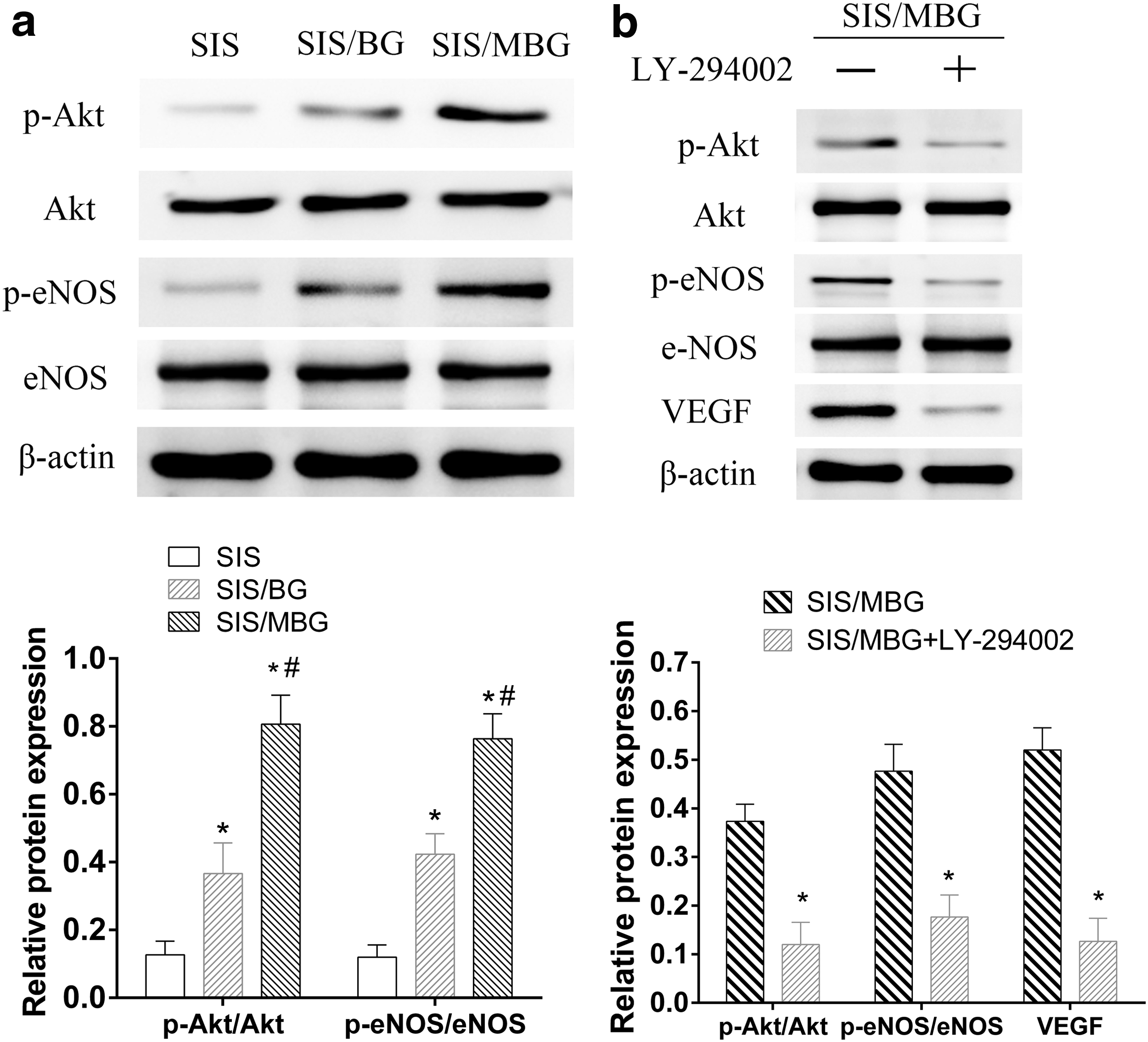
PI3k/Akt signaling pathway involved in the angiogenic differentiation of HUVEC cultured on scaffolds.
Evaluation of mineralized matrix and new bone formation in vivo
According to micro-CT analysis, the bone defects in the blank and SIS groups remained largely open. A few mineralized regions formed at the defect edges in the SIS/BG group (Fig. 8a). The defects filled with SIS/MBG displayed a much greater closure and its bone volume fraction (BV/TV%) was the highest (44.62% ± 6.77%) as result of the most efficient mineralized bone tissue formation (Fig. 8b). The mineralized density (BMD) of the SIS/MBG group (36.19 ± 4.99 mg/cm3) was almost 1.5-fold that of the SIS/BG group (25.28 ± 5.65 mg/cm3) and more than 3-fold (p < 0.05) that of the SIS group (10.15 ± 2.47 mg/cm3) (Fig. 8c).
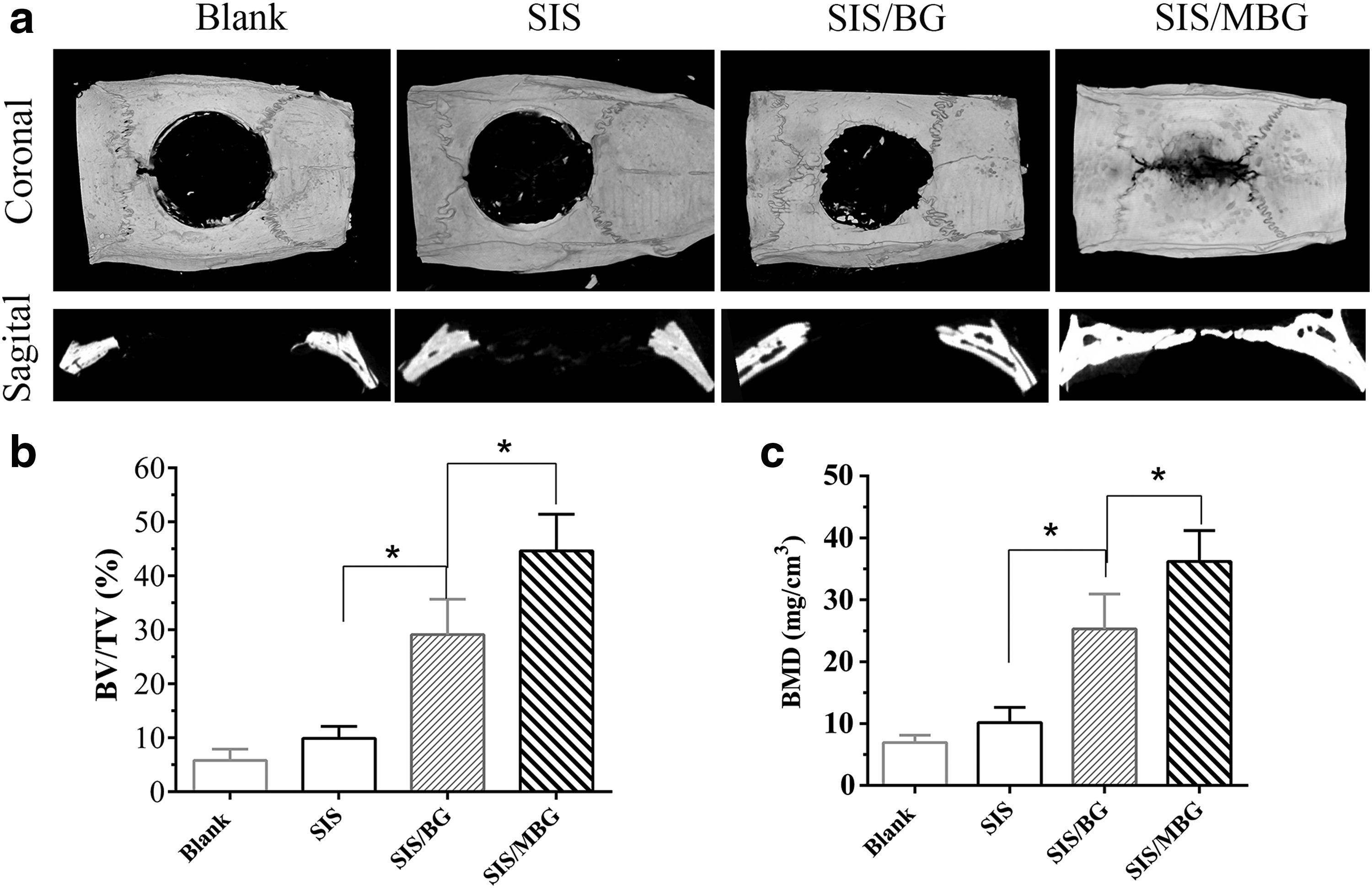
micro-CT evaluation of the repaired skull at 8 weeks after implantation.
H&E- and Masson-stained images (Fig. 9a–d) illustrate that the defect area in the blank group is filled with a thin connective fibrous tissue layer. The defect areas in the SIS group are bridged with a thick layer of fibrous connective tissue, composed mainly of collagenous tissue and newly formed vessels, although no newly formed bone was observed. In contrast, the morphological characteristics of active new bone formation are evident in the SIS/MBG and SIS/BG groups: the presence of osteocytes in a matrix of woven bone, and a layer of osteoblasts lining the boundaries of the newly formed bone. Newly formed bone stained with orange–red and collagenous tissue stained with blue are clearly seen in the Masson's Trichrome-stained image. Consistent with the micro-CT results, the histological analysis presented that SIS/MBG group had the highest new bone area fraction (60.92% ± 9.77%) compared with SIS/BG group (37.83% ± 8.74%) and pure SIS group (9.39% ± 2.60%) (Fig. 9b).

Histological analysis.
The osteogenic marker Ocn was detected by IHC staining. The Ocn was mainly expressed in cells with active osteogenic response. Ocn-positive staining is weak in the SIS group, but becomes apparent in the SIS/BG. The Ocn-positive staining is strongest in the SIS/MBG group (Fig. 10b).
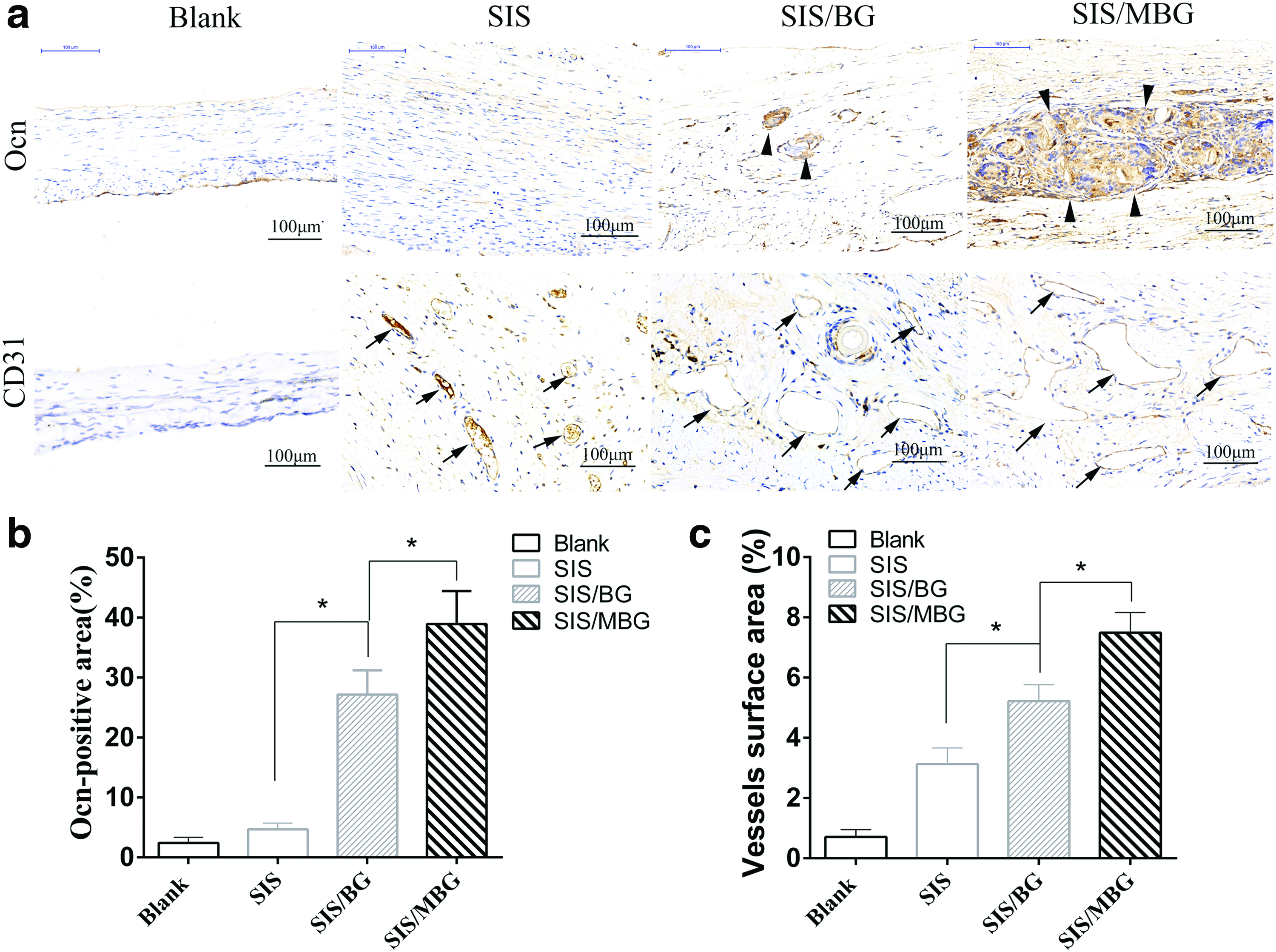
Immunohistochemical staining for Ocn and CD31 at 8 weeks after implantation.
Analysis of blood vessel formation in vivo
Massive blood vessels are seen in the SIS group at 8 weeks postimplantation (Fig. 9d), indicating a strong stimulatory effect of SIS on the ingrowth of blood vessels. Further enhanced angiogenesis resulted from incorporation of BG into SIS. New vessel density in the SIS/MBG group (51.12 ± 6.11 vessels/mm2) was 150% higher than that in the SIS group (37.02 ± 5.28 vessels/mm2) (p < 0.05) and 37% higher than that in the SIS/BG (20.92 ± 4.76 vessels/mm2) (p < 0.05) (Fig. 9c). Immunohistochemistry showed CD31-positive blood vessels (brown structures in Fig. 10a) in the specimen. The quantification of CD31-positve area showed that a higher fraction of vessel surface area was observed in the SIS/MBG group compared with that in the SIS/BG group or pure SIS group (Fig. 10c). Taken together, these results indicate that the MBG/SIS scaffolds had higher stimulatory effects on ingrowth of blood vessels than SIS/BG or SIS scaffolds.
Discussion
ECM-derived SIS highly retains the complex organization and composition of ECM that provide cells with the biological cues to proliferate and differentiate. 30 But the extracellular microenvironment of SIS is far away from the bony microenvironment. Moreover, the mechanical properties of pure SIS are reported to be considerably lower than those of native bone. 2 In this study, we tried, for the first time, to incorporate MBG into ECM-based material to develop composite SIS/MBG scaffolds with enhanced mechanical strength and capacity for osteogenesis and angiogenesis. MBG is extensively investigated to improve the osteogenic inductivity of bone grafts, because it could endow scaffolds with functional interface of well-ordered mesoporous structure and rapid apatite-formation ability. 14 This novel composite SIS/MBG harnessed the inherent advantages of SIS and MBG.
In the present study, we fabricated porous three-dimensional composite SIS/MBG by freeze-drying method. SIS is strengthened with bioceramic MBG, and SIS/MBG scaffolds showed a significant increase in mechanical property (∼0.87 MPa). From the perspective of bone tissue engineering, these scaffolds are within stiffness range (>30 kPa) that proved to favor osteogenic differentiation of stem cells. 31 In addition, the incorporation of MBG also may affect the topological properties and surface roughness, thereby regulating the cell–matrix interaction and the osteogenic commitment of stem cells. 1 The porosity and pore structure of scaffolds are another essential parameters that influence the in vitro and in vivo bone formation. 14 The highly porous structure with interconnected pores is effective for cell migration, nutrient delivery, bone ingrowth, and eventually vascularization.2,32 From the pattern of our results, SIS/MBG has higher porosity (76% ± 4%) and interconnected pore structure with pore size ranging from 20 to 150 μm.
Cell–biomaterial adhesion is crucial for cell–biomaterial interaction, which influences cellular function, including migration, proliferation, and differentiation. 1 In this regard, the ECM-derived SIS was expected as an excellent platform to support cell adhesion and proliferation. In this study, a favorable cell attachment and proliferation was observed in three scaffold types, exhibiting the tremendous advantage of SIS as matrix: SIS highly retains the cell-binding sites of ECM such as Arg-Gly-Asp (RGD), laminin, and fibronectin. 8 Furthermore, the composite scaffolds enhanced the proliferation and osteogenic differentiation of rBMSC as well as the proliferation and angiogenic differentiation of HUVEC compared with pure SIS. For one thing, it might be attributed to the fact that Ca and Si ions released from MBG or BG have a stimulatory effect on the proliferation and osteogenic differentiation of BMSCs, and the release of Si ions has a positive effect on the proliferation and angiogenic differentiation of HUVEC.33,34 For another, the incorporation of MBG or BG awards scaffolds with rigid and rough surface features. It has been documented that a more rigid or rough matrix enhances osteogenic differentiation through integrin-mediated mechanotransduction.35,36
Impressively, it was found that SIS/MBG scaffolds had significantly improved osteogenesis and angiogenesis compared with SIS/BG scaffolds, despite their having the same chemical composition, indicating that mesoporous channel structure and increased surface area on the scaffold are of great importance in improving their osteogenic and angiogenic activities. Furthermore, MBG has improved apatite mineralization ability compared with BG, 15 which may contribute to the osteogenic activity of the materials. Chou et al. also found that the biomimetic apatite layer has an active effect on osteoblast viability, and gene expression. 37 Another important property of MBG is the drug loading and release property. 38 Previous studies have shown that SIS is rich in growth factors, such as bFGF, VEGF, and TGF-β1, all strong inducers of angiogenesis.5,39 The angiostimulative factors would be released from SIS/MBG in a controlled pattern, thereby exerting a sustained stimulatory effect on HUVEC. Moreover, El-Fiqi et al. found that the incorporation of MBG into collagen could improve chemical stability by reducing enzymatic degradation. 28 It is reasonable to speculate that a more stable chemical interface of SIS/MBG as compared with SIS/BG would profit cell adhesion, proliferation, and differentiation.
For developing ideal bone graft, it is necessary to elucidate both materials specific effects and the molecular mechanisms directing cell differentiation. Wnt/β-catenin signaling pathway is involved in biomaterial-induced osteogenic differentiation.40,41 The canonical Wnt signaling pathway is transduced by stabilizing β-catenin protein through inhibition of GSK-3-mediated β-catenin phosphorylation. Nonphosphorylated β-catenin then translocates to the nucleus, where it activates downstream gene transcription, such as Runx2.42,43 Runx2 is a master regulator in the commitment of osteogenesis. 44 The western blot analysis exhibits that the activation of Wnt/β-catenin is involved in the stimulatory effect of SIS/MBG or SIS/BG on osteogenic differentiation (Fig. 7). It is consistent with the previous report that Si ions could induce the osteogenic differentiation by activating the Wnt-related signaling pathway of rBMSCs. 45 Particularly, higher protein expressions of Runx2 and β-Catenin were observed in SIS/MBG as compared with SIS/BG, suggesting that mesoporous structure on SIS/MBG has a positive effect on the protein expression of β-catenin and Runx2.
Many studies have shown that MBG-based materials stimulate angiogenesis.14,46,47 However, little is known about the mechanism of the stimulation process. The PI3-kinase/Akt/eNOS pathway has been demonstrated to exert a critical effect on angiogenesis. 48 The activation of PI3k and phosphorylation of Akt and eNOS lead to increased activity of eNOS and upregulated extracellular level of VEGF, which is critically involved in promoting endothelial cell proliferation and migration. 49 In the present study, SIS/MBG composite markedly augmented phosphorylation of Akt and eNOS. When treated with the PI3-kinase inhibitor LY294002, the expression of p-Akt, p-eNOS, and VEGF was downregulated. Collectively, these results underlie the importance of the PI3-kinase/Akt pathway in SIS/MBG-induced neovascularization.
Osteogenesis and angiogenesis are both critical process of bone regeneration. Until now, several strategies aim to accelerate osteogenesis and angiogenesis, including modification of the scaffold, transplantation of stem cell, and localized delivery of osteostimulative and angiostimulative growth factors. Biomaterial-based strategies for enhancing osteogenesis and angiogenesis possess some advantages compared with other approaches, such as favorable controllability, decreased complication risk, and low cost. Based on the inherent advantage of SIS or MBG, composite scaffold SIS/MBG was expected to accelerate osteogenesis and angiogenesis in vivo. At 8 weeks postimplantation, many new vessels (∼20.92 ± 4.76 vessels/mm2) formed in the entire defect area of pure SIS group despite minimal new bone formation observed at the edge of the defect. This was consistent with previous reports that SIS has a dominant stimulatory effect on angiogenesis.5,39 Consistent with the in vitro results, the in vivo results demonstrate that the incorporation of MBG or BG into SIS significantly increased the new bone fraction and new vessel numbers in the bone defect area. SIS/MBG scaffolds achieved the highest efficacy in bone regeneration and neovascularization, and the new bone area fraction and the vessels density were up to 60.92% ± 9.76% and 51.12 ± 6.11 vessels/mm2, respectively.
In summary, we attributed the high bone regeneration and neovascularization ability of SIS/MBG to the unique combination of the SIS and MBG composites: First, SIS highly retains multiple bioactive component of ECM, and growth factors such as VEGF or bFGF, stimulating cell proliferation differentiation, and the further tissue regeneration. 8 Second, MBG may enable controlled release of growth factors from SIS, such as VEGF and bFGF,24,38 thus enhancing angiogenesis. Third, MBG has an excellent apatite mineralization ability, 15 contributing to the osteogenic differentiation of stem cells. 14 Lastly, the physical features of MBG—superior specific surface area and nanopore structure—may have a positive effect on the osteogenic differentiation of stem cells and angiogenic differentiation of vessel endothelial cells.
Conclusions
In this work, we developed a novel biomimetic composite scaffold SIS/MBG with superior capacity for osteogenesis and angiogenesis. SIS/MBG possesses high porosity, excellent pore structure, and favorable mechanical properties. The in vitro studies show that the SIS/MBG not only enhances cell proliferation and osteogenic differentiation of rBMSCs, but also promotes angiogenesis in HUVECs. We confirmed that SIS/MBG induces rBMSCs differentiation through the Wnt/β-Catenin signaling pathway and promotes NO and eNOS protein synthesis in HUVECs through the PI3k/Akt pathway. SIS/MBG was shown to be capable of enhancing bone regeneration and neovascularization simultaneously in critical-sized rat calvarial defects. Therefore, the osteostimulative and angiostimulative biomimetic composite scaffold SIS/MBG represents an exciting biomaterial option for bone regeneration.
Footnotes
Acknowledgments
This work was financially supported by the National Natural Science Foundation of China (Grant No. 81672158 and 81371939), International Science & Technology Cooperation Program of China (2013DFG32690), and National Key R&D Program of China (2016YFC1100100).
Disclosure Statement
No competing financial interests exist.