
Abstract
Recombinant human bone morphogenic protein-2 (BMP-2)-loaded absorbable collagen sponges (ACS) have been successfully used to enhance bone formation and to induce spinal fusion in humans. However, side effects, such as soft tissue edema and inflammation, have been reported. NEMO binding domain peptide (NBD) inhibits activation of nuclear factor kappa-light-chain-enhancer of activated B cells (NF-κB), a central regulator of immune response. In this study, we investigated NBD's potential to reduce BMP-2-induced soft tissue inflammation without affecting BMP-2-mediated spinal fusion in rat. For evaluation of soft tissue inflammation, ACS containing BMP-2, BMP-2+NBD, NBD, or ACS only were implanted into intramuscular paraspinal sites of 32 rats. At day 2 postsurgery, edema formation at the implant sites was assessed using magnetic resonance imaging. T2-weighted relaxation time (T2-RT) values were increased in the BMP-2 group compared with BMP-2+NBD, NBD, and ACS groups. No difference in T2-RT values was detected between BMP-2+NBD versus NBD and ACS controls. Postsacrifice, histological analysis of the implant-surrounding zones showed increased mononuclear cell infiltration in the BMP-2 group compared with BMP-2+NBD and controls. The presence of BMP-2 increased relative NF-κB binding and gene expression of inflammatory markers, interleukin (IL)1β, IL6, IL18, and chemokine ligand (CCL)2 and CCL3 compared with controls. In the BMP-2+NBD group, cytokine expression was blocked. No differences were found between BMP-2+NBD and control groups. For evaluation of spinal fusion, posterolateral intertransverse lumbar fusion procedures were performed on 16 rats. ACS were loaded with BMP-2 or BMP-2+NBD. After sacrifice at week 12, microcomputed tomographic assessment of the fusion site detected a higher bone volume and reduced trabecular spacing in the BMP-2+NBD group compared with BMP-2. Histological analysis did not show any differences in newly formed bone microarchitecture. In summary, addition of NBD to BMP-2-loaded ACS reduces BMP-2-induced soft tissue edema formation and mononuclear cell infiltration, diminishes NF-κB binding, and thus blocks transcription of NF-κB-regulated cytokines in rat. Furthermore, NBD stimulates bone formation in BMP-2-mediated spinal fusion, possibly through crosstalk of the NF-κB pathway with other pathways. The results of this study might provide the basis to develop new therapeutic bone grafting approaches with combinatory administration of BMP-2 and NBD for spinal fusion.
Introduction
R
The inducible transcription factor, nuclear factor kappa-light-chain-enhancer of activated B cells (NF-κB), plays a crucial role in the inflammatory response. Activation of NF-κB has been shown to be important in many inflammatory diseases, including rheumatoid arthritis and intervertebral disc disease.15,16 It is induced by a variety of molecules, including TNF-α and IL-1β. 17 Activation of NF-κB results in the transcription of target genes, including the cytokines TNF-α, IL1β, IL6, and IL18, which can further increase the inflammatory response.18–20 Furthermore, CC-motif ligands such as CCL-2 (MCP-1) and CCL-3 (MIP-1α) are prominent target genes of the NF-κB signaling pathway. 21 An increased expression of these chemokines has been shown to attract proinflammatory cells such as macrophages, which further increase the inflammatory response. 22 Inhibition of NF-κB increased bone volume and bone mineral density (BMD) as a result of an increased osteoblast activity observed in mice, providing evidence for a role of NF-κB in inhibition of bone formation. 23
Corticosteroids are known to reduce soft tissue inflammation in spinal procedures, but have been associated with side effects, especially, when used in higher doses.24–27 NEMO binding domain peptide (NBD) is an attractive alternative to corticosteroids, since it has been shown to have anti-inflammatory effects and a low toxicity profile, as shown in vitro and in vivo,18,28 including a clinical phase I trial in dogs. 29 This cell-permeable peptide has been shown to block proinflammatory cytokine-induced NF-κB activation without affecting the basal activity of IκB kinase (IKK), the mediator of IκB phosphorylation/NFκB translocation into the nucleus. 18 Local treatment with NBD has anti-inflammatory effects on human rheumatoid arthritis and was shown to reduce osteoclastogenesis and bone erosion in inflammatory arthritis.30,31 Moreover, NBD has been shown to significantly inhibit carrageenan-induced edema formation and cellular inflammation in mouse paws. 18 However, there is no information on the effect of NBD on BMP-2-induced edema and inflammation to date. In this study, we hypothesized that inhibition of NF-κB using the NBD results in a reduction of BMP-2-induced inflammation in a rat model of paraspinal soft tissue inflammation. Furthermore, we hypothesized that combinatory administration of NBD/BMP-2/ACS has beneficial effects on spinal fusion outcomes.
The aims of this project were therefore to (1) demonstrate that addition of NBD to BMP-2/collagen sponges results in a reduction of BMP-2-induced edema and inhibition of NF-κB-regulated cytokine/chemokine levels, and to (2) demonstrate a solid spinal fusion when supplementing BMP-2/collagen sponges with NBD.
Materials and Methods
Study design
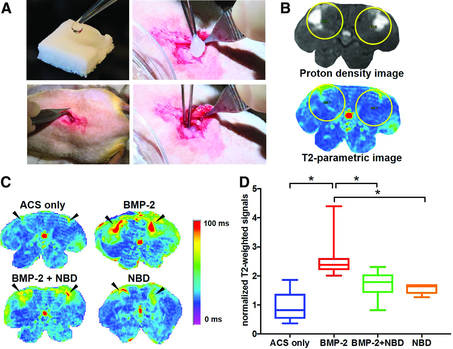
The Cedars-Sinai IACUC approved all procedures described in this study (protocol #006054 and #007328). To investigate the effect of NBD on BMP-2-induced soft tissue inflammation, 32 male adult Sprague Dawley rats (250–350 g), obtained from Charles River Laboratories, were divided randomly into four groups. Since a significant formation of soft tissue edema in response to 129 μg BMP-2 was previously demonstrated by our group on day 2 postsurgery, 14 Helistat® ACS (5 × 10 × 10 mm, Integra Lifesciences) was loaded with 129 μg BMP-2 in BMP-2 buffer solution (Medtronic) and implanted into paraspinal muscle to induce a strong inflammatory response in rat soft tissue (Table 1, group II). To evaluate the reduction of soft tissue inflammation in response to NBD (Anaspec), ACS containing 129 μg BMP-2 and 100 μg NBD were implanted into paraspinal muscle tissue (group III). The amount of NBD was selected based on the study of di Meglio et al. showing amelioration of acute inflammation by systemic administration of 100 μg of NBD per mouse. 18 In our study, 100 μg NBD was applied locally at each implant site. As controls, ACS loaded with 100 μg NBD (group IV) and BMP-2 buffer only (group I) were included. Two days postsurgery, micromagnetic resonance imaging was used to assess edema volume in vivo. To evaluate inflammatory responses, histological staining, quantitative reverse transcription polymerase chain reaction (RT-PCR), and an NFκB binding activity assay were conducted postsacrifice on day 2.
ACS, absorbable collagen sponges (5 × 10 × 10 mm); BMP-2, recombinant human bone morphogenetic protein-2; μCT, microcomputed tomography; NBD, NEMO binding domain peptide; NF-κB, nuclear factor kappa-light-chain-enhancer of activated B cells; RT-PCR, reverse transcription polymerase chain reaction.
To compare BMP-2-mediated spinal fusion in the presence and absence of NBD, eight male adult Sprague Dawley rats (250–350 g), obtained from Charles River Laboratories, underwent spinal fusion using BMP-2 (total dose of 30 μg/side/rat)- and NBD (100 μg)-loaded ACS (group V) and eight rats underwent spinal fusion with BMP-2 (30 μg)-loaded ACS (group VI). Based on a previous study of our group showing 100% spinal fusion when using a total dose of 30 μg BMP-2/ACS/per side/rat versus 0% fusion in 0 μg BMP-2/ACS-implanted rats, 4 the 30 μg concentration of BMP-2/ACS was chosen. After 12 weeks, ex vivo microcomputed tomography (μCT) was performed on rat spines to determine the structural properties of the fusion mass. For functional and quantitative assessment of spinal fusion, manual palpation testing was performed. Finally, histological analysis of spinal fusion sites was performed.
BMP-2 release profile in vitro
The kinetics of BMP-2 release from Helistat ACS (Integra Life Science) in the presence and absence of NBD was determined. ACS (10 × 10 × 5 mm) were loaded with either BMP-2 (129 μg) or BMP-2 (129 μg)+NBD (100 μg). The loaded ACS were incubated for 15 min at room temperature before being placed into a 2 mL tube containing 0.5 mL of BMP buffer (Medtronic). Tubes were incubated at 37°C with continuous agitation. After a period of 1, 2, 3, 6, 8, 10, and 14 days, the supernatant was collected, and fresh buffer was added. The amount of BMP-2 in the supernatant obtained at each time point was determined with the human BMP-2 Quantikine ELISA kit (R&D systems).
Animal surgery
Rats were anesthetized and maintained with inhaled isoflurane (2%) via the chamber of a rat anesthesia machine and weighted. For the procedure of intramuscular collagen sponge implantation, a 25 mm midline posterior longitudinal incision was made in the skin over the lumbar spine. Prepared solution was loaded onto ACS 15 min before intramuscular implantation into 10 × 10 mm pockets, which were created in the paraspinal muscle tissue at the incision site. For the single-level posterolateral intertransverse process fusion surgical procedure, a posterior midline longitudinal incision was made at the level of the lumbar spine. Dissection of the soft tissue was performed to expose the transverse processes of lumbar spinal segment of L4 and L5. The transverse processes were decorticated with a high-speed burr until punctate bleeding, whereas the lamina and facet joints were left intact. The loaded ACS implants were placed bilaterally in the paraspinal muscle bed, between and touching the transverse processes of L4 and L5. No internal fixation was used. After all surgical procedures, muscles and skin were closed with nonabsorbable sutures. The surgeon was blinded to the experimental treatment groups. The rats were medicated with subcutaneous injections of buprenorphine (0.05 mg/kg) to control pain. Rats were sacrificed at the individual study endpoints as detailed in the Study Design section.
Edema evaluation using μMRI evaluation
Micro-MRI was used to assess soft tissue edema volume as an index of inflammation in the presence and absence of NBD in groups I–VI on day 2. A small animal magnetic resonance imaging scanner, Bruker BioSpec 9.4T (94/20) with Avance III electronics, was used with technician support of the Research Imaging Core, Cedars-Sinai Medical Center. A quadrature 1H 65 mm coil was used to acquire all images. An multi-slice-multi-echo (MSME) sequence with an echo time of 11 ms with 16 echoes was used to collect data for T2 parametric images, 2 averages. Twenty contiguous axial slices with a field-of-view of 5.5 × 5.5 cm with a slice thickness of 2 mm and in-plane resolution of 430 μm were obtained encompassing the paraspinal tissues. The T2 relaxation time (T2-RT) in each implant was measured using MIPAV computer imaging software (Medical Image Processing, Analysis, and Visualization, NIH, Bethesda, MD). To define the region of interest (ROI), proton density images were used; first echo the T2 sequence. A circle of 0.9 cm2 was chosen to ensure that all implants fit into the ROI, thereby keeping the area size consistent across animals and between experimental groups. Next, with help of the imaging software, T2-parametric images of all 20 slices obtained per rat were analyzed for the defined ROI (Fig. 1B). The T2 relaxation time within the ROI was measured for each implant. MRI scans obtained from each rat before surgery were equally processed and used for background subtraction.
Histology
A hematoxylin/eosin (H&E) staining was performed to qualitatively analyze the tissue surrounding the implants in the presence and absence of NBD (groups I–IV). H&E is a widely used stain that allows for the visualization of mononuclear cell infiltration into muscle and other tissues.32,33 The tissue analysis was performed by two independent researchers, a histologist and a pathologist. In addition, H&E staining and Masson's trichrome staining were performed to assess new bone formation in the fusion sites across experimental conditions (groups V–VI). For analysis of groups I–IV, rats were sacrificed on day 2 postoperatively and soft tissue, including muscle and implants, was collected, fixed in 10% formalin, and paraffin embedded. For analysis of fusion groups V–VI, rats were sacrificed after 12 weeks, spines were resected, fixed with 10% formalin, and decalcified with 15% EDTA. For assessment of new bone formation, the specimens were sliced in the sagittal plane near the transverse processes at levels L4–5. 13
RNA isolation and gene expression analysis using quantitative RT-PCR
Total RNA was isolated from treated soft tissue using TRIzol® Reagent (Thermo Fisher Scientific) and subsequent homogenization. The quality and quantity of the RNA were determined by spectrophotometry. RNA was transcribed in cDNA using the High-Capacity cDNA Reverse Transcription Kit (Applied Biosystems). Levels of IL1β, IL6, IL18, TNF-α, CCL2, and CCL3 were detected in terms of commercially available TaqMan® Expression Assays as per the manufacturer's instructions using Bio-Rad CFX96 Touch™ Real-Time PCR Detection System (Bio-Rad, Hercules, CA). For the assay, the TaqMan™ Universal PCR Master Mix (4304437; Applied Biosystems) and the following primers were used: IL1β: RN00580432_M1, IL6: RN01410330_M1, IL18: RN01422083_M1, TNF-α: RN01525859_G1, CCL2: RN00580555_M1, and CCL3: RN01464736_G1; Applied Biosystems). Samples were assayed in triplicate. Expression levels of mRNA were normalized to 18S rRNA Endogenous Control (4333760T; Applied Biosystems). For data analysis, the 2-ΔΔCq (Livak) method was used.
NFκB p65 transcription factor assay
To demonstrate a reduced NF-κB DNA binding activity in soft tissue in the presence of NBD compared with controls, an NFkB p65 Transcription Factor Assay Kit (Abcam) was used as per the manufacturer's instructions. To obtain tissue nuclear extracts, soft tissue, including muscle and implants, was shock-frozen in liquid nitrogen. Immediately after thawing, the samples were suspended in ice-cold hypotonic lysis buffer and homogenized using a tissue homogenizer. The homogenates were chilled on ice and then treated with a Nuclear Extraction Kit (Abcam) as per the manufacturer's instructions. Protein concentrations in samples were equalized using a bicinchoninic acid assay (BCA assay; Thermo Scientific). Before NF-κB DNA binding activity assay performance, samples were 10-fold concentrated by using the Ultracel-3K Centrifugal Filter system for concentration and purification (Amicon, Ireland). Samples were assayed in duplicate.
Microcomputed tomographic scans
Ex vivo μCT (vivaCT 40, Scanco USA, Inc., Wayne, PA) was performed on explanted spines to determine the structural properties of the fusion mass. All μCT procedures used in this study were previously described in detail by Kallai et al. 34 Microtomographic slices were acquired using an X-ray tube potential of 55 kVp and reconstructed at a spatial nominal resolution of 35 μm. A constrained 3D Gaussian filter (σ = 0.8, support = 1) was used to partly suppress volume of interest (VOI) noise. The trabecular bone tissue was segmented from marrow and soft tissue by using a global thresholding procedure. 35 The researcher was blinded to the experimental groups. Histomorphometric 3D evaluation was performed on a cylindrical VOI, including the upper and lower vertebral bodies around the fusion bed at the transverse processes of L4–L5. Then, the following morphometric parameters were evaluated using ScancoMedical MicroCT Software (www.scanco.ch/en/systems-solutions/software/3d-analysis.html): volume of mineralized bone tissue (BV), connectivity density (Conn.D), BMD, trabecular number (Tb.N), trabecular thickness (Tb.Th), and trabecular spacing (Tb.Sp). To exclude BV of native vertebrae, BV of treated rats was normalized to BV of the same area of control rats (native, untreated), resulting in ΔBV.
Spinal fusion assessment using manual palpation
Fusion quality and quantity were assessed via manual palpation testing of spine segments by two independent observers, a method that has been described in detail in the published appendix of a prior study from our group. 4 Any motion detected between the L4 and L5 segment and adjacent segments, including the transverse process or vertebral bodies, was considered to indicate a fusion failure. Each side was tested separately; the observation of no motion on both the right and left sides indicated a successful fusion. In the figures, fusion rate refers to the number of animals showing successful fusion (unit of observation is “animal with at least one lumbar segment fused”). Number of fusion levels describes the number of bone segments (proximal vertebral body–disc–distal vertebral body) that were detected to show no motion between two vertebral bodies (e.g., one level, with fusion at L4 and L5). There could be more than one level fused within an animal (e.g., two levels, with fusion at L3–L4 and L4–L5).
Statistics
All statistical analyses were performed using SAS 9.3 and Prism 7; p < 0.05 was considered significant. The outcome measurements were (1) relaxation time (T2-RT), (2) levels of gene expression, (3) NFκB binding activity, (4) μCT measures, and (5) BMP-2 release kinetics. Separately for each dependent measure, analysis of variance, mixed model, was performed using mean values with grouping of implant group; appropriate post hoc tests for multiple comparisons using Tukey's or Sidak's honestly significant were applied. In figures, median values are shown as boxplots.
Results
NBD reduces BMP-2-induced edema formation in rat
To investigate the effect of NBD treatment on BMP-2-induced edema formation, μMRI analysis was performed in vivo on rat lumbar spines and surrounding tissue on day 2 postimplantation (Fig. 1A–D). Quantification of T2-RT of the ROI resulted in a 2.7-fold increase in the BMP-2 group compared with ACS-only group (group I vs. II: medianBMP-2: 2.4, min: 2.0, max: 4.4, medianACS: 0.8, min: 0.4, max: 1.9, p < 0.05). Addition of NBD to BMP-2-loaded collagen sponges reduced the BMP-2-mediated induction of edema formation (group II vs. III: medianBMP-2+NBD: 1.8, min: 0.8, max: 2.3, p < 0.05). To determine the effect of NBD only, ACS were supplemented with NBD in the absence of BMP-2 (group IV). No significant difference was detected between ACS-only and NBD-only groups (group I vs. IV), whereas the BMP-2 group had significantly higher T2-RT values compared with the NBD group (group II vs. IV: medianNBD: 1.6, min: 1.3, max: 1.7, p < 0.05). No differences in T2-RT values of the ROIs were observed before implantation. To investigate whether the addition of NBD to BMP-2-loaded ACS affects the BMP-2 release, a BMP-2 release profile was performed in vitro. No differences in the cumulative release of BMP-2 in solution were detected between the BMP-2 and BMP-2+NBD groups (Supplementary Fig. S1; Supplementary Data are available online at www.liebertpub.com/tea).
In the presence of NBD, endogenous mononuclear cell infiltration is reduced
H&E staining of tissue samples obtained from experimental groups I–IV indicated an increased mononuclear cell infiltrate in the area surrounding BMP-2-loaded implants compared with NBD- and ACS-only controls. The cell response was reduced in implants of experimental group III (BMP-2+NBD) compared with group II (BMP-2; Fig. 2).
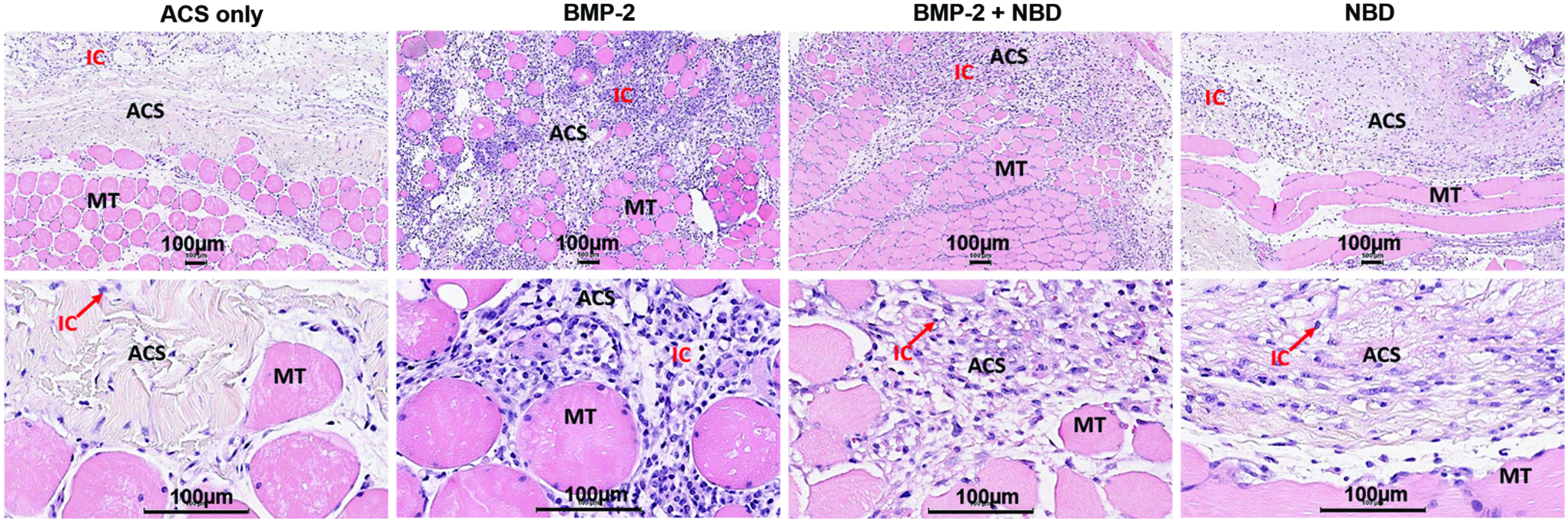
Histological evidence of mononuclear cell infiltration into implants/muscle tissue at day 2 postsurgery. Top: low-magnification histological images. Bottom: high-magnification histological images. H&E staining shows an increased cellular activity in the implant surrounding zone in the BMP-2 group, which is diminished in the BMP-2+NBD group. Images were obtained from n = 3 per condition. IC, infiltrating cells; MT, muscle tissue; ACS, absorbable collagen sponges; H&E, hematoxylin/eosin. Color images available online at www.liebertpub.com/tea
NBD blocks BMP-2-induced levels of inflammatory and chemotactic genes
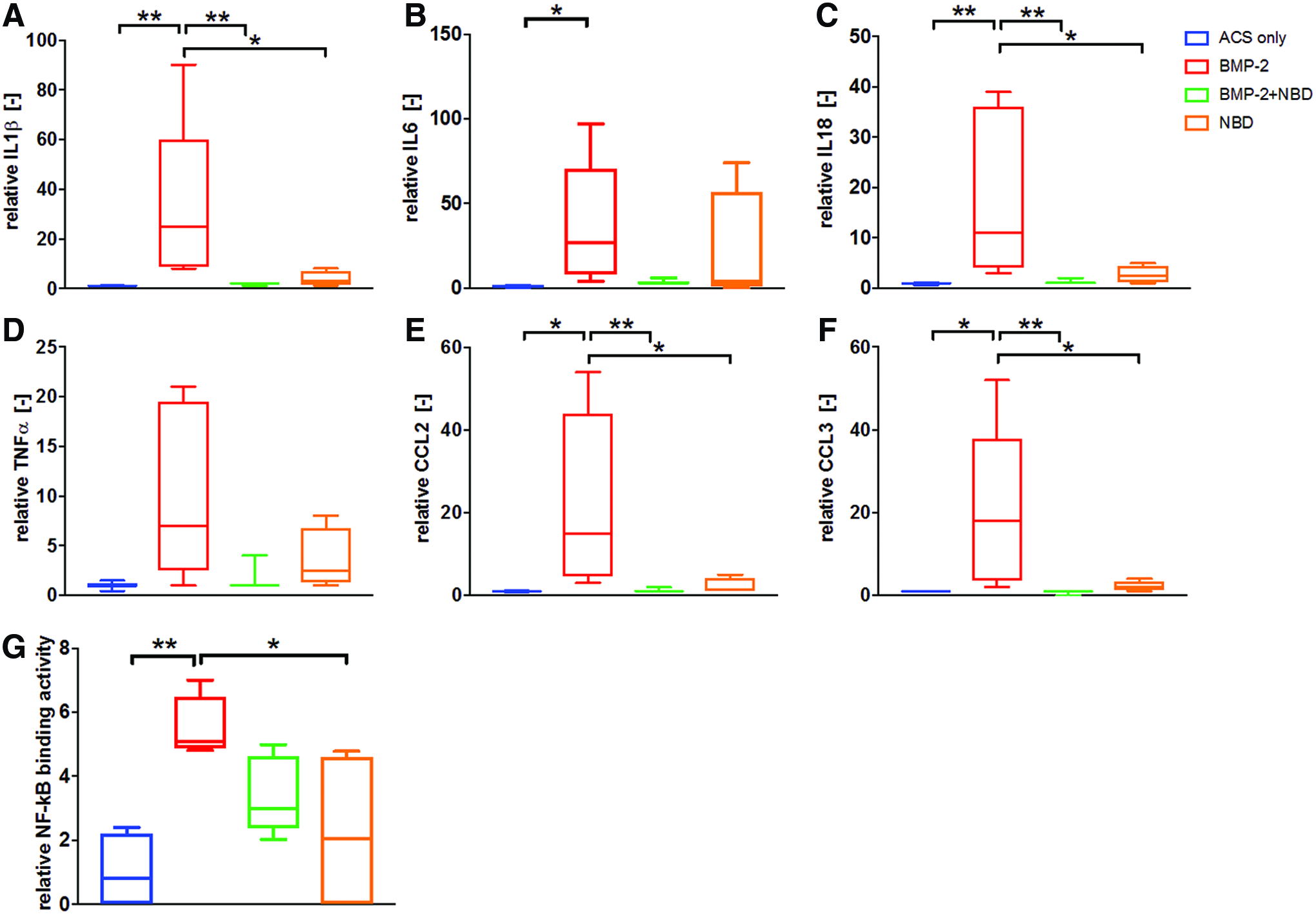
To evaluate whether treatment with NBD blocks NF-κB downstream target genes, implant-containing tissue samples were analyzed for expression of proinflammatory cytokines IL1β, IL6, IL18, TNF-α, and chemokines CCL2 (MCP-1) and CCL3 (MIP-1α) (Fig. 3A–F). Increased levels were observed in relative gene expression in the BMP-2 group compared with the ACS-only group for all genes except TNF-α (groups I vs. II; medianBMP-2: IL1β: 25.5, min: 8.4, max: 89.8, p < 0.01, IL6: 27.3, min: 4.0, max: 96.7, p < 0.05, IL18: 11.1, min: 3.2, max: 39.0, p < 0.01, CCL2: 14.8, min: 2.5, max: 53.9, p < 0.05, and CCL3: 18.2 min: 1.8, max: 51.9, p < 0.05). This BMP-2-mediated induction of gene expression levels was blocked in the presence of NBD (groups II vs. III; medianBMP-2+NBD: IL1β: 1.6, min: 1.4, max: 2.1, p < 0.01, IL18: 1.4, min: 1.3, max: 1.6, p < 0.01, CCL2: 1.1, min: 0.7, max: 1.9, p < 0.01, and CCL3: 0.6, min: 0.4, max: 1.4, p < 0.01) and showed a trend to be blocked for IL6: 2.6, min: 2.6, max: 5.8, p = 0.08. No induction of gene expression levels was measured in the NBD-only group, resulting in significant differences between the BMP-2 group and the NBD group (groups II vs. IV; medianNBD: IL1β: 3.1, min: 1.0, max: 8.1, p < 0.05, IL18: 2.3, min: 1.4, max: 4.6, p < 0.05, CCL2: 1.6, min: 0.6, max: 4.5, p < 0.05, and CCL3: 1.9, min: 0.9, max: 3.7, p < 0.05). There was no difference in IL6 levels between the BMP-2- versus NBD-only groups. No differences in gene expression levels of IL1β, IL6, IL18, TNF-α, and CCL2 and CCL3 were found between ACS-only versus NBD+BMP-2 and ACS-only versus NBD groups.
NF-kB DNA binding activity is increased in BMP-2-containing ACS
To investigate the NF-κB DNA binding activity in experimental groups I–IV, nuclear extracts were obtained from implant-containing muscle tissue samples and investigated in terms of the NF-kB p65 Transcription Factor Assay Kit (Abcam). Evaluation of the test results indicated a significant increase in NF-κB DNA binding activity in the BMP-2 group compared with ACS and NBD controls (groups II vs. I; medianBMP-2: 5.1, min: 4.8, max: 7.0, medianACS: 0.8, min: 0, max: 2.4, p < 0.01; groups II vs. IV; medianNBD: 2.0, min: 0, max: 4.8, p < 0.05). No difference was observed between BMP-2+NBD, NBD, and ACS groups (Fig. 3G).
NBD supplementation enhances BMP-2-induced spinal fusion
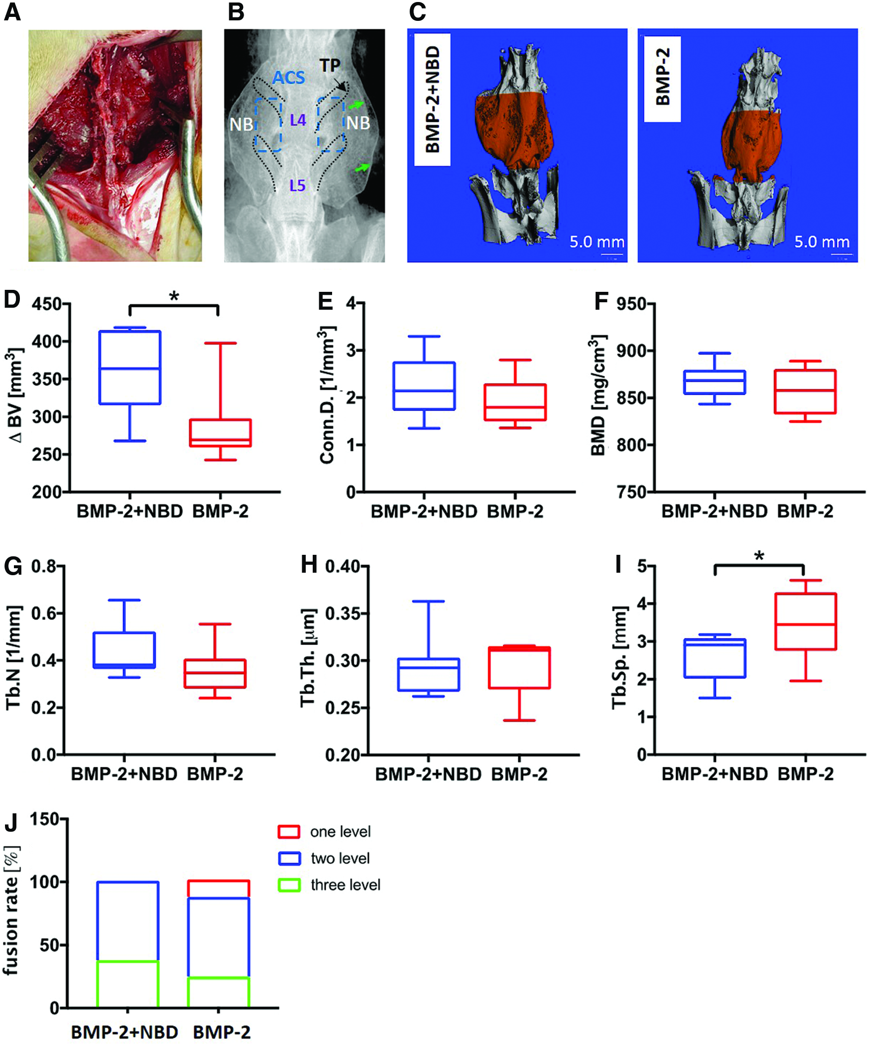
To evaluate the effect of NBD on BMP-2-induced new bone formation after the posterolateral intertransverse process fusion procedure (Fig. 4A, B), micro-CT analyses of explanted spines of experimental group I (BMP-2+NBD) and group II (BMP-2) were performed (Fig. 4C). Data evaluation showed a significant increase in the Δ bone volume ([bone volume at week 12]–[presurgical bone volume]) and a decreased trabecular spacing of group V compared with group VI (medianΔBV: group V: 363.9, min: 267.9.0, max: 418.7; group VI: 269.2, min: 242.5, max: 397.9, p < 0.05; median Tb.Sp: group V: 2.9, min:1.5, max: 3.1; group VI: 3.5, min: 2.0, max: 4.6, p < 0.05) (Fig. 4D and I). No differences were detected in the structural and qualitative parameters, connectivity density, BMD, trabecular number, and trabecular thickness (Fig. 4E–H). Manual palpation of freshly harvested spine explants showed 100% fusion in both experimental groups, with a higher number of levels fused in the BMP-2+NBD group (Fig. 4J). Qualitative analysis of histological sections of newly formed bone between the spinal processes L4–L5 did not indicate any differences in the microarchitecture of the newly formed bone mass (Fig. 5).
(
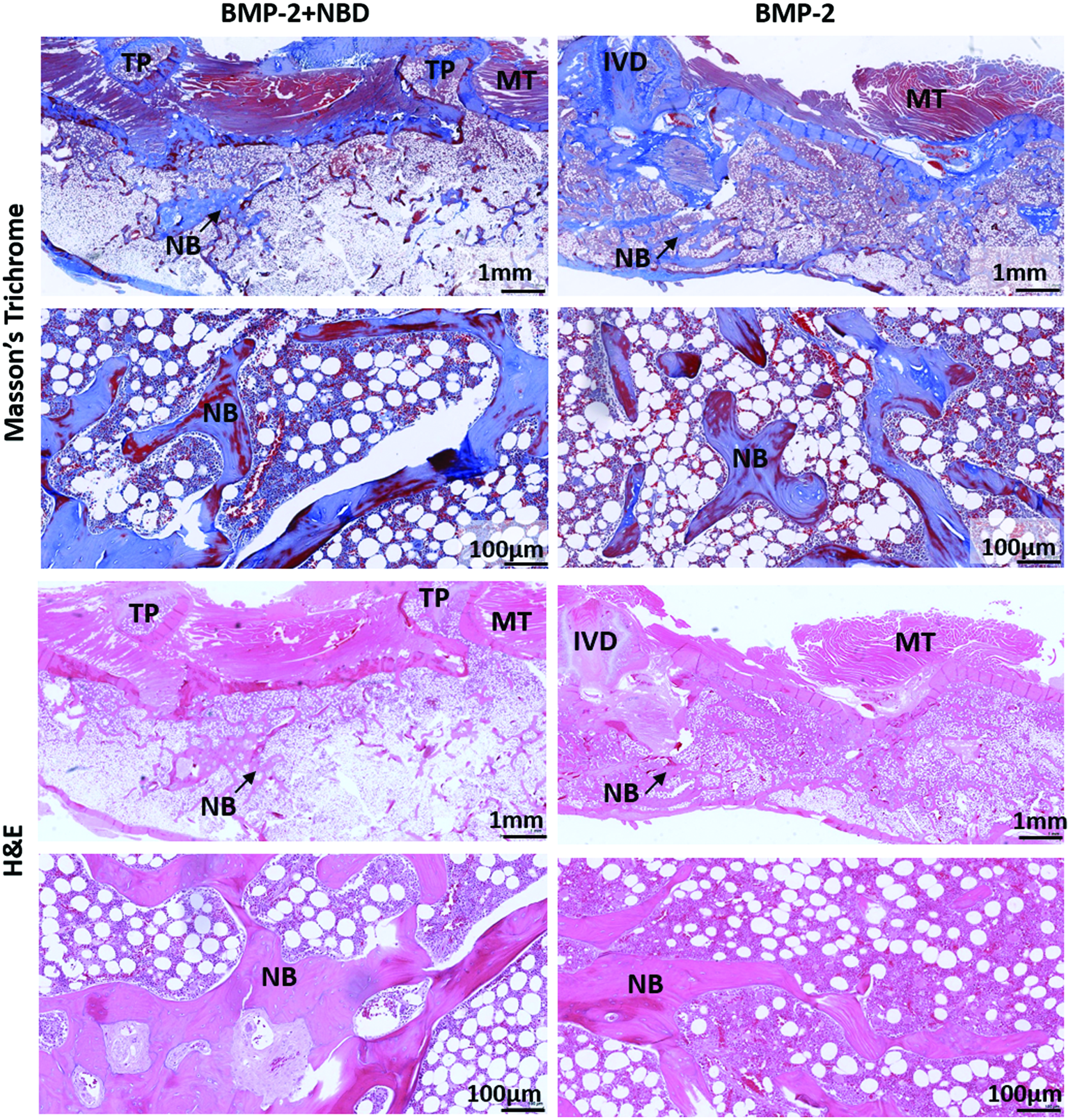
Representative images of fusion masses at the intertransverse sites. Masson's trichrome and H&E staining did not show any differences in new bone formation between BMP-2+NBD versus BMP-2 groups. Low (1 mm)- and high (100 μm)-magnification images are shown. TP, transverse processes; MT, muscle tissue; NB, new bone; IVD, intervertebral disc. Images were obtained from n = 3 per condition. Color images available online at www.liebertpub.com/tea
Discussion
Despite the well-known osteoinductive properties of BMP-2 and the protein's excellent fusion rates in the spine, adverse events related to exaggerated inflammatory responses and increased soft tissue edema adjacent to the implant sites have been reported.6,8,36 Corticosteroids, which have been shown to act at least partially through inhibition of the NF-κB activity in vitro and in vivo, are known to decrease edema formation and reduce cytokine levels.37,38 However, administration of corticosteroids has been associated with unwanted side effects, such as a higher risk of infectious complications.39,40 NBD has been shown to efficiently decrease NF-κB activity while sparing the protective functions of basal NF-κB activity,28,41,42 and may therefore have fewer undesired side effects.
In this study, by using a rat model of soft tissue inflammation, we demonstrated that addition of NBD (100 μg) to high-dose BMP-2 (129 μg)-loaded ACS results in a reduction of edema formation and mononuclear cell infiltration caused by BMP-2, suggesting a decreased inflammatory response. Indeed, an upregulation of proinflammatory cytokines and chemokines in response to BMP-2 was blocked by NBD in the treated soft tissues. Importantly, our study demonstrates that addition of NBD to BMP-2-loaded ACS does not change the BMP-2 release pattern (Supplementary Figure S1). Moreover, addition of NBD alone to ACS neither resulted in an induction of edema formation nor did it change the cytokine and chemokine levels, indicating a safe dose. Furthermore, we showed that a combinatory application of NBD (100 μg)/BMP-2 (30 μg)/ACS results in an increased bone volume and decreased trabecular spacing compared with BMP-2 (30 μg)/ACS only.
In a previous study of our group, we observed that addition of 129 μg of BMP-2 to ACS results in robust edema formation as well as immune cell infiltration 2 days after implantation. 14 Using the same BMP-2 dose for induction of inflammation, in the present study we confirmed those results. Moreover, we demonstrated a BMP-2-dependent upregulation of the inflammatory cytokines and chemokines, IL1β, IL6, IL18, CCL2, and CCL3, and an increased NF-κB binding activation in the implant containing muscle tissue 2 days after surgery. In line with our results, in a rat spinal arthrodesis model, spinal implantation of 100 μg BMP-2/ACS resulted in a systemic upregulation of IL-1β, IL-18, CCL-2, and CCL-3 levels, detected in the animals' serum. 12 In contrast to the literature, 12 we did not detect an upregulation of TNF-α in response to rhBMP-2. This might be due to the high variations in TNF-α expression levels in the rhBMP-2 treatment groups of our outbred rat animal model.
The observed activation of NF-κB in response to BMP-2 is consistent with a recent study demonstrating a BMP-2-dependent activation of NF-kB activity in fibroblast and macrophage cell lines. 43 Signaling transduction by BMPs occurs through canonical Smad-dependent and noncanonical signaling pathways, both known to be involved in cell differentiation.44,45 Both pathways have been shown to crosstalk with the NF-κB pathway.46,47 For example, a regulation of NF-κB by the noncanonical BMP pathway has been shown to signal via the XIAO-Tak1-Tab1 complex in endothelial cells and mouse embryonic fibroblasts (MEFs).47–49 A direct or indirect interaction between NF-κB and Smad signaling has been demonstrated in several studies.23,46,50 For example, TNF-α has been shown to suppress BMP signaling by interfering with DNA binding of Smads through activation of NF-κB p65 in MEFs. 46 Moreover, a recent study showed an inhibition of BMP-2-induced bone formation by the p65 subunit of NF-κB via an interaction with Smad4. 50 Therefore, the NF-κB pathway might be induced in response to high-dose BMP-2 via canonical or noncanonical BMP signaling.
In the present study, we demonstrated that addition of NBD to BMP-loaded ACS results in reduced edema formation, reduced cell infiltration, inhibition of cytokine and chemokine gene activation, and reduced NF-κB binding activity. These results are in line with the literature, showing the inhibition of NF-κB pathway and its downstream targets in response to NBD.18,28 Interestingly, we observed an increased bone volume and reduced trabecular spacing in response to addition of NBD to BMP-2/ACS, which were implanted into the lumbar intertransverse space. This might be a result of stimulation of osteoblast functions or inhibition of osteoclastogenesis.23,28 The balance between bone forming osteoblasts and bone resorbing osteoclasts is not only critical for bone homeostasis but also for bone healing. 51 It has been recently shown by Chang et al. that inhibition of NF-κB significantly increases bone formation by upregulating JNK/Fra-1, thereby preventing osteoporotic bone loss in adult mice. 23 In a mouse model of chronic inflammation, NBD was demonstrated to block RANKL-induced osteoclastogenesis and to lower bone loss. 28 In the present study, the spinal fusion procedures were performed using a BMP-2 dose that has been demonstrated to result in a 100% spinal fusion rate in rats without showing any evidence of complications. 4 Therefore, the positive effect of NBD on bone formation may not be a result of prevention of inflammatory bone destruction.
Taken together, based on our results and other works on the crosstalk between NF-κB and other pathways in bone and spine conditions, we propose that NBD may not only reduce BMP-2-induced inflammation and the likelihood of edema formation but also stimulates BMP-2-mediated spine fusion. Due to the low toxicity of NBD, as shown in a phase I clinical trial, 29 the concept of addition of NBD to BMP-2/collagen sponges for clinical use might be attractive. This may not only reduce the risk of inflammation but also allows for a dose reduction of BMP-2. However, further research is needed to elucidate the interplay between NBD, BMP-2, and NF-κB signaling in more detail and to demonstrate the efficacy of this peptide in larger animal models of musculoskeletal and spinal regeneration. Furthermore, the concentration dependence between BMP-2 and NBD, dosing-based release profiles from ACS or similar carriers, greater number of animals, and longer in vivo observation period should be investigated in future studies.
Footnotes
Acknowledgments
This study has been supported by Medtronic, Inc. We are grateful to Shawn Wagner, PhD, for support with MRI analysis and to M. Gerard O'Sullivan MVB, MSc, PhD, University of Minnesota, and Annunziata Nancy Crupi, PhD, for help with histological data evaluation.
Disclosure Statement
No competing financial interests exist.
References
Supplementary Material
Please find the following supplemental material available below.
For Open Access articles published under a Creative Commons License, all supplemental material carries the same license as the article it is associated with.
For non-Open Access articles published, all supplemental material carries a non-exclusive license, and permission requests for re-use of supplemental material or any part of supplemental material shall be sent directly to the copyright owner as specified in the copyright notice associated with the article.