
Abstract
Current delivery of chemotherapy drugs to osteosarcoma is limited by the difficulties in overcoming the solid tumor microenvironment and the cardiotoxicity of doxorubicin (DOX). In previous work we found that phase separation could generate microspheres with high porosity. In this study, we applied these phase-separated porous microspheres as carriers for long-term anticancer drug delivery. Novel poly(propylene fumarate)-co-poly(
Impact Statement
A composite hydrogel embedded with porous microspheres fabricated by phase separation methods was developed and showed excellent long-term anticancer drug delivery capability to cancer cells.
Introduction
D
Doxorubicin (DOX) is one of the most powerful anticancer chemotherapy drugs for osteosarcoma and many other types of cancers. However, DOX is dose-limited because of its cardiotoxicity and acute (e.g., tachycardia and myopericarditis) or chronic (congestive heart failure) adverse effects.6,8,9 To reduce these side effects, bolus injection of drug carriers such as liposomes and nanoparticles has been heavily researched to lower the risk of cardiomyopathy.10–14 While these carriers can capitalize on the disorganized and leaky vasculature of solid tumors, their clinical use has shown the risk of other unwanted reactions.11,15
Continuous infusion with dosages equal to bolus injections but given over an extended period of time has demonstrated reduced cardiotoxicity and yielded better treatment outcomes, especially for osteosarcoma patients.6,16–18 This method suggests that systems continuously releasing DOX may be a promising treatment of solid tumors by lengthened exposure and greater penetration into interior tumor cells after death of the outer ones.19,20
Polymer-based delivery systems are attractive for such purposes because of their versatility and ability to be porous.
21
Hydrogels and microparticles have been previously devised as porous systems for DOX release that display reduced systemic and cardiotoxicity as well as enhanced antitumor effects in osteosarcoma.22–24
Oligo(poly[ethylene glycol] fumarate) (OPF),
25
poly(propylene fumarate) (PPF),26,27 and poly(
In our previous work, we developed a novel PPF-co-PLLA block copolymer that can be formed into three-dimensional (3D) porous scaffolds by phase separation.31,32 During this process, polymer solutions are separated into distinct solid–liquid phases, and removal of the liquid phase by lyophilization creates an interconnected porous microstructure. By modulating the preparation parameters (e.g., polymer concentration and solvent content), a variety of structures can be produced, including microspheres. 32
In this study, we tried to fabricate an OPF composite hydrogel containing porous phase-separated PPF-co-PLLA microspheres loaded with DOX for osteosarcoma treatment, as schematically demonstrated in Figure 1a–d. The degradation and drug release in variable pH over a long-term period were evaluated. The finalized composite system was then tested against an osteosarcoma cell line to determine its antitumor capabilities.
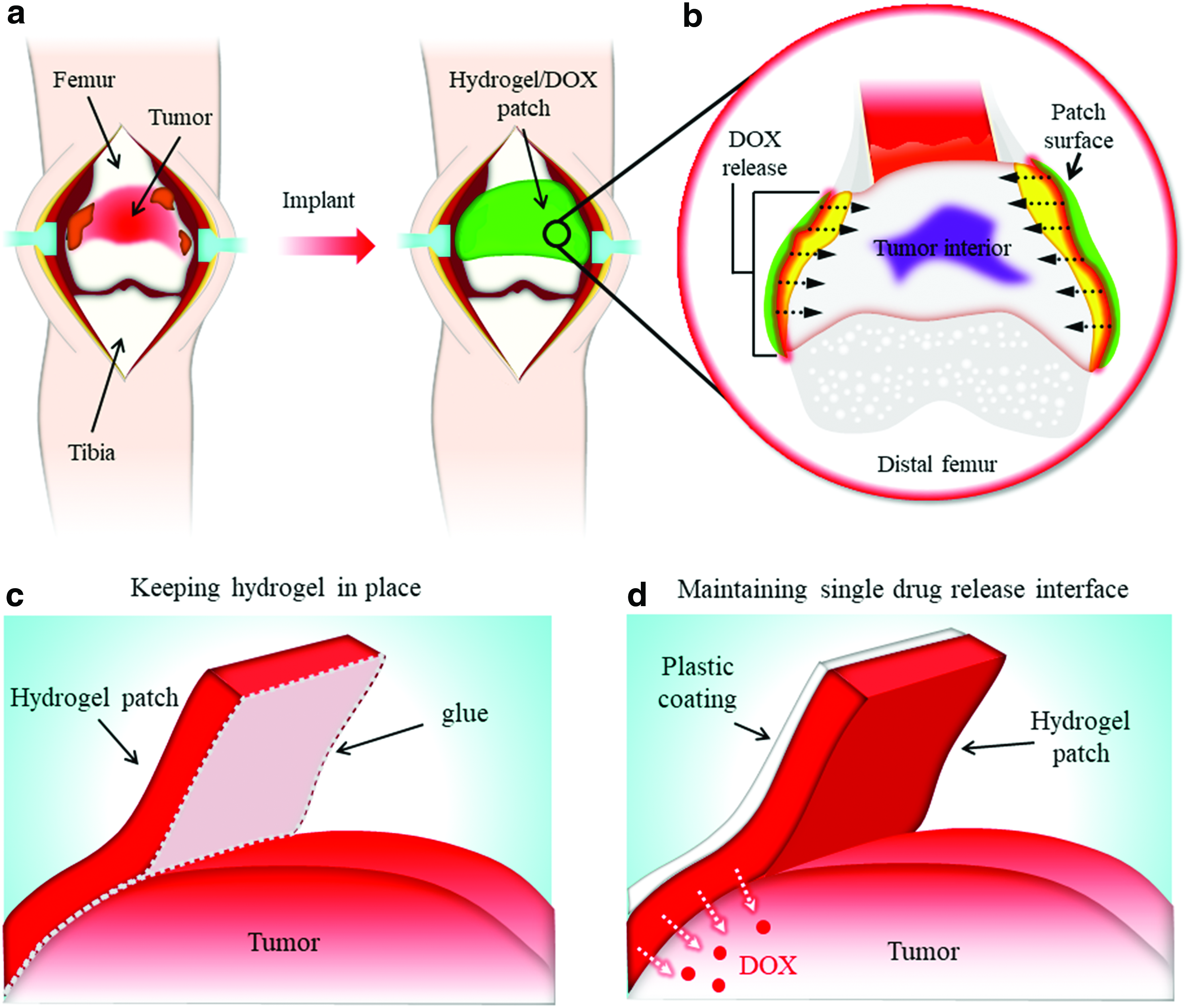
Potential application of P-Msp-DOX-embedded composite hydrogel for osteosarcoma treatment. Schematic demonstration of
Materials and Methods
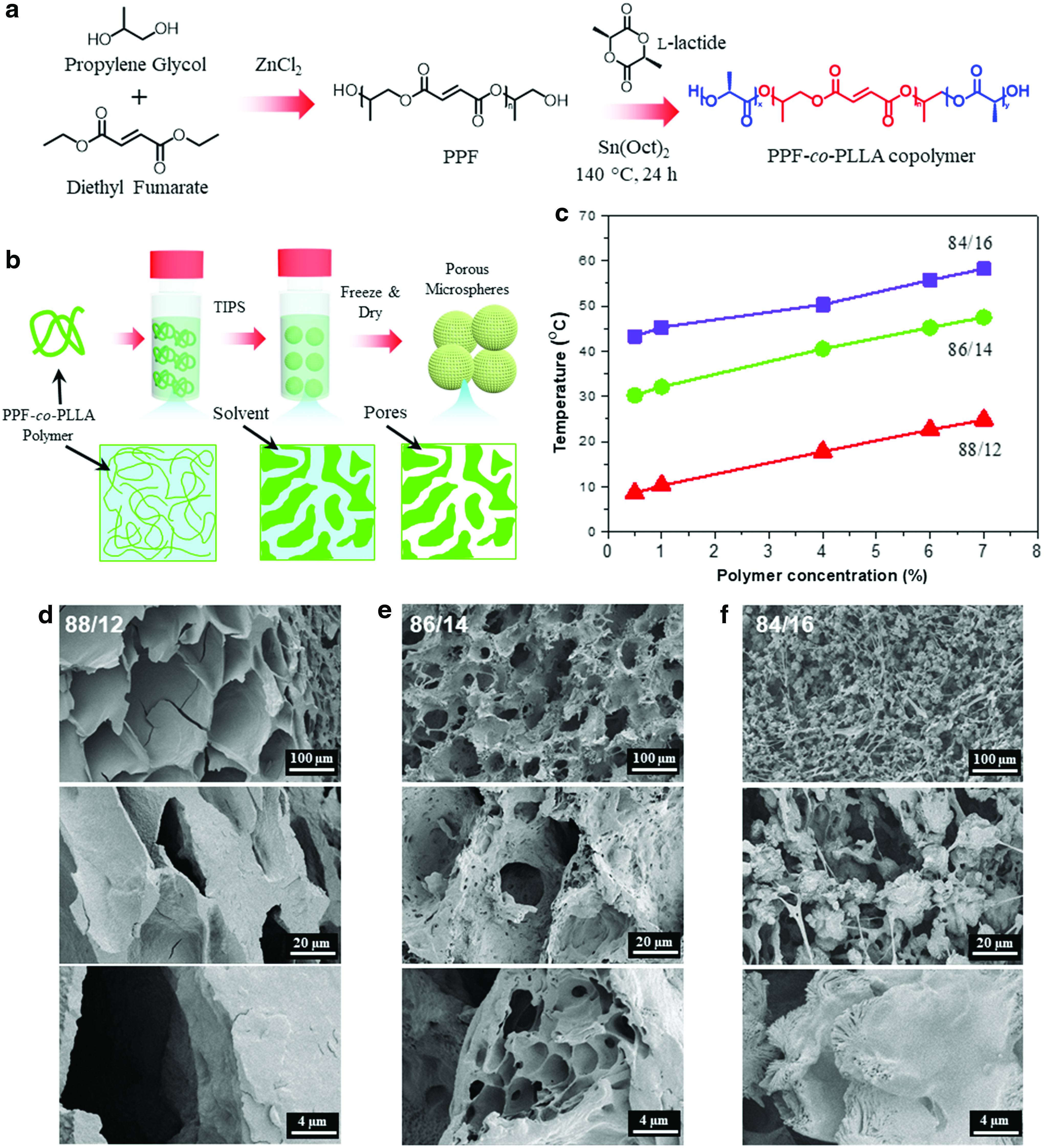
Synthesis of PPF-co-PLLA
PPF was synthesized from diethyl fumarate and propylene glycol according to an established method.
33
PPF-co-PLLA copolymers were then synthesized by ring-opening polymerization of
Fabrication of microspheres by thermally induced phase separation
The PPF-co-PLLA copolymers were fabricated into 3D porous morphologies by thermally induced phase separation (TIPS) as demonstrated in Figure 2b. To investigate the effect of preparation parameters on porous microspheres, variations in solvent and polymer content were examined. In our previous studies, we found that polymer concentrations between 3% and 9%, and 1,4-dioxane/water solvent ratios in the range of 83/17–87/13 wt/wt could generate porous scaffolds effectively.31,32 In this study, we explored varied polymer concentrations of 0.1%, 0.5%, 1%, 4%, 6%, and 7% and varied solvent ratios of 84/16, 86/14, and 88/12 wt/wt. The cloud points for various conditions were tested by visual turbidimetry according to a previously published method. 34 No obvious cloud point was determined at 0.1% polymer concentration due to low polymer content.
To explore the solvent effect, 0.07 g PPF-co-PLLA copolymer (7 wt%) was dissolved in 1 mL 1,4-dioxane/water mixture with varied solvent ratio of 84:16, 86:14, and 88:12 wt/wt. Systems were heated at 60°C for 30 min to allow the polymer to dissolve and then transferred immediately to −80°C for quenching followed by freezing for 1 h to achieve structure preservation. Frozen contents were transferred to 3 mL glass vials and lyophilized for 24 h to achieve porosity. Variance in polymer content (0.1, 0.5, 1, 4, and 6 wt%) was also investigated in 1,4-dioxane/water (85:15, wt/wt) by similar procedures. Dried product was then vortexed at a high speed for 30 s to maximize physical separation and stored at −20°C. All samples were placed in liquid nitrogen for 1 min and broken with forceps for observation of internal morphology by scanning electron microscopy (SEM; S-4700; Hitachi).
Drug loading into microspheres
DOX (2 mg) was mixed into a system of PPF-co-PLLA (0.03 g, 6 wt%) and 0.5 mL 1,4-dioxane/water (85:15, wt/wt). After homogenous mixing, the drug/polymer/solvent system was then heated to 60°C for 30 min to allow for polymer dissolution. The solution was then frozen at −80°C for 1 h for structural preservation. The frozen system was lyophilized with protection from light for 24 h to remove solvent and obtain DOX-loaded porous microspheres (P-Msp-DOX). Dried product was vortexed for 30 s to maximize physical separation and obtain independent structures. To evaluate the drug loading ability, microspheres containing 2 mg DOX were placed inside transwell chambers (mesh size 3 μm) in six-well tissue culture polystyrene (TCPS) plates, then washed at least three times with 5 mL distilled water. The washing medium was collected and DOX concentration was determined using a ultraviolet and visible (UV-Vis) absorbance microplate reader (490 nm). Four tests were conducted and averaged. Loading efficiency was calculated as:
All contents were stored at −20°C before usage.
Degradation of microspheres
Degradation of DOX-loaded structures was evaluated in four solutions: 0.1 M HCl, 0.01 M HCl, phosphate-buffered saline (PBS) adjusted to pH 5.5, and PBS pH 7.4. For samples in 0.1 and 0.01 M HCl, the remaining mass of the microspheres was measured until the structures disappeared completely. For samples in the PBS solutions, the mass lost over 70 days was recorded.
Composite hydrogel fabrication
Two types of OPF hydrogels were fabricated, one with DOX-loaded microspheres (OPF/P-Msp-DOX) and the other with free DOX (OPF/DOX). For the OPF/P-Msp-DOX composite, DOX was first loaded into microspheres by the TIPS method as described above. Afterwards, hydrogels were prepared by combining the obtained P-Msp-DOX with 1 g OPF (Mn = 2000 g mol−1), 36 mg of the crosslinking agent N,N′-methylenebisacrylamide, and 2 mL photoinitiator Irgacure® 2959/water (0.1%) in a glass vial. The solution was then vortexed at a high speed for 20 s to allow for polymer dissolution, producing a final mixture of volume ∼2.8 mL. The total solution volume was added to 28 wells in rubber molds made using a cork borer to create openings of 1 cm diameter. Each well held ∼100 μL of hydrogel solution with ∼160 μg DOX. The filled molds were set on glass plates and exposed to 365 nm UV light (Blak-Ray Model 100AP, Upland, CA) at an intensity of ∼8 mW cm−2 at room temperature for ∼10 min, allowing photo-crosslinking of the OPF polymer. Upon gelation, hydrogels were carefully removed from their molds and stored at −20°C. Meanwhile, the OPF/DOX hydrogel was prepared in the same way by combining free DOX at the same concentration into the described hydrogel solution. This produced a composite hydrogel with the drug directly incorporated into the hydrogel network.
Drug release
The release profiles of DOX from OPF hydrogels were determined. Two release media conditions were created by adjusting buffer solution to pH 7.4 using phosphate buffer and to pH 5.5 using acid buffered solution. For each hydrogel type, 10 disk samples (∼1 mL) were placed into 20 mL of either pH 7.4 or pH 5.5 solution. The release system was placed at 37°C with constant shaking. At given time points, 1 mL of the release medium was extracted and read on a UV-Vis absorbance microplate reader (490 nm). The DOX concentration was calculated using standard calibration curves established by known DOX release profiles in buffer at the respective conditions. After removal, equal volumes of fresh buffer were immediately added to replenish the release media. Each test was conducted in triplicates over 720 h (30 days), and average values with standard deviations were calculated.
Osteosarcoma bone tumor inhibition
Osteosarcoma 143B cell line was used as a model bone cancer cell for cytotoxicity studies. Before usage, the cells were cultured in Dulbecco's modified Eagle's medium/F-12 (DMEM/F-12) supplemented with 2 mM
Trypsinized 143B cells were seeded at 10,000 cells/cm2 onto 48-well TCPS plates and further cultured for 24 h in DMEM/F-12 medium for attachment. For cell viability test, the DMEM/F-12 medium was removed and replaced with equal amount of collected DOX release media. After 3 days of co-culturing, the relative cell densities in each well were tested by MTS assay (CellTiter 96 Aqueous One Solution; Promega, Madison, WI). Wells seeded with 143B cells at the same density but not treated with release media were used as positive control (set as 100%).
Results and Discussion
Porous microsphere fabrication
The cloud points of PPF-co-PLLA solutions at various polymer concentrations and 1,4-dioxane/water solvent ratios were studied. Results showed that by increasing the PPF-co-PLLA polymer concentration from 0.5% and 1% to 4%, 6%, and 7%, the cloud points of the solutions increased drastically, as displayed in Figure 2c. Meanwhile, an increase in 1,4-dioxane content in the binary system resulted in a decrease in cloud points. To study the effect of solvent content, PPF-co-PLLA (7 wt%) was dissolved in three different 1,4-dioxane/water solutions (88:12, 86:14, and 84:16).
SEM micrographs of the resulting phase-separated structures are shown in Figure 2d–f. Porous interconnected structures were observed for all solutions and porosity increased with solvent amount. The 88:12 system produced a large-scale porous structure with a smooth and thick solid surface populated by a highly organized arrangement of deep pores (Fig. 2d). Cavities shared walls with one another and were pseudo-polygonal in shape, loosely resembling a honeycomb. Polymer chains here would be highly solvated and elongated since this condition contained the largest proportion of solvent. Decreasing the solvent content in the 86:14 system led to large pores with additional smaller micropores within cavity walls (Fig. 2e). Compared to the previous condition, polymer chains would form increased associations with another and tighten. The 84:16 system contained the least amount of solvent, leading to structures forming a collection of microscopic clumps connected by an abundant network of thin branches (Fig. 2f). Higher magnification revealed an interior characterized by nanoscale fibers and pores.
Based on these observations, it was determined that a solvent ratio between 84:16 and 86:14 could produce structures that were both porous and displayed particle morphology. This was completed by testing the effect of polymer content by dissolving PPF-co-PLLA at varying amounts (0.1, 0.5, 1, 4, and 6 wt%) in 1,4-dioxane/water (85:15, wt/wt). SEM micrographs of the structures obtained following phase separation are shown in Figure 3a–e. All systems produced 3D porous structures composed of interconnected microparticle networks. Particle size was dependent upon the amount of polymer where diameter increased with weight percent. At 0.1%, small droplet-shaped particles with smooth surfaces formed (Fig. 3a). They were densely packed, and many were merged together in clusters.

SEM images of PPF-co-PLLA microstructures fabricated with different copolymer concentrations:
Increasing the content to 0.5% led to slightly larger particles that were more spherical in shape (Fig. 3b). Their smooth surfaces were shown to contain narrow surface cracks at higher magnification. Several particles exhibited indents and small pores that could have formed during the breaking process before SEM imaging, revealing a possibly hollow interior. Particles of similar and slightly elongated shape were formed from the 1% system, yet were larger in size and regularly porous (Fig. 3c). They displayed smooth surfaces and many were merged together. Solid microspheres covered in shallow-surface depressions were observed at 4% (Fig. 3d). Some of them were irregularly shaped, even displaying fan-like projections. Just as in the 0.5% condition, these patterns could have formed during SEM preparation and might indicate a fibrous, radiating interior structure.
Massive and highly porous microspheres formed from the 6% system (Fig. 3e). Their diameters were magnitudes greater than those from other systems despite being more variable. Surface structure was microfibrous and mesh-like with occasional indents. The size distribution of these microspheres (P-Msp) was between 10 and 70 μm with a peak value of ∼30 μm (Fig. 3f).
Drug loading and microsphere degradation
The addition of DOX into the initial mixture of copolymer and solvent allowed for facile drug loading of porous microspheres using the TIPS method (Fig. 4a). Drug loading did not negatively affect the appearance of porous microspheres, as confirmed by SEM imaging at various magnifications (Fig. 4b). Similar to the empty 6 wt% microspheres shown in Figure 3e, the microspheres with drugs were also porous and displayed excellent interconnectivity. The loading efficiency of DOX was determined to be 46.4 ± 14.6%. The size distribution of P-Msp-DOX was quite broad with diameters in the range of 14–40 μm and an average diameter of 26 μm (Fig. 4c). A closer view of the microspheres' interior showed interconnected pores (Fig. 4d), which could allow liquid solutions to enter with greater contact area for drug release and degradation.

Degradation of P-Msp-DOX was observed across four different conditions (0.1 M HCl, 0.01 M HCl, PBS pH 5.5, and PBS pH 7.4) with an emphasis on conditions below physiological pH due to the acidic tumor microenvironment. Microspheres degraded rapidly in the strongly acidic conditions with complete disappearance of structures within 6 days in 0.1 M HCl and 14 days in 0.01 M HCl (Fig. 4e). Degradation in pH 7.4 PBS solutions was slower than in others with 88.9 ± 4.7% mass remaining even after 70 days in solution. At pH 5.5, the mass remaining was 52.7 ± 12.3% of the original, showing an enhanced and preferential breakdown of microspheres in acidic conditions versus those at physiological pH. This low pH-responsive characteristic may be due to acidic hydrolysis of PPF-co-PLLA copolymer chains, where multiple ester bonds inside the polymer chain can be acid-hydrolyzed and break apart into smaller fragments. The acidic hydrolysis of ester bonds and potential degradation points in the PPF-co-PLLA copolymer are schematically illustrated in Figure 4f.
Composite hydrogel with long-term drug release
The hydrogels prototyped in this study can be characterized as composite systems made of either free drug (DOX) or drug carriers (P-Msp-DOX) set within an OPF hydrogel. Their fabrication is assisted by their ability to gelate at room temperature by photo-crosslinking of fumarate bonds found in OPF (Fig. 5a). The additional presence of these bonds in PPF-co-PLLA microspheres contributes to the structural integrity of all components and thereby creating a more stable drug delivery system. For OPF/P-Msp-DOX specifically, it can be envisioned as a porous drug vessel both housed by and crosslinked to the vast hydrogel network that forms an encasing mesh around them. Samples that were produced were around 1 cm in diameter, cylindrical in shape, and orange-red in color (Fig. 5b).
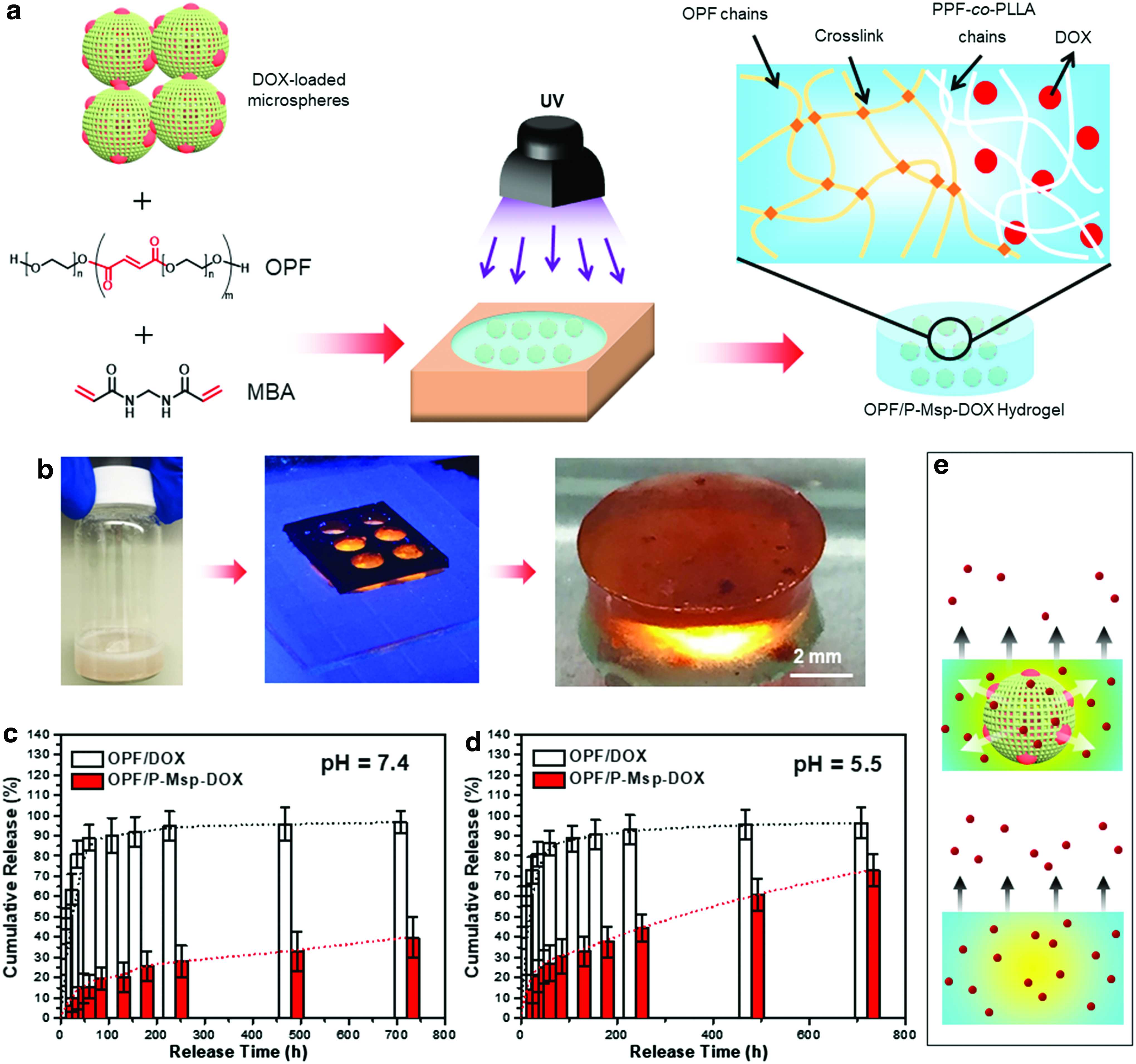
The release profile of DOX at pH 7.4 mimicking the healthy physiological environment was determined. As demonstrated in Figure 5c, the OPF/DOX hydrogel produced a release curve that plateaued after 48 h, indicating a full release in short term. For the OPF/P-Msp-DOX hydrogel, the release was continuous with 39.8 ± 9.9% by 720 h. When immersed in pH 5.5 mimicking the acidic microenvironment in tumor sites, the release profile of OPF/DOX hydrogel also reached a plateau after 48 h (Fig. 5d).
For the OPF/P-Msp-DOX hydrogel, the release profile was continuous at pH 5.5. However, the released anticancer drug contents reached 72.8 ± 7.9%, almost double compared to the pH 7.4 environment. This trend indicated that the composite hydrogel had a pH-responsive release profile, resulting from hydrolytic degradation of microsphere polymer chains, as evidenced by accelerated degradation profile in 0.01 and 0.1 M HCl solution (Fig. 4e). Under pH 7.4, the mass loss of microspheres at 30-day point was not obvious, while only slight weight loss <10% under pH 5.5 (Fig. 4e). This may result from the slow degradation of polymer chains under mild conditions.
During early stages (e.g., 30 days), it is believed that the polymer chains experience partial acidic degradation. However, this incomplete bond breaking does not result in massive bulk degradation of the microspheres. The remaining bonds could still maintain the overall mass, resulting in only light weight loss. Nevertheless, the breakdown of partial polymer chains could facilitate the release of DOX. Therefore, drug release from the OPF/P-Msp-DOX hydrogel is in a continuous, long-term manner due to encapsulation by porous microspheres, as schematically demonstrated in Figure 5e.
Osteosarcoma bone tumor treatment
Both empty microspheres (P-Msp) and crosslinked pure OPF hydrogels were first determined to be biocompatible (Fig. 6a). To study the long-term anticancer effects, OPF/DOX and OPF/P-Msp-DOX hydrogels loaded with DOX were placed in transwells and immersed in six-well TCPS plates. The medium with released DOX was collected every 3 days, as demonstrated in Figure 6b, c. The osteosarcoma 143B cell viability after 3 days of co-culturing with release media was tested using MTS assay and normalized to the positive control.
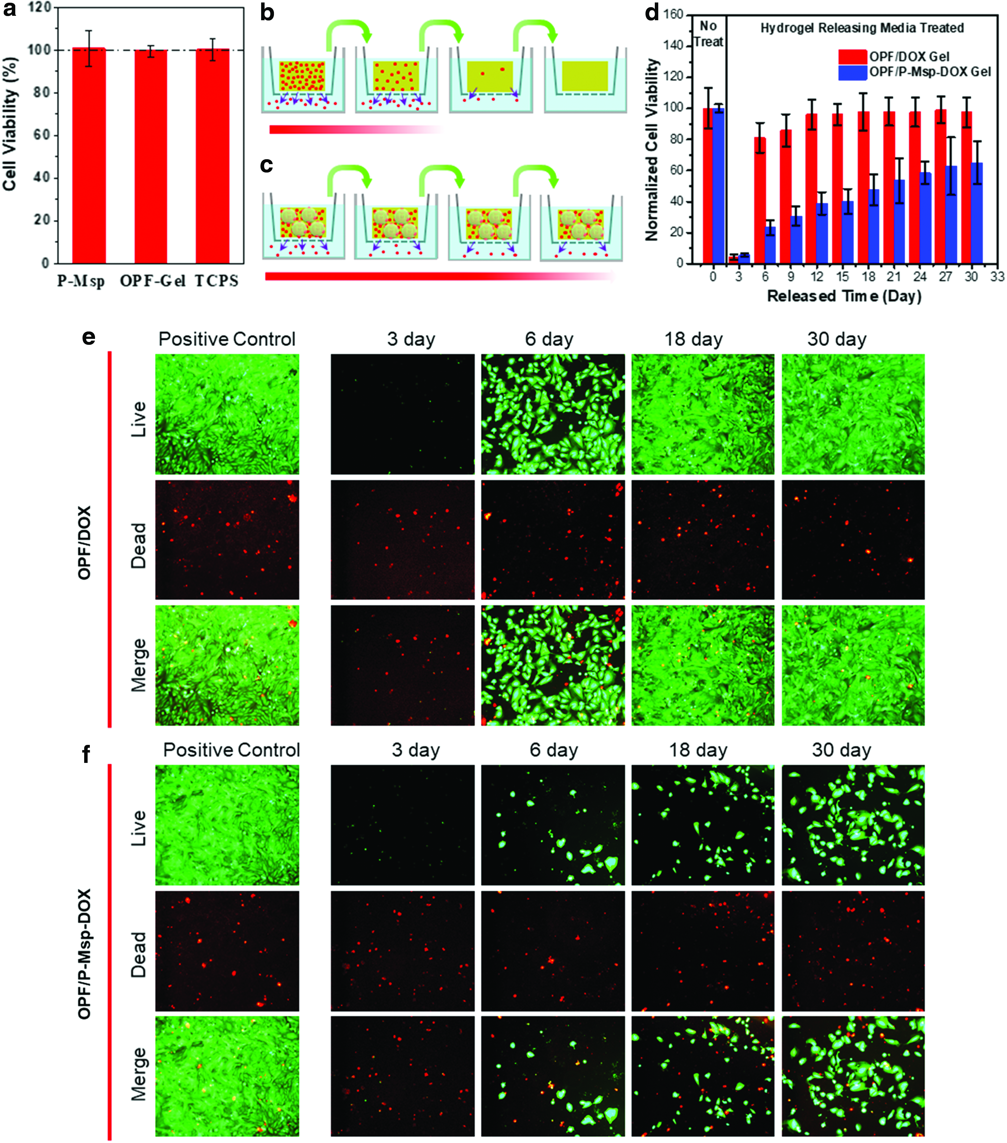
As demonstrated in Figure 6d, the nontreated wells without drug treatment served as positive control and set as 100%. For the OPF/DOX hydrogel, the cell-killing effect was robust for the media collected during the first 3 days with a cell viability of 4.4 ± 1.8%. However, the tumor cell-killing effect dropped drastically for media collected at 6 and 9 days with cell viability determined as 81.1 ± 9.6% and 85.9 ± 10.4%, respectively. After 9 days, the media collected from the OPF/DOX hydrogel no longer exhibited a tumor-killing effect with cell viability approaching the value of positive control without drug treatment.
For the OPF/P-Msp-DOX hydrogel, the drug release was continuous. For the media collected during the first 3 days, the killing effect was as robust as the OPF/DOX hydrogel, showing cell viability of 5.4 ± 1.1%. Under the long-term drug release profile, a series of media was collected at 3-day intervals until 30 days, and it was observed that the tumor-killing effect remained, eradicating a substantial portion of cells. As demonstrated in Figure 6d, the osteosarcoma cell viability after co-culturing with release media collected at 6, 9, 12, 15, and 18 days was 23.3 ± 4.7%, 30.6 ± 6.2%, 38.9 ± 7.3%, 40.2 ± 8.1%, and 47.5 ± 10.0%, respectively, less than half of that seen for positive controls.
After 18 days, the release media collected from OPF/P-Msp-DOX hydrogel still exhibited tumor-killing effect, as indicated by a low cell viability of 53.6 ± 14.3%, 58.5 ± 7.3%, 62.9 ± 18.5%, and 65.0 ± 13.9% after co-culturing with media collected at 21, 24, 27, and 30 days, respectively. Consequently, the OPF/P-Msp-DOX hydrogel exhibited anticancer activity for a much longer period than the plain OPF/DOX, as indicated by live/dead test shown in Figure 6e, f.
The potential use of the OPF/P-Msp-DOX hydrogel for treating osteosarcoma is by surgical implantation at the tumor site, such as distal femur (Fig. 1a, b). This procedure is envisioned to proceed similar to a knee replacement surgery to first expose the tumor-containing bone (Fig. 1a) and then wrap the hydrogel around it (Fig. 1b). This will ensure the entire tumor site is surrounded by the hydrogels for localized and directed delivery of DOX, as shown by the cross-sectional view.
The often-friable nature of osteosarcoma tissue demands careful means of attachment such as polymer-based thermosensitive adhesives35,36 or nanoparticle glues 37 that allow hydrogel–tissue binding without comprising the patient or the system (Fig. 1c). Concurrently, maintaining a single drug release interface is important to prevent unwanted damage to surrounding tissues and introduction of drug into circulation, which can cause systemic side effects. Impermeable films composed of cellulose acetate propionate 38 or electrospun layers of poly(ɛ-caprolactone) and PLLA 39 are desirable options in maintaining unidirectional DOX delivery toward the tumor (Fig. 1d).
The hydrogel would thus serve as a localized and long-term preoperative chemotherapy device that could reduce the chance of DOX-induced cardiomyopathy while assisting in reducing tumor mass for easier resection. Thereafter, the resulting bone defect that would normally be replaced by a prosthesis could now be resolved by techniques focused on bone regeneration. 40 Overall, these phase-separated porous microspheres show a great potential to serve as long-term drug delivery vehicles for osteosarcoma and may also have broad applications in other types of cancers that require localized long-term drug delivery.
Conclusions
This study presents the prototyping of a long-term, localized, and pH-sensitive drug delivery system for DOX comprising phase-separated, porous PPF-co-PLLA microspheres housed in an OPF hydrogel with significant applications for use in osteosarcoma microenvironment. DOX was successfully incorporated into porous microspheres by a phase separation method, and the microspheres were then embedded into the OPF hydrogel. Degradation and drug release studies showed a system that preferentially broke down and released DOX at a sustained rate under acidic conditions. Long-term anticancer effects against osteosarcoma 143B cells were observed for up to 30 days. In summary, the OPF/P-Msp-DOX composite hydrogel reported in this study holds a great promise as a carrier for long-term localized drug delivery to osteosarcoma and other cancers.
Footnotes
Acknowledgment
This work was supported by the NIH grant R01 AR56212.
Disclosure Statement
The authors declare no competing financial interest.