
Abstract
Iliac crest autograft (AG) is the gold standard for bone grafting. Due to the limited supply of autograft, synthetic materials such as ceramics and polymers have been proposed as AG extenders to minimize the volume of AG required for induction of new bone formation. However, the feasibility of reactive polymers for use as settable AG extenders has not been previously investigated. In this study, a reactive oxygen species-degradable poly(thioketal urethane) (PTKUR) was evaluated as a settable AG extender. AG was anticipated to enhance infiltration of cells into the defect and induce new bone formation. Histological analysis of a preliminary study in a rat femoral segmental defect model showed that cells infiltrated PTKUR/AG implants at 4 weeks. In a second experiment, implantation into an intertransverse process model of bone formation showed bone remodeling from the superior and inferior transverse processes. Histological analysis combining data from stains and fluorochrome injections showed lamellar bone formation ongoing near the base of the transverse processes after 8 weeks. Similar findings were observed for a second group, in which 35% of the AG was replaced with calcium phosphate granules. These observations highlight the potential of PTKUR for use as a settable AG extender.
Impact Statement
The development of autograft extenders is a significant clinical need in bone tissue engineering. We report new settable poly(thioketal urethane)-based autograft extenders that have bone-like mechanical properties and handling properties comparable to calcium phosphate bone cements. These settable autograft extenders remodeled to form new bone in a biologically stringent intertransverse process model of bone formation that does not heal when treated with calcium phosphate bone void fillers or cements alone. This is the first study to report settable autograft extenders with bone-like strength and handling properties comparable to ceramic bone cements, which have the potential to improve treatment of bone fractures and other orthopedic conditions.
Introduction
The American Academy of Orthopedic Surgeons reports that over half a million bone grafting procedures are performed every year in the United States. 1 Iliac crest autograft (AG) is the gold standard for bone grafting of open fractures and has utility in extremity, spine, and craniomaxillofacial bone regeneration.2–5 In addition to the well-known limitations of available quantity and donor site morbidity, AG lacks mechanical integrity and generally requires the additional implantation of a retainer to maintain graft placement throughout healing. 6
Local delivery of growth factors, such as recombinant human bone morphogenetic protein-2 (rhBMP-2), from synthetic scaffolds is used clinically and new carriers are being investigated,6–9 but the risks of adverse events associated with these growth factors have been reported to be higher than those associated with iliac crest AG.10,11 AG extenders have been proposed to combine the osteoinductivity of AG with the osteoconductivity and mechanical integrity of established bone graft substitutes. 12 In addition, AG extenders may facilitate the use of AG in large defects that require more AG than can be harvested or enable the conservation of AG for future use.
Another alternative to AG or growth factor delivery from scaffolds is the use of injectable ceramic bone cements, which remodel to form new bone in patients with distal radius and proximal tibia fractures. 13 However, osteoinductive ceramics are currently not available, 14 and thus ceramics must be combined with autologous tissue, such as bone marrow aspirate and/or AG, to induce new bone formation in biologically stringent applications such as posterolateral fusion. In a recent study, 88.9% of patients showed successful fusion at 2 years when treated with ChronOS strip (DePuy Synthes) combined with bone marrow aspirate, local AG, and interbody support. 15 Other ceramics such as hydroxyapatite, β-tricalcium phosphate (β-TCP), and calcium sulfate have been investigated as AG extenders to minimize the volume of AG required to induce new bone formation.16–22
Clinical studies have shown that blending AG with ceramic particles at ratios up to 1:1 leads to comparable or improved new bone formation than achieved with AG alone.16,17,22,23 However, their particulate form results in low cohesive and mechanical properties, potentially resulting in implantation and fixation challenges that may limit their application.24–27 While a nonsettable ceramic/collagen putty AG extender improved new bone formation in rabbits at ratios of 1:1 AG:ceramic/collagen, 21 a settable AG extender with mechanical properties comparable to those of trabecular bone has not been previously reported.
Poly(thioketal urethane)s (PTKURs) set to yield bone cements with mechanical properties approaching those of trabecular bone. 28 PTKUR degrades in the oxidative microenvironment generated by osteoclasts and macrophages present during wound healing and bone remodeling.28–31 Previous studies have reported that nonsettable polymeric AG extenders promote bone healing in small animal models.12,32,33 Thus, the favorable handling, mechanical, and resorptive properties of PTKUR render it a promising candidate for use as a settable AG extender that can be molded to conform to the geometry of the defect where it cures in situ.
An AG extender with handling properties comparable to calcium phosphate (CaP) bone cements and sufficient biological activity to induce new bone formation could potentially improve outcomes for a number of orthopedic procedures, but these materials are currently not available. In this study, we tested the hypothesis that settable PTKUR-based AG extenders would induce new bone formation in a stringent intertransverse process (ITP) bone formation model in rabbits. The reactive PTKUR component was anticipated to provide clinically relevant (10–20 min) setting times and set to yield implants with bone-like mechanical properties. Handling properties were measured by direct measurement of setting and hardening times, and mechanical properties were assessed by compression testing. Cellular infiltration and new bone formation were evaluated in two stringent models of bone regeneration: a critical-size femoral segmental defect model in rats and a posterolateral ITP bone formation model in rabbits. To our knowledge, this is the first report of a settable AG extender investigated in this challenging, noninstrumented model in rabbits.
Materials and Methods
Experimental design
PTKUR/AG was formulated to incorporate the minimum amount of PTKUR (31 vol%) that yields a cohesive paste. PTKUR/AG and an AG control were first implanted in a critical-size segmental femoral defect model in rats to test the hypothesis that cells infiltrate PTKUR/AG. After confirming cellular infiltration, PTKUR/AG and an AG control were implanted in a posterolateral ITP model in rabbits to test the hypothesis that PTKUR-based AG extenders induce new bone formation in a biologically stringent model. 34 A second experimental group (PTKUR/CaP/AG) was also tested, in which a portion of the AG was replaced by osteoconductive CaP granules, considering a previous study reporting that implants with 50% AG induce new bone formation in the rabbit ITP model. 35 Thus, two AG concentrations were evaluated: 69 vol% (PTKUR/AG) and 44 vol% (PTKUR/CaP/AG).
Materials
Materials for thioketal diol (TK) synthesis, including solvents, were purchased from Sigma-Aldrich. Lysine triisocyanate-polyethylene glycol prepolymer (LTI-PEG) was acquired from Ricerca Biosciences LLC and used as received. Iron (III) acetylacetonate (FeAA) catalyst was purchased from Fisher Scientific. CaP ceramic particles (CaP, MASTERGRAFT®: 85% β-TCP/15% hydroxyapatite) were acquired from Medtronic, ground using a mortar and pestle, and screened using 100–300 μm sieves. The mean particle size was 238.1 ± 0.49 μm.
Synthesis of TK
A TK diol (MW = 196 g/mol) was synthesized as described previously. 28 In brief, thioglycolic acid and 2,2-dimethoxypropane were reacted in the presence of bismuth (III) chloride for 24 h at room temperature. The intermediate product was filtered and dried for 24 h under vacuum. The intermediate was then dissolved in 100 mL of tetrahydrofuran and slowly added to lithium aluminum hydride (LiAlH4) in diethyl ether at 0°C. After all the intermediates were added, the reaction was refluxed at 52°C overnight. Excess LiAlH4 was quenched by adding water dropwise. The product was then filtered from the by-products and extracted using aqueous NaOH and diethyl ether. The aqueous layer was removed using a separation funnel and sodium sulfate. After filtering, solvent was removed from the product using rotary evaporation, and the product was dried under vacuum at least 24 h. FeAA catalyst was dissolved directly in the dry TK diol (0.5% FeAA in TK) by stirring in a closed vial overnight.
Fabrication of PTKUR/AG and PTKUR/CaP/AG
PTKUR/AG and PTKUR/CaP/AG were prepared by adapting a reactive liquid molding technique. Two volume fractions of AG were investigated, and the volume fraction PTKUR was selected as the minimal amount required to provide desirable handling properties. The compositions of PTKUR/AG and PTKUR/CaP/AG are listed in Table 1. The AG extender incorporated 69 vol% AG, which was the highest concentration that could be achieved to maintain a cohesive paste when mixed with the reactive PTKUR (31 vol%). PTKUR/CaP/AG incorporated 44 vol% AG, which is at the low end of the range that has been previously reported to be effective for bone regeneration.16,17,22,23,33 CaP particles (17 vol%) were added to enhance the osteoconductivity while maintaining a cohesive paste when mixed with the reactive PTKUR (39 vol%). AG was weighed and (when appropriate) mixed with CaP before reacting with the polymer components.
Composition and Handling Properties of Autograft Extenders
p ≤ 0.05, **p ≤ 0.01.
AG, autograft; CaP, calcium phosphate; PTKUR, poly(thioketal urethane).
Separately, TK diol (with catalyst) and LTI-PEG prepolymer (NCO = 21%) were hand mixed together for 45 s. An NCO:OH index of 140 was used for all extenders. AG or CaP/AG was then added to the reactive polymer and mixed vigorously until homogeneous. The exothermic reaction resulted in a less than 15°C increase in temperature as reported previously for LTI-based poly(ester urethane) (PEUR) foams. 36 The tack-free time was reported as the time when a metal spatula no longer stuck to the composite material. Hardening time was measured by hand as the time after which the material could no longer be compressed by hand. The morphology of PTKUR/AG and PTKUR/CaP/AG was visualized by scanning electron microscopy (SEM; Zeiss Merlin) of gold-coated specimens.
Degradation of PTKUR
The degradation characteristics of PTKUR were investigated in accelerated oxidative conditions to estimate degradation in vivo.28,29 PTKUR films, without AG or CaP, were cast in cylindrical tubes and cured overnight. The material was cut into small discs (50 mg) using an IsoMet Low Speed Saw. The initial mass of each sample was recorded before immersing the samples in oxidative media (20 wt% hydrogen peroxide in 0.1 M cobalt chloride) or phosphate-buffered saline (PBS; hydrolytic control). Samples were incubated at 37°C on a shaker table for 72 h. They were then washed 3 × in 1 mL of deionized water and dried under vacuum for 48 h. The mass of the dry sample was compared with the initial mass to determine extent of degradation in oxidative and hydrolytic conditions. The degradation characteristics of the PTKUR films were compared to those of an LTI-based PEUR that has been shown to degrade hydrolytically.37,38
Cytotoxicity of PTKUR/AG and PTKUR/CaP/AG
The cytotoxicity of the extracts from the materials was measured using MC3T3-E1 embryonic mouse osteoblast precursor cells in vitro. In brief, 1-g specimens of PTKUR/AG and PTKUR/CaP/AG were incubated in 5 mL α-minimum essential medium (α-MEM) with 10 vol% fetal bovine serum and 1 vol% penicillin/streptomycin for 24 h at 37°C. The extracts were collected and used for subsequent cytotoxicity experiments. Cells were seeded in a 96-well plate at a density of 5 × 10³ cells per well and cultured in α-MEM with 10 vol% fetal bovine serum and 1 vol% penicillin/streptomycin in a CO2 incubator with 5% CO2 at 37°C. Medium was removed from cells and replaced with the collected extracts.
Cells were analyzed for viability using a live/dead viability kit (Invitrogen) after 24 h exposure to the extract solution. The assay was completed as recommended in the manufacturer's instructions. ImageJ was used to asses cell viability of each group and control group treated with PBS (n = 5).
Mechanical properties of PTKUR/AG and PTKUR/CaP/AG
Compressive testing was performed to characterize the mechanical properties of the materials. PTKUR/AG and PTKUR/CaP/AG were synthesized as described above and molded into cylindrical tubes with a diameter of 6 mm (n = 3). The specimens were compressed under a weight (0.96 kg) for 2 h to ensure cohesion and mimic forces of surrounding musculature in vivo.28,39 Polyurethane composites based on LTI-PEG prepolymers require 24 h to fully harden, and thus the mechanical properties were measured after 24 h, as we have reported previously. 40 Specimens were removed from the molds and hydrated overnight with wet gauze at 37°C. Hydrated samples were cut to a height of 12 mm (2 × diameter) before mechanical testing.
Prepared specimens were compressed between flat platens to a preload of 5 N and then to failure at a rate of 25 mm/min (MTS 858 Bionix Servohydraulic Test System). The strain and load were recorded throughout the test and used to calculate engineering stress and engineering strain based on the specimen geometry. The compressive modulus, ultimate strength, and yield properties (0.2% offset method) were calculated from the resulting stress-strain curve. The modulus of resilience (i.e., the elastic energy absorbed) was calculated as the area under the stress–strain curve from the initial zero strain to the yield point. 41
In vivo analysis
All animal procedures were approved by the Institutional Animal Care and Use Committee of the U.S. Army Institute of Surgical Research (Fort Sam Houston, TX) and were conducted in compliance with the Animal Welfare Act, implementing Animal Welfare Regulations, and the principles of the Guide for the Care and Use of Laboratory Animals.
Evaluation of PTKUR/AG in a rat model
PTKUR/AG and an AG control were implanted in critical-size segmental defects in athymic nude rat femurs to test the hypothesis that cells would infiltrate PTKUR/AG at an early time point (4 weeks). Fresh iliac crest bone graft (ICBG) was harvested from donated New Zealand white (NZW) rabbit cadavers and used in place of “autograft” for the rat study. For purposes of clarity, we refer to the xenograft ICBG as autograft in this study and the use of athymic rats prevents the immune rejection.42–44 Autograft from the same animals and syngeneic grafts were not used because of the lack of cancellous bone in rats. 45 Following humane euthanization, the NZW rabbit iliac crests were exposed and AG aseptically removed using an oscillating saw. The AG was scraped of soft tissue and processed using a bone mill (R. Quentin) to yield particulated bone, ∼3 mm in diameter. Bone was patted dry with sterile gauze and set aside until use.
Immediately following AG harvesting, a 6-mm segmental defect was created in the right femurs of six anesthetized athymic nude rats (341 ± 8.5 g; Harlan Laboratories, Houston TX). Animals were premedicated with SR buprenorphine (1.2 mg/kg SC) and anesthetized with 1–3% isoflurane in oxygen. The right hindlimb was shaved and prepped for sterile surgery with alternating 70% ethanol and betadine scrubs. The right femur was exposed, a stabilizing polyacetal plate affixed with four threaded K-wires, and a 6 mm mid-diaphyseal segment of the bone removed.46,47
Animals received one of two treatment groups: AG control (n = 3) or PTKUR/AG (n = 3). PTKUR/AG was mixed as described above with the composition of 69 vol% AG/31 vol% PTKUR (60 wt% AG/40 wt% PTKUR) described above. PTKUR/AG was loaded into a blunted 1-cc syringe 5 min before use, injected into the defect space, and positioned using a small surgical elevator to ensure contact with the bone on each side. The materials cured for 10 min in situ before wound approximation and closure. Similar volumes of AG only (no PTKUR) were used as clinical controls. Four weeks postoperative, animals were sedated, euthanized, and the femurs harvested and placed in 10% neutral-buffered formalin for histology.
Assessment of outcomes: histology
Bone regeneration and material degradation were assessed via nondecalcified histology. Formalin-fixed femurs were dehydrated in ethanol and embedded in poly(methyl methacrylate) (PMMA) for histological analysis. Sagittal sections were taken from the center of the defect and stained with Sanderson's Rapid Bone Stain and Van Gieson to qualitatively assess cellular infiltration, bone regeneration, and polymer degradation. For quantitative analysis, new bone, infiltrating cells/matrix, and residual PTKUR were measured from the histological sections using Metamorph software (Version 7.0.1, Waltham, MA). For each section, the area of interest was defined as the 6 mm defect region where the material was implanted. Metamorph was utilized to color threshold the residual PTKUR (gray), infiltrating cells/matrix (blue), and residual AG and new bone (red) and quantify the area of each component within the area of interest.
Evaluation of PTKUR/AG and PTKUR/CaP/AG in a biologically stringent ITP model in rabbits
After confirming cellular infiltration at early time points in the rat model, PTKUR/AG, PTKUR/CaP/AG, and an AG control were implanted in a posterolateral ITP model in 13 NZW rabbits (4.0 ± 0.06 kg) to test the hypothesis that PTKUR-based AG extenders induce new bone formation. 34 A no-treatment negative control or CaP alone group was not included in the study design because no bone growth is observed in the absence of an osteoinductive component (e.g., AG, bone marrow aspirate, or rhBMP-2) as reported previously.15,34,48 A PTKUR/CaP/AG group was included in the study design to test the hypothesis that the osteoconductive CaP would support bone formation comparable to PTKUR/AG based on a previous study reporting that AG concentrations as low as 50% induce bone formation in the ITP model. 35 PTKUR/CaP/AG comprised 44 vol% AG, 17 vol% CaP, and 39 vol% PTKUR compared with 69 vol% AG and 31 vol% PTKUR for PTKUR/AG. All graft components were gamma-irradiated at ∼25 kGy before surgery.
Animals were premedicated with SR buprenorphine (0.5 mg/kg SC). Anesthesia was induced with ketamine/xylazine (25/5 mg/kg IM, respectively) and maintained with 1–3% isoflurane via a laryngeal mask airway. Animal dorsums were shaved and prepped for sterile harvest and surgery with alternating 70% ethanol and betadine scrubs. The AG harvest and placement of bone graft procedures were accessed from a single cutaneous midline incision from L4 to L7. First, 3 cc of AG was harvested from each iliac crest (IC). In brief, sharp dissection was used to access the IC through the posterior musculature, and blunt dissection used to elevate the muscle off the IC. Using an oscillating saw, the dorsal third of each IC was removed followed by closure of the musculature over the remaining IC.
AG harvested from the IC was morselized as described above using a bone mill and set aside until implantation of the bone grafts near the processes. Following AG harvest, two paramedian incisions were made through the fascia and musculature over the L5–L6 transverse processes. Using blunt dissection, the plane between the multifidus and longissimus muscles was developed to expose the transverse processes as well as the intertransverse membrane. The dorsolateral two-thirds of the transverse processes were decorticated using an electric burr to enhance cellular activity at the tissue/implant interface.
Materials were prepared as described above. AG and CaP were mixed by hand until homogeneous before adding to the polymer mixture. Materials were loaded into a blunted 5 cc syringe and delivered on the paraspinal bed, between the decorticated transverse processes. A surgical elevator was used to adjust positioning and ensure contact between the material and the transverse processes. Once the material was placed, the surrounding musculature was sutured and the wound closed. Aqueous fluorochromes were injected subcutaneously at 2 (calcein green, 10 mg/kg) and 6 (xylenol orange, 90 mg/kg) weeks as a complementary tool to investigate the dynamics of new bone formation. 49
Assessment of outcomes: computed tomography and microcomputed tomography
In vivo computed tomography (CT) scans (Prime Aquilion TSX-303A; Toshiba) were acquired at 4 and 8 weeks postoperatively to investigate the progression of mineralization. The DICOMs were converted to 16-bit BMP files, normalized to the 500-μm slice thickness of the clinical CT, and measured using CTAn (Bruker Skyscan). Regions of interest were interpolated every five slices (2.5 mm) to include the entire implanted graft. Both sides of each animal were first averaged before averaging across the group.
Eight weeks postimplantation, the animals were anesthetized and euthanized with sodium pentobarbital (Fatal Plus®), the spinal segments (L4–L7) harvested, and the soft tissue removed. Spinal segments were cut in half longitudinally then immersed in 10% formalin for microcomputed tomography (μCT) and nondecalcified histology. μCT data were acquired using a Scanco μCT 50 at an isotropic voxel size of 24.2 × 24.2 × 24.2 μm, and reconstruction and visualization were performed using the accompanying Scanco software. Images of 2D sagittal sections were acquired from coronal and sagittal perspectives to visualize new bone growth. Serial sagittal sections were acquired from the lateral-most point of the graft medially until the vertebral body was approached to distinguish growth patterns from the processes.
Assessment of outcomes: histology
After the μCT scans were complete, specimens were dehydrated and embedded in PMMA for histological analysis. Longitudinal histological sections were made to assess bone formation between the L5 and L6 transverse processes. Serial sagittal sections (30–70 μm), with both L5 and L6 in view, were cut from the lateral most incidence of the implant, until the spinal column was reached. Every other section was stained with Sanderson's Rapid Bone Stain/Van Gieson to visualize cellular activity and neotissue formation. Quantitative measurement of infiltrating cells and residual PTKUR and CaP in the histological sections was performed using Metamorph software (Version 7.0.1). The area of interest was defined as the area between the L5 and L6 processes where the material was implanted. Metamorph was utilized to color threshold the residual PTKUR and CaP (gray/black) and infiltrating cells (blue) and quantify the area of each phase within the area of interest.
The remaining sections were used for fluorochrome analysis. An Olympus BX60 microscope equipped with a Luminera Infinity 2 camera and an Olympus Reflected Light Fluorescence attachment was used to image sections at 2 × magnification. 2 × images were stitched together to visualize the continuity of new bone and fluorescence throughout the entire section. Magnified images were also acquired for cellular detail. Stained and fluorescent images were overlaid to provide an understanding of the mechanism of bone regeneration from the transverse processes and implanted AG. For each specimen, the section with the largest area of new bone without reaching the vertebral column was selected for further analysis.
ImageJ was used to quantify the front of new bone growth and ITP space dimensions from the fluorochrome data at 2 weeks (green) and 6 weeks (orange) and from brightfield images of histological sections stained with Sanderson's Rapid Bone Stain at 8 weeks (red). To make consistent measurements between groups, the transverse processes were included when measuring bridging distances in both the fluorescent and brightfield images. The normalized bridging (% bridging) was calculated as the bridging distance divided by the entire process-to-process distance for each sample (illustrated in Fig. 7A).
Statistical methods
Unpaired t-tests were used to compare the handling and mechanical properties of PTKUR/AG and PTKUR/CaP/AG, and the data are reported as the mean ± standard deviation. A linear regression of the % bridging versus time data was performed to calculate the normalized bridging rate of the processes (% bridging/week). The data are reported with standard error of the mean. A two-way ANOVA (GraphPad Prism) was used to compare the statistical significance of the graft volume (measured by CT) and % bridging measurements between groups and time points. A post hoc Tukey's multiple comparison test compared the differences in % bridging between PTKUR/AG, PTKUR/CaP/AG, and the AG control at each individual time point. A second post hoc Tukey's test compared the increase in % bridging over time for each individual group.
Results
Cytotoxicity of FeAA catalyst
The FeAA catalyst has been used to prepare settable PTKUR and PEUR bone cements with no observable cytotoxic effects in rabbit 28 and sheep 41 models of bone regeneration. The cytotoxicity of the FeAA catalyst was measured by treating MC3T3 murine osteoprogenitor cells with extracts obtained by incubating PTKUR/AG and PTKUR/CaP/AG in medium for 24 h. 50 The viability of cells cultured in the PTKUR/AG and PTKUR/CaP/AG extracts were 94.8% ± 2.4% and 82.9% ± 2.7%, respectively. Neither of the groups showed cytotoxicity, which is defined by the ISO 10993:5 Cytotoxicity standard as <70% viability.
Handling properties of PTKUR/AG and PTKUR/CaP/AG
While the FeAA catalyst has a high selectivity for the gelling reaction (gelling reaction rate constant/blowing reaction rate constant = 6.4),28,51 the AG particles contained a significant amount of moisture. PTKUR/AG and PTKUR/CaP/AG were prepared by modifying the fabrication technique to initiate the polymerization reaction before introducing the AG component. The prereaction time of 45 s allowed the TK diol/LTI-PEG reaction to attain partial conversion before addition of AG. After prereacting about 1 min, the material was too crosslinked to homogeneously mix the AG or CaP/AG components into the polymer phase. The setting and hardening times measured in vitro are reported in Table 1 (since the materials were partially cured before implantation, in vivo setting times were comparable to those measured in vitro). The tack-free time (defined as when the material no longer stuck to a spatula) is a measure of the setting time of the bone graft. 28 PTKUR/CaP/AG (11:27 ± 2:23 min) set faster than PTKUR/AG (18:29 ± 0:42). The setting times of both grafts were comparable to the setting times reported for CaP cements (10 min). 14
Representative photographs of PTKUR/AG (Fig. 1A, left) and PTKUR/CaP/AG (Fig. 1A, right) show the appearance of the implants when cured in vitro. SEM images of PTKUR/AG (Fig. 1B, left) indicate the irregularly shaped pores <500 μm in size (white arrows). The AG particles are not readily detectable likely due to their small size and the adhesion of PTKUR to the AG surface. The CaP particles (black arrows, Fig. 1B, right) can be observed in PTKUR/CaP/AG, which also showed evidence of spherical pores (white arrows) <500 μm in diameter associated with gas blowing due to the water reaction. 52
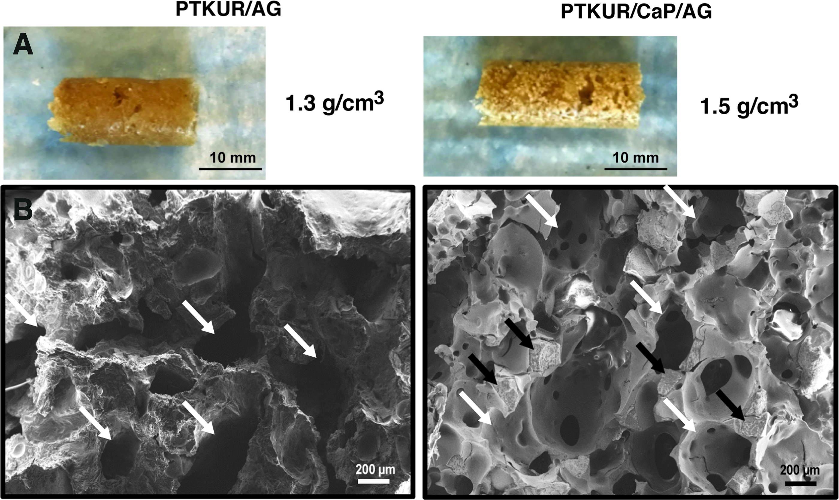
In vitro characterization of PTKUR/AG and PTKUR/CaP/AG.
Degradation of PTKUR
PTKUR films were immersed in oxidative media to evaluate degradation in oxidative conditions simulating the wound healing microenvironment. Degradation of the PTKUR was compared to that of a degradable PEUR composed of a 300 g/mol polyester triol [poly(ɛ-caprolactone-co-glycolide-co-
Mechanical properties of PTKUR/AG and PTKUR/CaP/AG
Compressive modulus, yield strength, and yield strain were calculated from the engineering stress-strain curve (Fig. 2A). The yield point (intersection between the dotted line and the PTKUR/CaP/AG curve in Fig. 2A) was calculated based on the 0.2% offset method, and the ultimate point (intersection between the dashed line and the PTKUR/CaP/AG curve in Fig. 2A) was defined as the maximum stress. The stress–strain curve was divided into the two zones shown in Fig. 2A: (i) elastic zone (from the initial to the yield point), and (ii) postyield zone (from the yield to the ultimate point). 54 The compressive modulus (12.0 ± 1.9 MPa, Fig. 2B) and ultimate strength (2.05 ± 0.13 MPa, Fig. 2C) of PTKUR/CaP/AG were significantly higher than that of PTKUR/AG (6.08 ± 0.87 and 1.17 ± 0.095 MPa, respectively). This difference can be attributed to the higher modulus and strength of the CaP particles compared to AG. Despite the higher yield strength of PTKUR/CaP/AG (Fig. 2D), the yield strain for both implants was 15% (Fig. 2E). PTKUR/CaP/AG absorbed significantly more energy than PTKUR/AG as indicated by the higher modulus of resilience, which represents the elastic energy absorbed in zone 1 calculated as the area under the curve (Fig. 2F).
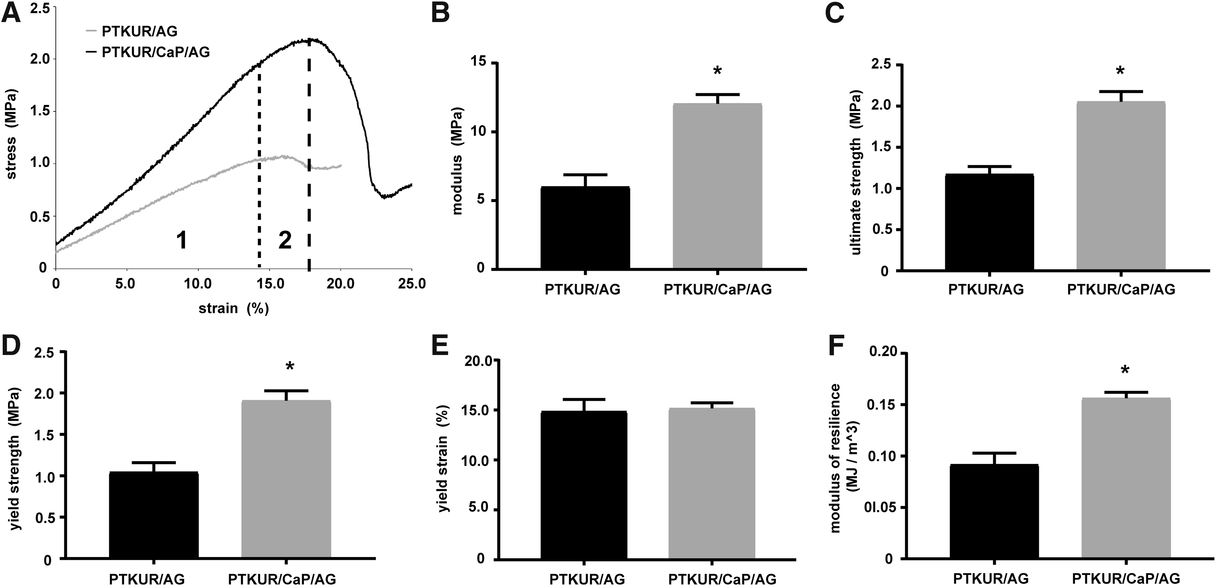
Mechanical properties of PTKUR/AG and PTKUR/CaP/AG determined from quasistatic compression testing.
Cellular infiltration in a rat model
In vivo cellular infiltration of PTKUR/AG was first assessed in femoral segmental defects in athymic rats using rabbit AG to eliminate graft volume limitations. Histological sections of the PTKUR/AG showed evidence of cellular infiltration throughout the implant after 4 weeks (Fig. 3A–D). Bone-forming cells were identified at the center of the defect area in both PTKUR/AG (Fig. 3B, arrows) and the AG control (Fig. 3D, arrows) groups. Both residual AG and new bone stained red in the histological sections and could not be distinguished from each other.
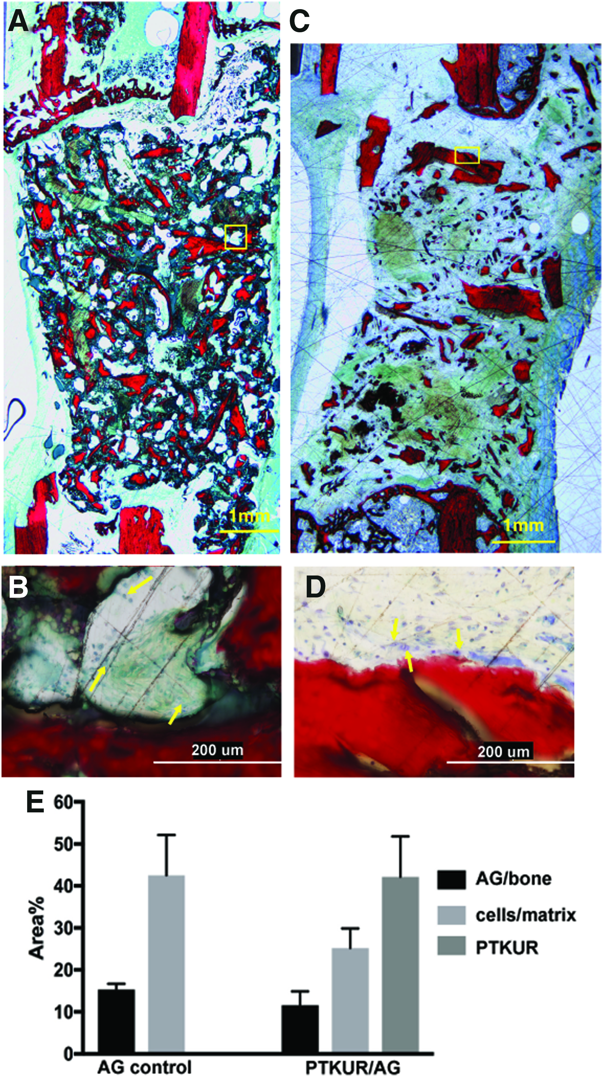
Images of histological sections of femoral segmental defects in rats at 4 weeks postimplantation showing areas of cellular activity (arrows) and new bone formation near edges of implanted AG.
The area% AG and new bone, area% cells and matrix, and area% residual PTKUR were measured by histomorphometry at 4 weeks (Fig. 3E). Both PTKUR/AG and the AG control showed 10–15% AG/new bone, which suggests that cells infiltrated the implants by migrating into pores and resorbing the AG particles. Residual PTKUR was 40 area%. The area% cells and matrix trended higher for the AG control (42 area%) compared to PTKUR/AG (26 area%) but the differences were not significant. These findings suggest that cells infiltrated the implants primarily through resorption of the AG component and that the persistence of the PTKUR component delayed cellular infiltration.
Implantation of bone grafts in an intertransverse process model of bone formation in rabbits
The rabbit spine model requires 2 mL graft (total volume) to bridge L5 and L6 (Fig. 4A) on each side of the vertebral column (4 mL total/animal). Photographs of the implanted bone grafts are shown in Figure 4B. PTKUR/AG (69 vol% AG) and PTKUR/AG/CaP (44 vol% AG) received 1.38 and 0.88 mL AG particles, respectively. Although in vivo CT images cannot provide sufficient resolution to differentiate between specific components of the graft, they enabled assessment of changes in total graft volume over time (Fig. 4C). CT images at the time of surgery could not be reliably reconstructed due to attenuation of the signal caused by blood present at the surgical site. At both 4 and 8 weeks, the volume of PTKUR/CaP/AG was significantly higher than that measured for the AG control and PTKUR/AG (p < 0.01, Fig. 4D). The volumes of the bone grafts did not change over time.

Implantation of AG extenders.
Induction of new bone growth in the intertransverse space in rabbits
Coronal sections of μCT images showed trabecular bone formation in the ITP space from the transverse processes for all groups (Fig. 5A). Serial 2D sagittal sections exemplified the healing pattern throughout the graft. Residual implant material was observed outside the ITP space (green arrows, Fig. 5B). The AG control, PTKUR/AG, and PTKUR/CaP/AG groups showed new bone growing from the surfaces of the transverse processes (white arrows, Fig. 5B). Newly formed bone grew from both processes toward the center of the ITP space in the plane of the transverse processes, with superior and inferior growths approaching complete bridging near the vertebral body.
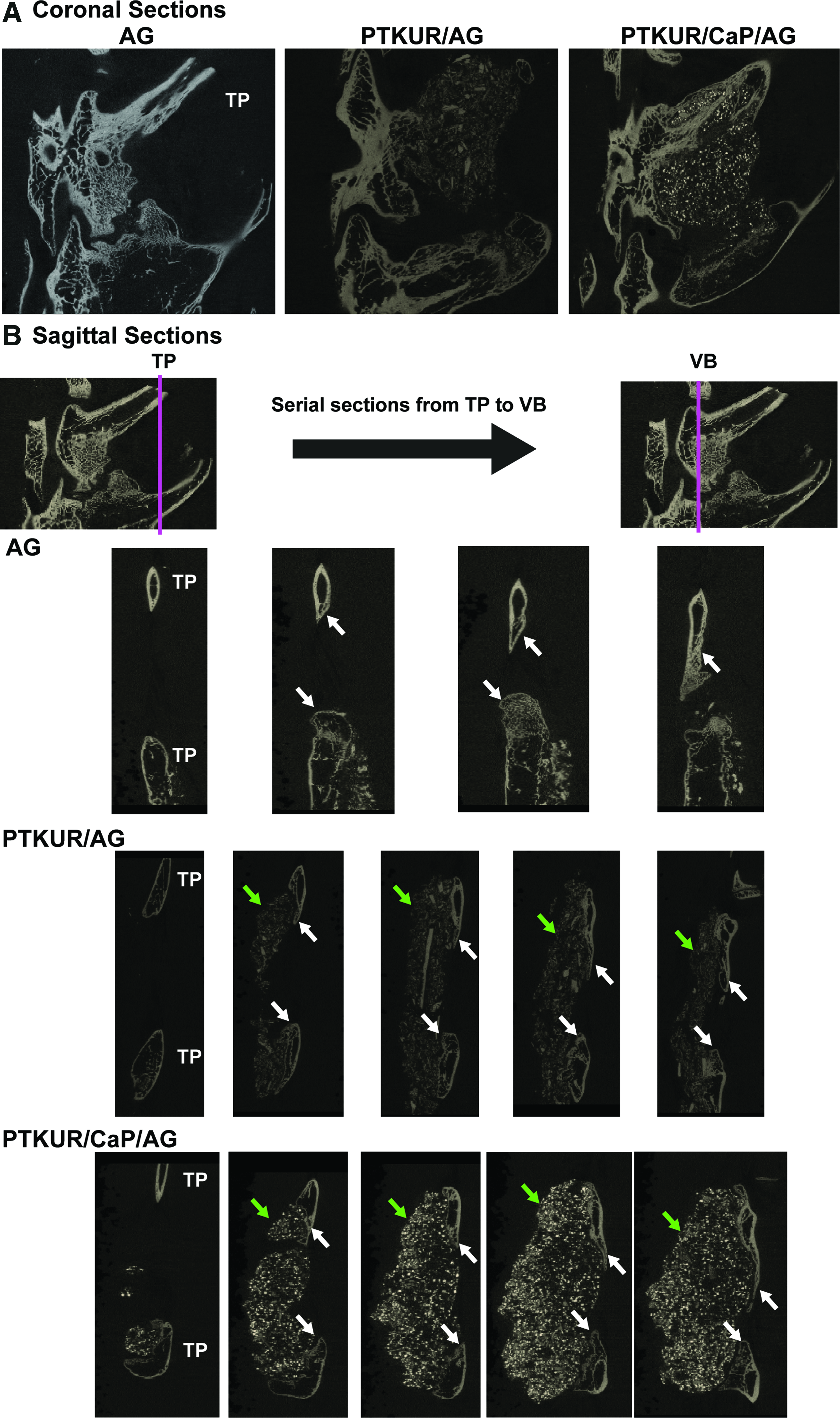
μCT images of spines cut down the spinal column.
Serial sagittal sections were cut throughout the defect to assess bone growth patterns histologically, which is shown schematically in Figure 6A. Stained histological sections indicated small gaps of connective tissue where specimens were close to bridging of the processes with new bone (Fig. 6B, D, F—single arrows: ongoing lamellar bone formation, double arrows: new bone). Fluorescent images of unstained sections demonstrated the chronological patterns of osteogenesis for each graft type. The AG control showed mineralization throughout the graft at 2 weeks postimplantation (Fig. 6C, green). There was some continuation of growth at 6 weeks (orange-red fluorescence) within the graft; however, the perimeter of bone evident in the stained section was produced between 6 and 8 (double black arrows, stained histology) weeks.

Images of histological sections of the AG control, PTKUR/AG, and PTKUR/CaP/AG implanted on the L5–L6 lateral processes.
Bright-field and fluorescent images of adjacent slides were compared side-by-side to differentiate implanted AG from new bone. Fluorescent images of PTKUR/AG and PTKUR/CaP/AG revealed autofluorescence of the polymer. Thus, fluorescence in the ITP space far from the processes may represent residual PTKUR or new bone formation originating from either the implanted AG or migrating from the transverse processes. To eliminate the need to discriminate between autofluorescent polymer and new bone formation within the graft, analysis was focused on the area of interest spanning the base of the processes where there was minimal residual polymer. The fluorescent images of PTKUR/AG showed initial bone growth near the processes at 2 weeks (green) followed by a semilinear continuation along the base of the ITP space from process to process at 6 weeks (orange-red fluorescence, Fig. 6E).
PTKUR/CaP/AG showed a similar growth pattern from process to process (Fig. 6G). The area% cells and matrix area% residual PTKUR and CaP measured at 8 weeks are plotted in Figure 6H. Cellular infiltration trended higher in the AG Control and PTKUR/CaP/AG compared with PTKUR/AG, but the differences were not significant. Both PTKUR/AG and PTKUR/CaP/AG showed 30–35 area% residual PTKUR/CaP, which is comparable to the initial concentrations and suggests that the degradation rate of PTKUR was relatively slow.
Images of histological sections stained with Sanderson's Rapid Bone stain at 8 weeks are shown in Figure 7. Magnified images of histological sections in the plane of the processes (yellow boxes in the 2 × images) of the AG control (Fig. 7A), PTKUR/AG (Fig. 7C), and PTKUR/CaP/AG (Fig. 7E) show collagen fibrils that are visible as bundles with their long axes aligned (yellow arrows) at the base of the ITP space extending from process to process. This pattern is characteristic of lamellar bone structures. 55 New bone (denoted by *) was also observed, suggesting that bone formation is ongoing between the processes. Cells were also identified within the remodeled graft space of the AG control (Fig. 7B). In the AG control, large voids were also observed, where there was no evidence of residual AG or new bone, suggesting that the AG had resorbed. In the interior of the ITP space far from the processes (orange boxes in the 2 × image), PTKUR/AG (Fig. 7D) and PTKUR/CaP/AG (Fig. 7F) showed evidence of residual AG particles (denoted by AG) embedded in residual polymer (P). This observation suggests that the polymer delayed the resorption of the AG in the interior of the ITP space.
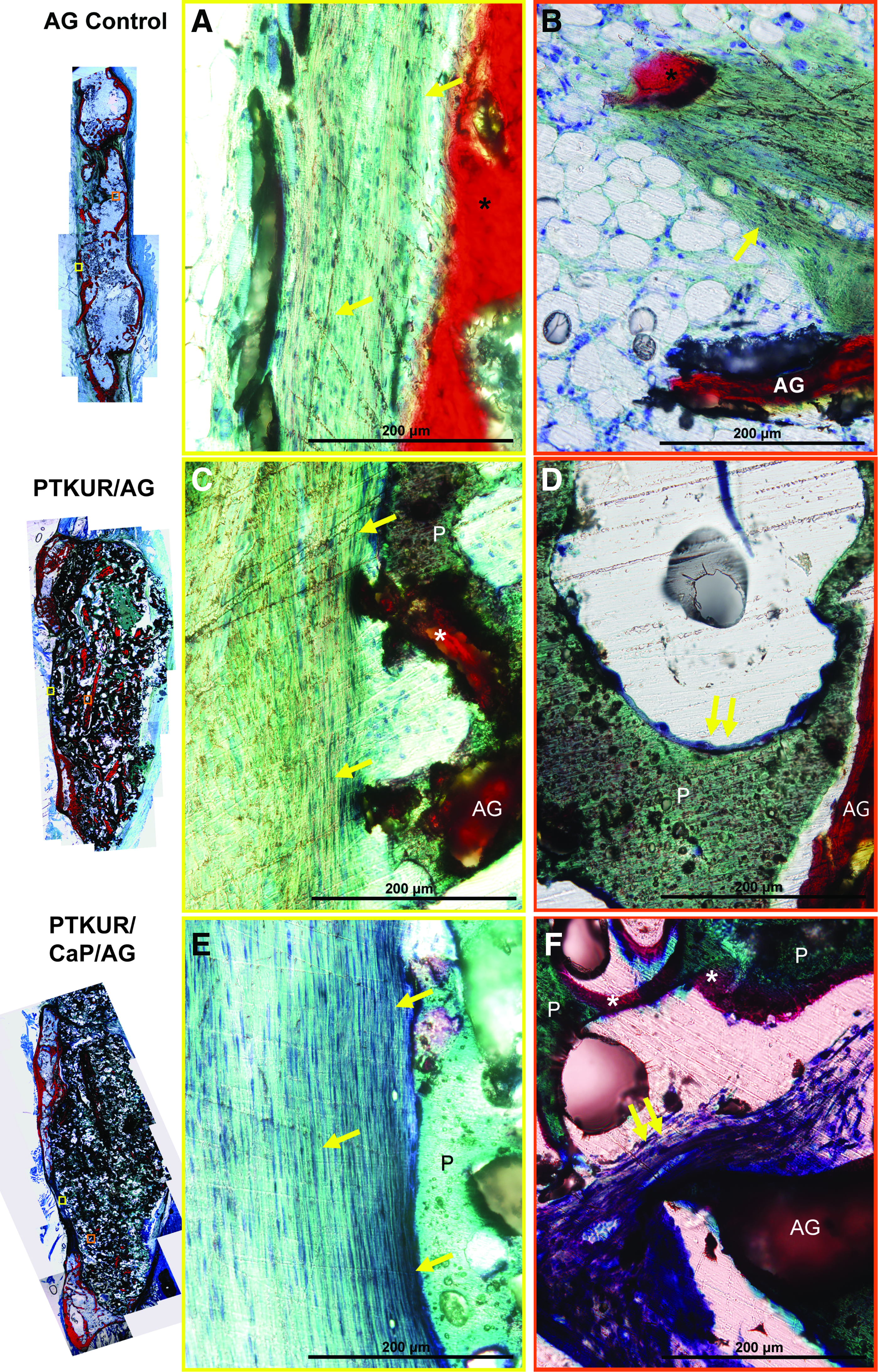
Magnified images (40 × ) of histological sections of AG control (top row), PTKUR/AG (middle row), and PTKUR/CaP/AG (bottom row).
Combining data from the fluorescent dyes (2 and 6 weeks) and histological stain (8 weeks) and focusing on the plane of the processes allowed for temporal investigation of the mechanisms of neo-osteogenesis. Because osteogenesis generally started at the processes, the processes were included in the calculation of the osteogenesis front (% bridging) to eliminate bias associated with the diameter of the processes (Fig. 8A). At 2 weeks, there were no significant differences in % bridging between any of the groups, while at 6 weeks the AG control showed significantly higher % bridging compared with PTKUR/AG and PTKUR/CaP/AG (Fig. 8B). There was a significant increase in % bridging from 2 to 8 weeks for all groups, indicating that osteogenesis was ongoing at the base of the ITP space. The % bridging rate was calculated from the data plotted in Figure 8B. PTKUR/AG and PTKUR/CaP/AG showed similar bridging rates of ∼2% per week (PTKUR/AG: 2.2% ± 0.0076%/week, PTKUR/CaP/AG: 2.4% ± 0.35%/week). The AG control exhibited a significantly higher bridging rate compared with that calculated for PTKUR/AG and PTKUR/CaP/AG (6.4% ± 0.23%/week). These data suggest that while persistence of the PTKUR polymer delays bone bridging, osteogenesis in PTKUR/AG and PTKUR/CaP/AG is ongoing for both groups (R2 > 0.98).

Quantification of bridging of lateral processes.
Discussion
It is widely accepted that AG is the gold standard in bone grafting. AG processed using a bone mill retains its osteoinductivity and growth factors that aid in bone regeneration.56,57 The importance of osteocytes in orchestrating bone regeneration has recently been highlighted,58,59 and a recent study has shown that milled AG releases signaling molecules that direct the activity of osteoblasts and osteoclasts. 59 The finite availability, invasive harvesting methods, and unpredictable resorption of AG have stimulated interest in synthetic AG extenders that reduce the volume of AG required to promote bone healing.6,12 In this work, settable PTKUR-based AG extenders were evaluated in an intertransverse bone formation model in rabbits.
PTKUR has been reported to undergo cell-mediated oxidative degradation to noncytotoxic breakdown products in rat,29,30 rabbit, 28 and pig 31 models of tissue regeneration. We hypothesized that PTKUR-based AG extenders would set within clinically relevant setting times to form bone grafts with sufficient strength to resist mechanical forces from the muscles posterior to the lumbar spine. 60 The ability of the graft to resist these mechanical forces and maintain space is critical, as demonstrated by insufficient bone induction when rhBMP-2 was delivered from a compliant collagen sponge in an ITP model in nonhuman primates. 61 Thus, the goal of the present study was to design new bone grafts with handling and mechanical properties approaching those of bone cements and osteoinductivity comparable to AG. PTKUR/AG was first evaluated in an athymic rat model to test the hypothesis that cells would infiltrate the graft. The rabbit xenograft was well tolerated by the rats, and there was no evidence of graft rejection. Histomorphometric analysis showed that cellular infiltration at 4 weeks trended higher in the AG control compared with PTKUR/AG, but the differences were not significant. These observations supported more extensive assessment of bone induction in the rabbit spine model.
The posterolateral ITP bone formation model has been used to evaluate bone induction in a biologically stringent environment. 34 In this model, host cells migrate into the graft from the transverse processes, and no bone induction is observed in the absence of an osteoinductive component, such as autologous tissue or rhBMP-2.15,34,48 ChronOS strip (DePuy Synthes), a composite of β-TCP granules and poly(lactide-co-ɛ-caprolactone), showed no new bone formation when implanted in the paraspinal muscle of beagles. 62 However, ChronOS strip augmented with bone marrow aspirate and local AG showed fusion rates of 88.9% in patients undergoing posterolateral spinal fusion. 15
Similarly, extending AG with other ceramics, such as β-TCP,16,19,23 hydroxyapatite, 18 and β-calcium pyrophosphate, 17 has shown comparable outcomes to AG controls at 1–3 years postoperatively in patients.63,64 These previously reported AG extenders often comprise blends of ceramic particles with local and/or ICBG AG, which lack mechanical integrity. Injectable biphasic CaP bone cements have been reported to promote bone healing and resorb in patients with distal radius and proximal tibia fractures 13 and bone cysts and tumors. 65 However, while these materials are effective for filling bone voids, there are no currently available bone cements that induce new bone formation in the more demanding ITP environment. 14
In contrast, PTKUR/AG and PTKUR/CaP/AG exhibited setting times comparable to CaP bone cements and induced bridging of the ITP space with new bone. After injection, these materials hardened to form a graft with sufficient mechanical properties to maintain space in the presence of mechanical forces from the muscles posterior to the lumbar spine. However, the rabbit ITP model is not suitable for assessing the effects of mechanical forces on bone induction, as evidenced by earlier studies reporting that local delivery of rhBMP-2 from a compliant collagen sponge induces complete bridging in rabbits, but not in nonhuman primates.34,61 The effects of the mechanical properties on space maintenance and bridging will be assessed in a more mechanically stringent nonhuman primate model in future studies.
Fluorochromes allowed for the differentiation of implanted AG and new bone growth and also provided insight into the regeneration patterns of PTKUR/AG and PTKUR/CaP/AG compared with the AG control. The distance of the osteogenesis front for each fluorochrome and the final stain were compared to investigate phases of remodeling. Areas of fluorescence within the graft were colocalized with more darkly stained areas of stained sections. There was evidence of endochondral bone formation in these areas where a cartilaginous phase (stained dark blue-purple) was actively being calcified into bone (Fig. 6).
Stained histology slides showed evidence of possible pseudarthrosis in specimens from all groups (Fig. 6, single arrows). This “false joint” of cartilaginous tissue centrally located between the two processes may have had sufficient mechanical stability to simulate bridging of the processes, which has been described as the “reparative phase” characteristic of weeks 4–6 postimplantation when there is a lag between cartilage formation and ossification.3,66 This reparative phase was delayed for PTKUR/AG and PTKUR/CaP/AG, which could be due to the persistence of PTKUR. Bone grew around the entire implanted graft in the AG controls, whereas a planar growth from process to process was ongoing in PTKUR/AG and PTKUR/CaP/AG (Fig. 7).
Enhanced new bone formation compared to an AG control has been reported for a ceramic extender (25% or 75% MASTERGRAFT granules) in the same rabbit model used in the present study. 21 When a MASTERGRAFT strip was used, the 50% MASTERGRAFT AG extender group exhibited new bone formation superior to the autograft control, while the 75% MASTERGRAFT group generated less bone than the AG control. 35 However, neither of these grafts was settable in vivo. In the present study, new bone formation was observed in PTKUR/AG and PTKUR/CaP/AG, but complete bony bridging of the processes was not observed (Fig. 8). These findings suggest that while the settable polymer enhanced the handling and mechanical properties of the AG extender, the persistence of the PTKUR component at 8 weeks reduced cellular infiltration and new bone formation.
When implanted alone, AG undergoes rapid and unpredictable resorption compared to some synthetic grafts.5,67 Premature graft resorption could result in incomplete healing and the formation of fibrous tissue. 40 A nonsettable polymeric AG extender comprising poly(lactide-co-glycolide) (PLGA)/hyaluronic acid (50 vol% AG) has been investigated in the same rabbit model used in this study. 33 Progression was noted from 3 to 6 months and a radiographic bone fill score of 4.5 (0–5 scale, 5 = 81–100% bone fill) reported for the AG extender 6 months postimplantation. Nonsettable PLGA/AG exhibited a score of 4.0 at 3 months compared with 4.75 for the control animals. The authors also observed residual PLGA at the implantation site at 3 months. These observations are consistent with our findings that persistence of the polymer delays new bone formation.
In a previous study, local delivery of rhBMP-2 from a PEUR scaffold showed complete bridging of the processes and nearly complete degradation at 8 weeks. 68 While a slowly degrading settable polymer reduces the induction of new bone formation, it may also protect the implant against premature AG resorption and loss of mechanical integrity. The effects of the polymer degradation rate on new bone formation will be assessed in future studies.
Conclusion
In this study, settable and cell-degradable PTKUR-based AG extenders were formulated and tested in preclinical models of bone regeneration. PTKUR/AG and PTKUR/CaP/AG exhibited working times of 10–20 min and compressive strengths of 1–2 MPa. When implanted in a posterolateral ITP bone formation model in rabbits, bone growth along the posterior plane of the processes was consistent from 2 to 8 weeks for PTKUR/AG and PTKUR/CaP/AG. New bone formation was also evident within the ITP space away from the processes. The inclusion of PTKUR/CaP/AG established that AG content can be reduced to ∼50% without significantly compromising bone healing. This work provides evidence for the potential of PTKUR-based AG extenders for treatment of bone defects.
Footnotes
Acknowledgments
This work was funded by the National Institutes of Health (NIH) (R01AR064772 and T32DK101003), a National Science Foundation Graduate Research Fellowship to Madison McGough (Grant No. 1445197), and the United States Army Institute of Surgical Research. Any opinions, findings, and conclusions or recommendations expressed in this material are those of the authors' and do not necessarily reflect the views of the National Institutes of Health or the National Science Foundation.
Disclosure Statement
No competing financial interests exist.