
Abstract
Hyaluronic acid (HA) is an important component of the extracellular matrix (ECM), and electrospun scaffolds have the three-dimensional porous network structure characteristics similar to those of the autologous ECM. However, scaffold fabrication of water-dissolved HA without surfactant by electrospinning is difficult. This study reports the fabrication of HA-coated biomimetic nanofiber scaffolds through coaxial electrospinning of poly(
Impact Statement
The extracellular matrix (ECM)-inspired electrospinning scaffolds have a good cytocompatibility. Hyaluronic acid (HA) is one of the main components in the bladder ECM, but it is hard to be made by electrospinning alone. In this study, it is shown that coaxial electrospinning can fabricate poly(
Introduction
Advanced bladder cancer, neurogenic bladder dysfunction, and bladder exstrophy are the most common acquired and congenital urinary tract diseases and often require bladder reconstruction. 1 However, treatment is difficult owing to many complications. 2 Thus, development of innovative methods for bladder reconstruction is urgently needed. Biological materials have the potential to minimize these complications and show great promise in bladder repair and regeneration. Tissue-engineered biomaterials have been used to regenerate bladder tissue in various animal models, 3 and scaffolds constructed from synthetic polymers and naturally derived materials have been widely used for bladder reconstruction.4–7
Electrospun nanofiber meshes could be applied to regenerative medicine and tissue engineering, because the meshes have structures similar to that of the natural extracellular matrix (ECM), 8 which is a three-dimensional (3D) network comprising many protein fibers and biomolecules. Electrospun nanofiber meshes have features such as large surface-to-volume ratio, high porosity, and spatial interconnectivity, which benefit cell communication as well as metabolic waste and nutrient transport. 9
We previously utilized poly(
Thus, we chose hyaluronic acid (HA), which is a naturally linear polysaccharide that consists of alternating disaccharide units of β-1,3-N-acetyl-
Some researchers have found that crosslinking HA with small intestinal submucosa (SIS) enhances angiogenesis in a partial cystectomy canine model. 15 These findings encouraged modification of electrospun fiber surfaces with HA in bladder tissue engineering. However, electrospinning of HA aqueous solutions is very difficult, mainly due to their high surface tension and viscosity, even at low concentrations.16,17 Resolving these complications in the electrospinning of HA alone usually requires complicated electroblowing systems or harsh solvents, which complicate HA nanofiber fabrication. 16
Coaxial electrospinning is an effective means of functionalizing nanofiber surfaces by generating core–shell structured nanofibers. Previous research has revealed that liquids with low electrical conductivities, traditionally considered unsuitable in electrospinning, can be readily stretched into thin filaments by coelectrospinning with a highly spinnable solution. 18
In this study, we aimed to use HA as the shell and PLCL as the core to enable production of uniform HA-functionalized core–shell structured nanofibers for tissue engineering applications, which was more controllable fabrication and bionic structure, as schematically shown in Figure 1. Because no surfactant was added in the HA emulsification process, the approach avoided the complications related to surfactant removal, such as chemical crosslinking. Furthermore, PLCL core blending also maintained the structural stability of the nanofiber scaffolds.
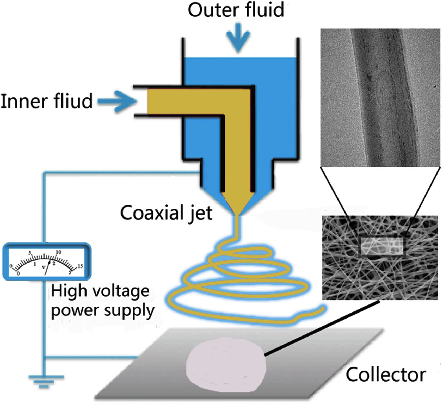
Schematic illustration for coaxial electrospinning nanofiber preparation.
To our knowledge, this research is the first, evaluating PLCL/HA core–shell nanofibers for bladder tissue engineering. Thus, we thoroughly characterized the physicochemical properties of PLCL/HA nanofibers with scanning electron microscopy (SEM), transmission electron microscopy (TEM), attenuated total reflection Fourier transform infrared (ATR-FTIR) spectroscopy, and water contact angle (WCA) measurements. Morphological features, proliferation, and migration were analyzed to assess cytocompatibility of the scaffolds in vitro. Anatomical and functional outcomes of scaffold implantation in a rat cystoplasty model were analyzed to evaluate in vivo biological performance.
Experimental Procedures
Materials
Poly(e-caprolactone) (PCL) (Mw = 1000) was purchased from Solvay Co., Ltd. (Korea). Poly(lactic acid) (PLLA) (Mw = 100,000) was purchased from Guanghua Co. Ltd., (China), and HA powder was purchased from Chemgreatwall Co., Ltd. (China). The two solvents, 1,1,1,3,3,3-hexafluoro-2-propanol (HFIP) and formic acid (FA), were analytical grade and used without further purification to dissolve PCL, PLLA, and HA.
Preparation of electrospun PLCL/HA core–shell nanofibers
PLCL was prepared by mixing PLLA and PCL as previously described. 10 Briefly, PLLA and PCL were dissolved in HFIP at a 1:1 ratio, and the total concentration of mixed polymer was 10% (w/v). HA was added to the HFIP/FA (70/30 vol%) solution at 2% (w/v). All solutions were mixed with magnetic stirrers for 12 h at room temperature (25°C).
The coaxial electrospinning method was employed to produce the core–shell nanofibers. Two-fluid concentric nozzles comprised the coaxial spinneret. The outer and inner needle had diameters of 1.07 and 0.25 mm, respectively. The outer capillary was loaded with HA solution pushed at a flow rate of 0.15 mL/h, and PLCL solution was delivered to the inner coaxial capillary at a flow rate of 0.6 mL/h. The electrospinning system was similar to that reported previously with some improvement. 10
Briefly, a DC voltage of 16 kV was supplied by a high-voltage power supply. The distance between the grounded collector and the coaxial syringe tip was 15 cm. For comparison, we exchanged the core and shell compounds for HA/PLCL core–shell nanofibers, and solely PLCL nanofibers were also fabricated. All nanofibers were prepared under similar conditions. The electrospun nanofiber mats were placed in a vacuum oven for ∼24 h to remove any residual organic solvent before further use. For bladder regeneration, we used a 3D dome-shaped collector to fabricate domed electrospun nanofiber scaffolds.
Characterization of the HA/PLLA core–shell nanofibers
Topography and composition analysis
Morphology of the electrospun nanofibers was investigated by SEM at an accelerating voltage of 15 kV. Before imaging, the samples were fixed on metal stubs and sputter coated with palladium–platinum–gold for 120 s to increase conductivity. The core–shell structure was observed by TEM, and the samples were prepared by depositing the nanofibers onto a rectangular frame of copper meshes directly, then drying them in the vacuum oven for 48 h at room temperature. The average fiber diameter was measured from the SEM images using ImageJ (NIH, Bethesda, MD) software. For each sample, an average of 50 nanofiber filaments was counted. We performed ATR-FTIR spectroscopy to examine the composition of the nanofibers. The spectra were recorded in the wavelength range of 1000–4000 cm−1.
Contact angle and swelling
The WCAs of the electrospun scaffolds were measured to determine hydrophilicity. Images were acquired by photographing water drops on the sample surface, and WCA was measured in directions parallel and perpendicular to the nanofiber axis direction. Three samples were measured in each group. For the swelling studies, we immersed the scaffolds into phosphate-buffered saline (PBS) at 37°C for 1 h, removed them, and quickly wiped the residual liquid from their surfaces. All samples were embedded into a cryomatrix (Jung Tissue Freezing Medium; Leica Microsystems, Wetzlar, Germany) at −20°C for 30 min. Sections of 10 mm thickness were examined within 2 min without any fixation. Images were acquired by inverted light microscopy.
Cell–scaffold interaction in vitro
Rat bladder SMCs were obtained from rat urinary bladders as previously described. 19 Briefly, the luminal surface layers of bladders were removed mechanically by scraping with a scalpel blade. The tissues were washed three times in PBS containing penicillin–streptomycin and minced for digesting by 30 min at 37°C in PBS containing elastase, papain, and collagenase type II. The harvested cells were cultured in Dulbecco's modified Eagle's medium, 10% fetal bovine serum at 37°C in 5% CO2, with medium changes twice weekly. The passage-3 cells were used in the experiments. The animal experiments were approved by the Ethics Committee of Tongji Medical College, Wuhan, China (Permit Number: TJ-A20141214).
SMCs seeded onto scaffolds were cultured for 1, 3, or 5 days for the cell morphology study. The methods to characterize the SMCs and cell morphology analysis are in Supplementary Data. A noninjury cylinder assay was performed to evaluate cellular migration, which was in Supplementary Data for the detail. Outward migration percentage (OMP) was calculated by normalizing this area to the cell occupied area (CA) at the 0-h migration time point (CA0), according to the following equation: OMP = (CA − CA0)/CA0 × 100%. Three samples were measured in each group. Cell proliferation was analyzed in triplicate using the MTT (Beyotime Biotech, Shanghai, China) assay at the predetermined time points of 1, 3, and 5 days, according to the manufacturer's manual.
Animal implantation, urodynamic test, and histological and immunohistochemical staining evaluation
Adult Sprague-Dawley rats (Tongji Laboratory Animal Center, Wuhan, China) weighing 200–250 g underwent bladder augmentation cystoplasty by implantation of the PLCL/HA (group 1, H) or PLCL nanofiber (group 2, P) domed grafts, each group had eight rats. Rats were anesthetized by intraperitoneal injection of chloral hydrate (30 mg/kg), and an ∼1 cm longitudinal incision was made to explore the bladder. A 40–50% supratrigonal cystectomy was then carried out in the bladder dome to create the bladder defect. The domed nanofiber scaffolds were anastomosed to the cystectomized defect. Eight rats without grafted nanofiber scaffold implants were set cystotomy controls (Group 3). Moreover, eight normal rats receiving sham surgery were maintained (Group 4).
At postoperative week 4 and 10, the bladder urodynamics of each group were monitored. The animals were anesthetized, and the bladders were emptied by manual abdominal pressure. We retrogradely advanced a sterile 2F transurethral polyurethane catheter into the bladder and connected it to the infusion pump for continuous infusion of saline solution into the urinary and bladder pressure transducer. The physiological saline infusion rate was 100 μL/min, and intravesical pressure and saline flow measurements were continuously recorded.
The volume of infused saline that triggered the first leakage of urine was recorded as maximal bladder capacity (DV, mL), and the bladder pressure that triggered voiding minus resting bladder pressure was defined as threshold pressure (DP, cm H2O). Bladder compliance was assessed by volumetric pressure ratio (Vpr), defined in the following equation: Vpr = DV/DP. After scheduled euthanasia, full-thickness bladder tissue was excised according to standard procedures. 20 For immunohistochemical analyses, we chose alpha-smooth muscle actin (α-SMA) as the contractile smooth muscle marker. 21 Specimens were visualized and imaged by microscope.
Statistical analyses
All data are presented as mean ± standard deviation. Quantitative measurements of urodynamic parameters were analyzed by independent Student's t-tests; other statistical analyses were carried out using one-way analysis of variance. Differences were considered statistically significant at p < 0.05. All experiments were carried out at least three times.
Results and Discussion
Morphology of the electrospun nanofibers
The morphological characteristics of electrospun PLCL/HA and PLCL meshes were evaluated by SEM. SEM images revealed that the electrospun PLCL/HA core–shell nanofibers (Fig. 2A) were highly uniform and morphologically comparable to similarly obtained PLCL fibers (Fig. 2B). Quantitatively, the average diameters measured for the aligned HA/PLCL and PLCL fibers were in the range of 784.2 ± 138.2 and 730.9 ± 137.2 nm, respectively, and were not significantly different. When exchanging the constituents of the core and shell and using HA as the core and PLCL as the shell, we observed the lower spinnability in the coxial system due to droplets and inefficiency. The average diameter of HA/PLCL core–shell fibers was 501.2 ± 101.5 nm (Supplementary Fig. S1).

Characterization of PLCL/HA and PLCL nanofibers:
We investigated the newly prepared nanofibers by TEM to explore the core–shell structures. The interface between the core and shell was observed in the TEM images of PLCL/HA nanofibers, and the contrast from the core to the shell revealed that the density of the nanofibers had changed (Fig. 2C). In contrast, when HA was used as the core component, the scaffold surface was irregular because of droplets and crosslinking between nanofibers. HA microspheres were randomly dispersed throughout the PLCL nanofibers of the HA/PLCL group (Supplementary Fig. S1B). As we expected, the composition of PLCL nanofibers was single with no inner contrast (Fig. 2D).
Generally speaking, the nanofibers had a core–shell microstructure which comprised two different materials. HA coaxial electrospun fibers have been demonstrated to be fabricated effectively and suitably for engineering structurally anisotropic tissues.22,23 Furthermore, electrospun fibers appear to be more uniform, efficient, and produced fewer droplets in PLCL/HA core–shell nanofibers than do HA/PLCL core–shell ones.
The PLCL/HA core–shell fiber matrices can be successfully fabricated by coaxial electrospinning, although HA aqueous solution has a high surface tension and viscosity, which are obstacles to electrospinning. We demonstrated that PLCL as a spinnable solution facilitated the stretching of HA into a shell coating on the PLCL core. However, we also observed that the core–shell structure did not exist in HA/PLCL core–shell nanofibers. This may have been because when the Taylor cone tips were significantly elongated, the HA core was more likely to form microspheres, since the PLCL shell failed to provide friction to resist the surface tension of HA. Nevertheless, some studies have claimed that HA/PLCL core–shell nanofibers can be fabricated.22,24
We noticed that the HA concentration used in these studies were low and located in the core within nanofibers, which may reduce the biocompatiblity of the scaffolds. Moreover, HA as a biomaterial is better placed in the shell in direct contact with cells. It is worth noting that increasing HA concentrations reduces the density of other solutions in the coaxial electrospinning system, making the core–shell structure harder to generate. 15 The results from studies using lower concentrations of HA as the core are consistent with our own findings. There were also some studies, where coating HA simply upon a scaffold got satisfactory regeneration, which was a simple and effective method.25,26 But coaxial electrospinning was more controllable; it could endow a thin HA-coated layer atop each PLCL nanofiber.
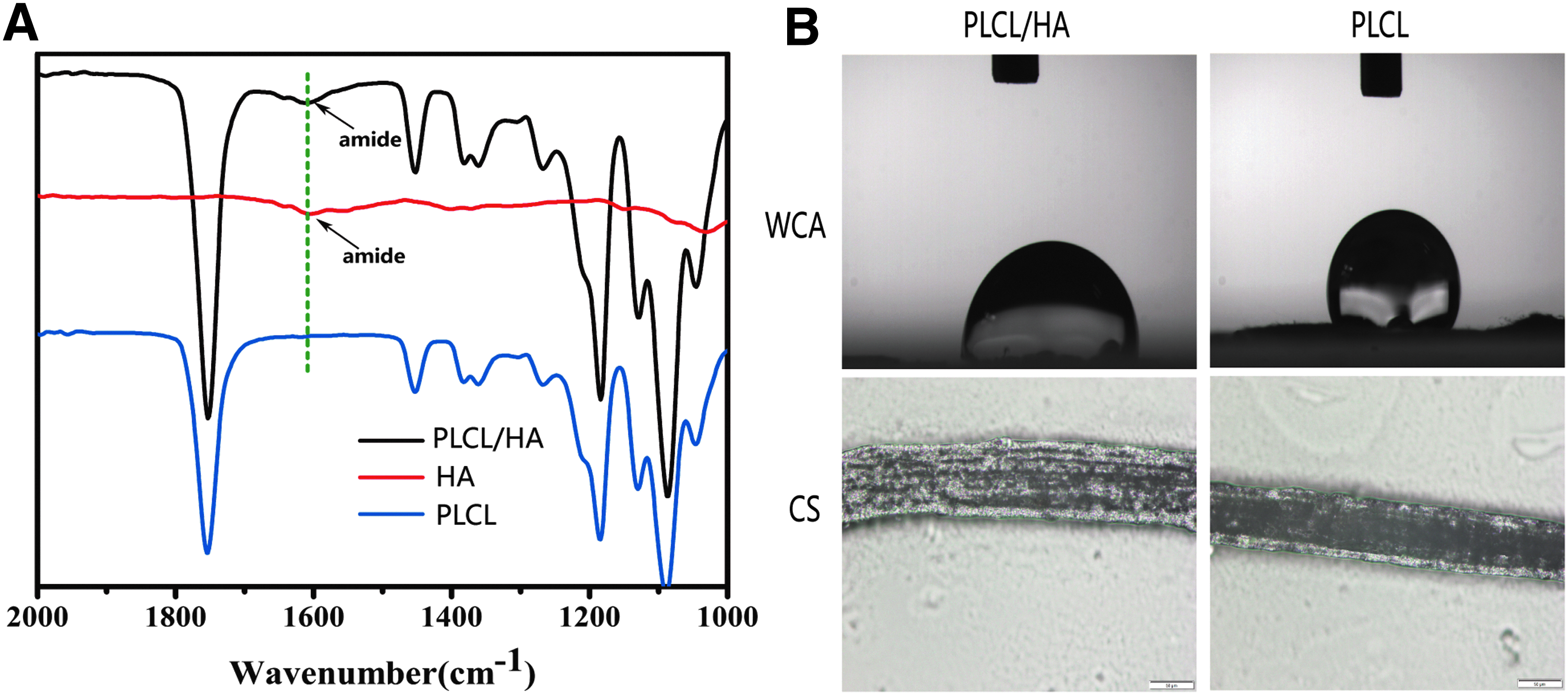
The nanofibers were examined by ATR-FTIR spectroscopy to determine the presence of PLCL and HA (Fig. 3A). In the ATR-FTIR spectra of the nanofibers, the peak at 1730 cm−1 corresponded to the C = O stretching band absorption, which could be observed in both the PLCL and PLCL/HA samples. 27 The characteristic peak in HA at 1630 cm−1, which corresponds to the amide group, was only observed in PLCL/HA and HA powder samples confirming the presence of the HA coating. 28
Wettability and water retention capacity
We used HA as a highly hydrophilic biopolymer to modify PLCL nanofiber surface wettability properties, which are important factors for cell adhesion, growth, and tissue regeneration. Hydrophilicity of nanofibers was determined by WCA measurement, and we further tested the swelling properties of nanofibers to reveal the water absorption capability. Figure 3B shows a WCA of 87.2 ± 1.8° for PLCL/HA and 128.8 ± 1.5° for PLCL, which are significantly different (p < 0.05). Typically, electrospun PLCL fibers are hydrophobic polymers, and the WCA is further significantly decreased by the HA coating, which offers indirect evidence of the PLCL/HA core–shell structure of the nanofibers.
Observation of frozen cross-sections revealed that HA-coated scaffolds exhibited an internal structural change and thickness increases (Fig. 3B). HA, as a naturally occurring linear polysaccharide, has strong retention capacity and immobilizes water in human tissues. Additionally, swelling of the HA shell could induce layered distribution and volume expansion, which could present a more suitable 3D structure for cell proliferation. 29 Thus, it is reasonable to conclude that the enhanced wettability and enlarged nanotopography of PLCL/HA core–shell structures provide biochemical and physical cues for cell orientation along the nanofiber axis.
Assessment of cell morphology and proliferation
Scanning electron micrographs and confocal laser scanning microscopy with cytoskeleton staining of SMCs cultured in vitro on the PLCL/HA and PLCL nanofiber scaffolds for 12 h, 1, and 3 days are shown in Figure 4. The SMCs on PLCL/HA scaffolds exhibited greater cell elongation, spreading morphology, and proliferation rate than did those on PLCL scaffolds. After 5 days, SMCs had almost grown to confluency on the scaffolds (Supplementary Fig. S2). Intriguingly, we found that more filopodial protrusions were observed in PLCL/HA scaffolds after 2 days culture, which suggests that HA promotes filopodial outgrowth (Fig. 5A, B).

Cell morphology changes and proliferation in different scaffolds.
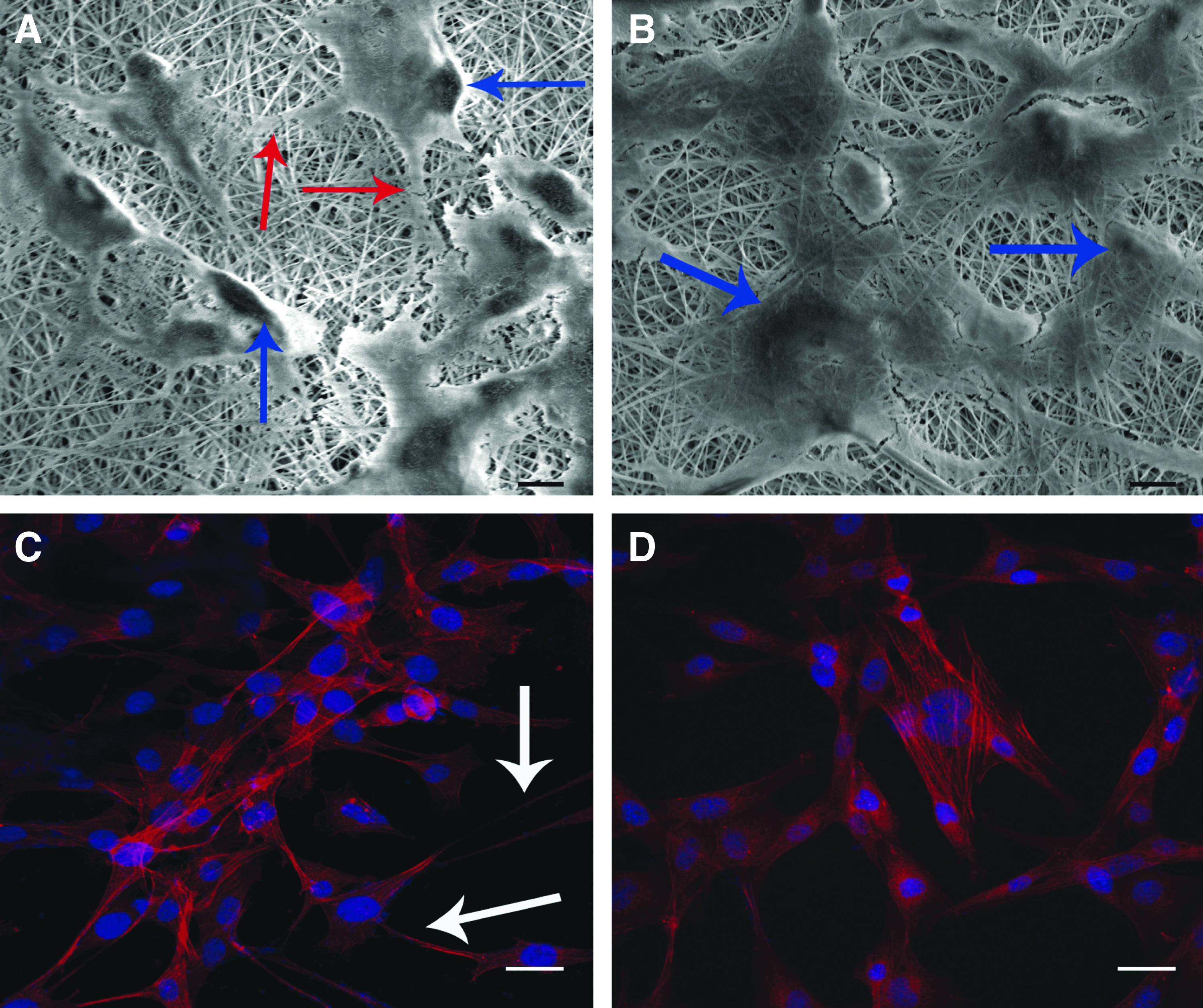
Effect of PLCL/HA scaffolds on cellular filopodia formation.
Protrusion activity is an important component of efficient cell migration. The cell filopodial extensions can bridge the gaps between neighboring cells, and enhance cellular interactions. 30 Such morphological changes are often reported when cells are cultured on 3D ECM matrices reflecting a more physiological cellular environment. 31 HA is crucial in maintaining tissue hydration and matrix stability, which can regulate cell functions. Therefore, supplementing the electrospun nanofiber scaffolds with HA improves the cellular 3D microenvironment, enabling cells to adopt more physiological morphologies. 15
To investigate the migration of SMCs on the scaffolds, we used a noninjury cylinder assay. The results suggested that SMCs continued outward migration on the nanofiber scaffolds (Fig. 6A). The PLCL/HA scaffolds showed higher migration of SMCs than did the PLCL scaffolds. Briefly, the scaffolds showed similar outward migration rates before 12 h. After 24 h of migration, the PLCL/HA scaffolds showed a higher outward migration rate than did PLCL scaffolds (Fig. 6B).

Effect of different scaffolds on SMC migration and proliferation.
Quantitative MTT analysis indicated that cells proliferated better on the PLCL/HA nanofibers. SMCs cultured on both nanofiber scaffolds exhibited similar growth patterns with time-dependent cell number increases during the incubation, as shown in Figure 6C. There was no significant difference between the proliferation of SMCs on the two scaffolds at day 1, but proliferation was higher on PLCL/HA than on PLCL scaffolds at days 3 and 5, suggesting a better biocompatibility of PLCL/HA than of PLCL. The observed promoting effect on proliferation can be ascribed to the presence of HA in the PLCL/HA scaffolds. The SMCs expressed muscle contractile protein markers and maintained the contraction function (Supplementary Fig. S3).
Obtaining the desired cellular morphology and proliferation through manipulation of material surface characteristics is still a challenge in biomaterials. Nanoscale-structured polymer scaffold surfaces have been introduced to functionalize surfaces and improve interactions between cells and scaffolds, cell adhesion, proliferation, migration, and ECM production.32–34 In addition, nanostructured scaffolds modified by biochemical moieties to provide biochemical stimuli can be crucial for regeneration processes. Many studies have tried to immobilize biomolecules for different targets directly on a biomaterial surface.35,36 HA maintains the stable and hydrated extracellular space, and previous studies have found that cellular migration and adhesion in the ECM are highly correlated to HA.37,38
In our study HA coating applied onto PLCL surfaces, and we found that this coating facilitated cell elongation and migration owing to the extraordinary regulation properties of HA in cells and the ECM. Thus, the introduction of HA as a shell for PLCL nanofibers was beneficial.
Rat bladder augmentation, histological analysis, and physiological bladder testing
A dome-shaped scaffold (inner diameter = 15 mm; thickness = 1 mm; concavity = 5 mm) was used in cystoplasty (Fig. 7A). After implantation, the grafts retained good continuity with the native tissue without urinary leakage or breakdown of the implanted scaffolds (Fig. 7B). Ten weeks after implantation, gross examination revealed that the tissue-engineered urinary bladder wall had rich vascularization, the suture line was not visible, and the augmented urinary bladder tissue was integrated with the host bladder wall, as shown in Figure 7C and D (Supplementary Fig. S4). In addition, we found some urinary crystals and stones in all three operated groups by macroscopic examination after euthanasia.

Masson's trichrome staining showed that some infiltrating cells had formed a better linear distribution along the circumference of the PLCL/HA grafts than along that of the tubular PLCL grafts within the regenerated tissues after 4 weeks in vivo, likely due to the enhanced hydrophilicity of PLCL/HA nanofibers (Fig. 8A). By the 10th postoperative week, the de novo bladder walls involved highly fragmented portions of the nanofiber scaffolds embraced by ECM-rich lamina propria. To confirm the regeneration of SMCs, α-SMA staining of the infiltrating cells was performed.
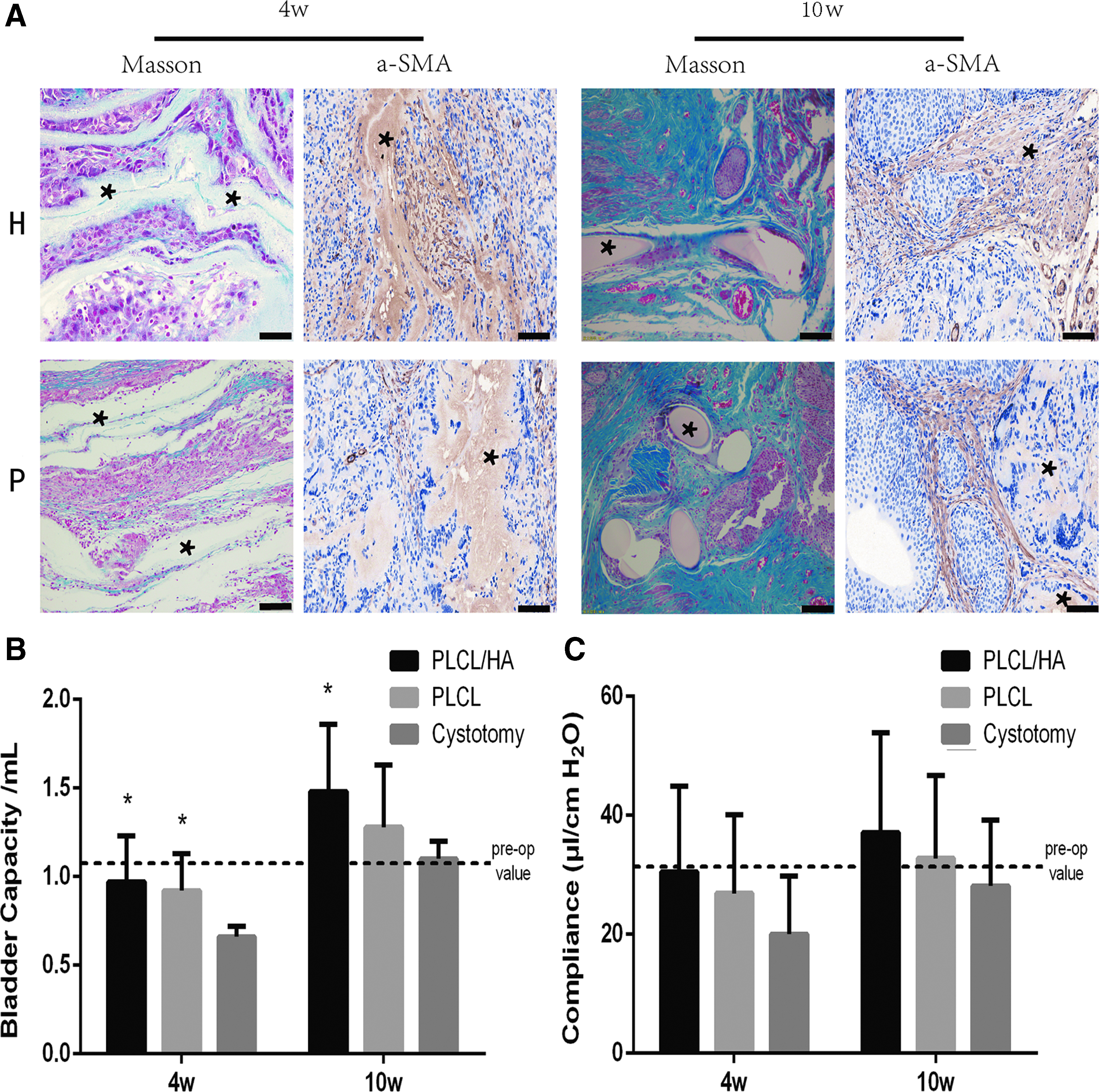
A large number of α-SMA-positive cells was observed around the PLCL/HA graft wall relative to that on the shallow surface cell layer formed on the PLCL scaffolds, suggesting that contractile SMCs were achieved in vivo on the PLCL/HA nanofiber grafts. These staining results indicated that the PLCL/HA grafts, and not their PLCL counterparts, were prone to recruit and transform more infiltrating perivascular cells into the contractile phenotype of SMCs, which is consistent with our in vitro observations (Fig. 8A).
In this study, we used dome-shaped scaffolds to augment the bladder capacity, which led to significant improvements in bladder capacity compared with that in the control group after four postoperative weeks. All three groups exhibited capacity comparable to that of normal bladders after 10 postoperative weeks. The partial cystectomy and PLCL groups exhibited decreased bladder compliance, but the difference was not significant. This result is inconsistent with previous bladder augmentation studies, wherein scaffolds were able to augment organ compliance.39,40 This phenomenon may be explained by the stones present in these groups, which may have masked these parameters. Further investigation is needed to confirm this assumption.
Appropriate scaffolds play a crucial role in bladder regeneration, and previous uses of simple acellular scaffolds, such as SIS or ABM, failed to induce fast SMC regeneration in large-area defects.41,42 Scaffolds designed for bladder reconstruction should focus on compatibility and promoting the growth of SMCs, because SMCs are the main components that determine the capacity and compliance of the bladder. 43 As one of the major components of the ECM, HA contributes significantly to cell proliferation and migration, and supports bladder regeneration. 14
Therefore, we chose the PLCL/HA core–shell scaffolds, which significantly improved bladder capacity by promoting smooth muscle tissue proliferation in our study. It is much anticipated that nanostructured biomaterials will be the next-generation scaffolds for bladder tissue engineering since nanostructured biomaterials can be fabricated to mimic the microenvironments of the tissues or organs that need to be regenerated.
Conclusion
We successfully fabricated PLCL/HA core–shell nanofibers that possessed the advantages of both HA and PLCL, including wettability, high swelling ratios, and spinnablility. In addition, PLCL/HA scaffolds improved the growth of SMCs in vitro; we observed that the number of cellular filopodia increased, potentially benefiting migration. The animal study indicated that the dome-shaped scaffolds comprising PLCL/HA nanofibers also simultaneously promoted regeneration of SMCs and enhanced bladder capacity. These results suggest a better role of HA coating of PLCL nanofibers for SMC regeneration in vitro and in vivo. These findings may lead to more advanced in vivo tests and clinical applications of PLCL/HA grafts.
Footnotes
References
Supplementary Material
Please find the following supplemental material available below.
For Open Access articles published under a Creative Commons License, all supplemental material carries the same license as the article it is associated with.
For non-Open Access articles published, all supplemental material carries a non-exclusive license, and permission requests for re-use of supplemental material or any part of supplemental material shall be sent directly to the copyright owner as specified in the copyright notice associated with the article.