
Abstract
Severe infection and mechanical injury of the uterus may lead to infertility and miscarriage. Currently, there is a lack of effective treatment modality for functional repair of uterine injury. To address this clinical challenge, this study aimed to develop a chemotactic composite scaffold by incorporating recombinant human stromal cell-derived factor-1α (rhSDF-1α) into a silk fibroin-bacterial cellulose (SF-BC) membrane carrier. A rat model of uterine injury was utilized for this study, which was composed of three groups as follows: blank control, implantation with SF-BC only, or SF-BC loaded with rhSDF-1α. The tissue regeneration efficacy of the three groups was analyzed and compared. The results showed that SF-BC loaded with rhSDF-1α significantly enhanced endometrial regeneration and arteriogenesis of the injured rat uterus, which led to improved pregnancy outcomes, thus indicating much promise for functional uterine repair and regeneration.
Impact Statement
In this study, we demonstrated that the silk fibroin-bacterial cellulose (SF-BC) membrane possessed good physical, chemical, and biocompatibility properties in vitro. The in vivo study showed that the incorporation of recombinant human stromal cell-derived factor-1α (rhSDF-1α) within the SF-BC membrane promoted regeneration of full-thickness uterine injury and also improved the pregnancy outcome of the damaged uterus. The results thus suggest that SF-BC loaded with rhSDF-1α has good potential in future clinical applications for the repair of uterine injury.
Introduction
The uterus is an important hormone-responsive organ in female mammals and is essential for the implantation and development of embryos. However, severe infection and certain serious uterine injuries, such as curettage, may cause uterine adhesion, while cesarean section may result in scar formation, infertility, recurrent miscarriages, and other obstetrical complications, such as placenta previa and placenta accreta. Therefore, it is essential to repair the damaged uterus to enable a normal pregnancy. 1 Functional repair and regeneration of the injured/damaged uterus still remains a formidable challenge in clinical practice.
Tissue engineering and regenerative medicine could be a promising way to solve this problem. Biomaterials used in uterus repair and regeneration should have strong water absorption to deliver bioactive molecules to promote regeneration, with good biocompatibility to support endometrial cell attachment and proliferation, as well as excellent mechanical properties to support the expansion of uterus during pregnancy. 2
Bacterial cellulose (BC) is produced by certain types of bacteria and is characterized by good biocompatibility, porosity, and strong water absorption, making it suitable for tissue engineering of bone and nerve, repair of tympanic membrane perforation, and vascular grafts.3,4 The dense structure of BC networks would display a pore size not large enough to allow migration; supplementation of other materials like silk fibroin (SF) would increase the pore size and, subsequently, induce a significant increase in cell adhesion and higher cell viability in comparison to pure BC. 5 SF based scaffolds have been widely utilized in bone repair, bladder regeneration, and wound healing due to its good biocompatibility, great mechanical properties, and biodegradability. 6 Recently, we have fabricated a composite scaffold of SF and BC that combined the advantages of both SF and BC to possess the properties of good biocompatibility, proper pore size, mechanical properties, and water absorption. 7
Migration of endogenous cells to the injury site and adequate vascularization are crucial to tissue regeneration. Stromal cell-derived factor-1α (SDF-1α), also known as CXCL12, is a well-known homing factor for certain stem/progenitor cells. It can recruit tissue resident stem cells such as mesenchymal stem cells (MSCs) through the CXCL12-CXCR4 signaling axis. 8 SDF-1α has already been applied in the regeneration of the liver, heart, skin, and nervous system.9–11 Hence, the delivery of exogenous SDF-1α to the injury site of the uterus might promote tissue regeneration by inducing migration of endogenous cells.
In this study, we used the fabricated composite SF-BC membrane as a carrier to incorporate the recombinant human stromal cell-derived factor-1α (rhSDF-1α) and investigated the effects of the rhSDF-1α on the functional regeneration of the uterine injury.
Materials and Methods
Physical and chemical properties of SF-BC membrane
Production of SF-BC composite scaffold
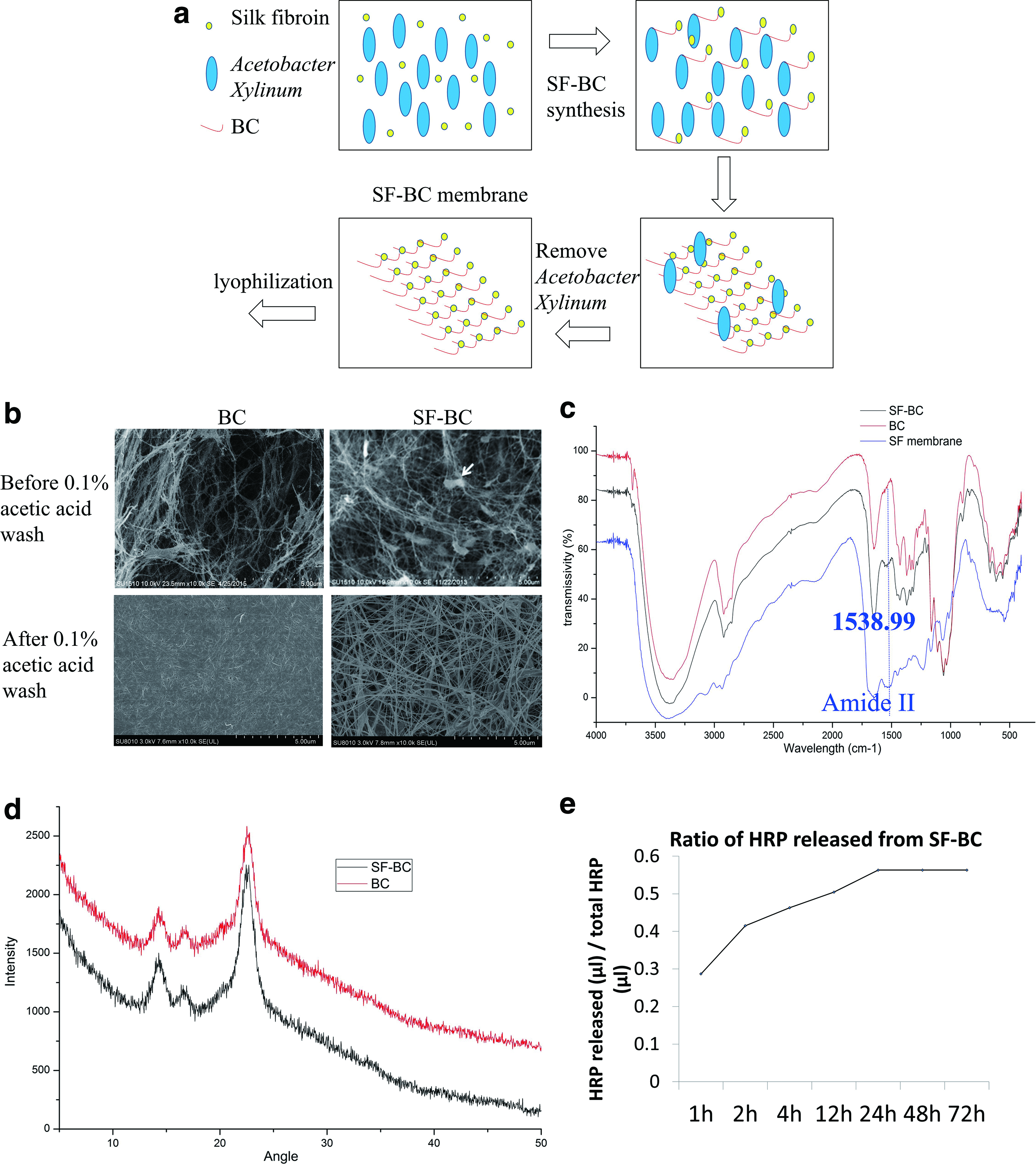
The SF-BC composite scaffold was prepared according to previous methods. 7 Briefly, the SF was prepared by dissolving degummed silk fiber in CaCl2-CH3CH2OH-H2O (1:2:8 in mole ratio) at 60°C for 40 min and dialyzing against distilled water.12,13 Then, 1 g/100 mL SF was added in fermentation medium (50 g/L glucose, 10 g/L peptone, 1 g/L citric acid, 1 g/L Na2HPO4·12H2O, 3 g/L KH2PO4, and 1.46 g/L MgSO4·7H2O) and cultivated with activated Acetobacter xylinum (1.1812), purchased from the Institute of Microbiology Chinese Academy of Science, at 30°C for 10 days without shaking the incubator. The SF-BC membrane thus obtained were first washed in distilled water to remove fermentation medium and then immersed in 1% (w/v) NaOH solution at 80°C for 3 h, with several rinses in distilled water every hour. Subsequently, 1% (w/v) NaOH solution was neutralized by 0.1% acetic acid, and the SF-BC membrane was then rinsed with distilled water, followed by lyophilization; then SF-BC was sterilized by autoclaving (Fig. 1a).
Physical and chemical analysis of the SF-BC membrane.
Scanning electron microscopy imaging of the SF-BC membrane
The BC and SF-BC membrane without cells were mounted on aluminum stubs and coated with gold directly. Human uterine cells cultured on SF-BC membrane for 7 days were fixed with 0.25% glutaraldehyde solution for 24 h. After rinsing with phosphate-buffered saline (PBS) thrice, the samples were soaked in OsO4 (Ted Pella) for 1 h and washed again thrice with PBS. The samples were then dehydrated with a concentration gradient of acetone (30%, 50%, 70%, 80%, 90%, 95%, and 100%, v/v), followed by drying. Finally, the samples were placed on aluminum stubs and coated with gold. The cells cultured on the SF-BC membrane were visualized by scanning electron microscopy (SEM) (Hitachi SU-8010N, Japan) at an accelerating voltage of 15 kV.
Fourier transform infrared spectroscopy of the SF-BC membrane
The BC, SF, and SF-BC membranes were air dried and were subjected to Fourier transform infrared (FTIR) spectroscopy with a Nicolet AVA TAR370 spectrometer (Thermo Fisher Scientific). The scanning range was from 4000 to 400 cm−1 with a resolution of 4 cm−1.
X-ray diffraction analysis of the SF-BC membrane
The SF-BC and BC membranes were air dried, and X-ray diffraction (XRD) analysis was measured with an X-ray diffractometer at 2.7 kW (Shimadzu XRD-6000, Japan), with the tested angle being from 0° to 40°.
Mechanical properties of the SF-BC membrane
Mechanical properties of BC and SF-BC membrane were tested using an Instron tension/compression system with Fast-Track software (Model 5543; Instron, Canton, MA) under hydrated condition. The membranes were cut into a rectangular piece of 4 mm in width and 13 mm in length; after being saturated with 0.9% saline for 12 h at 37°C, 2 mm in length at both sides was clamped by hemostatic forceps. The membranes were then stretched at a speed of 5 mm/min. At least four replicate samples of each membrane were measured; data were presented as mean ± standard error of the mean. The experiment was performed at room temperature.
Protein release of SF-BC membrane
SF-BC membrane was soaked with 1:20 Horseradish peroxidase (HRP) labeled goat anti-mouse immunoglobulin G (IgG) solution for 12 h at 37°C. Then SF-BC membrane was taken out and input in 500 μL PBS and incubated at 37°C. Subsequently, 50 μL (10 μL was put in each well in 96 well) supernatant solution was removed and supplemented with 50 μL PBS at 1, 2, 4, 12, 24, 48, and 72 h, respectively. Next, 50 μL TMB (MultiSciences (Lianke) Biotech Co. Ltd., China) was added to each well, and this was followed by addition of stop solution (MultiSciences(Lianke) Biotech Co. Ltd.) into it 10 min later. Concentration of released HRP labeled goat anti-mouse IgG was measured by SpectraMax (Molecular Devices) at a wavelength of 405 nm.
Biological properties of the SF-BC membrane
Cytotoxicity evaluation of the SF-BC membrane
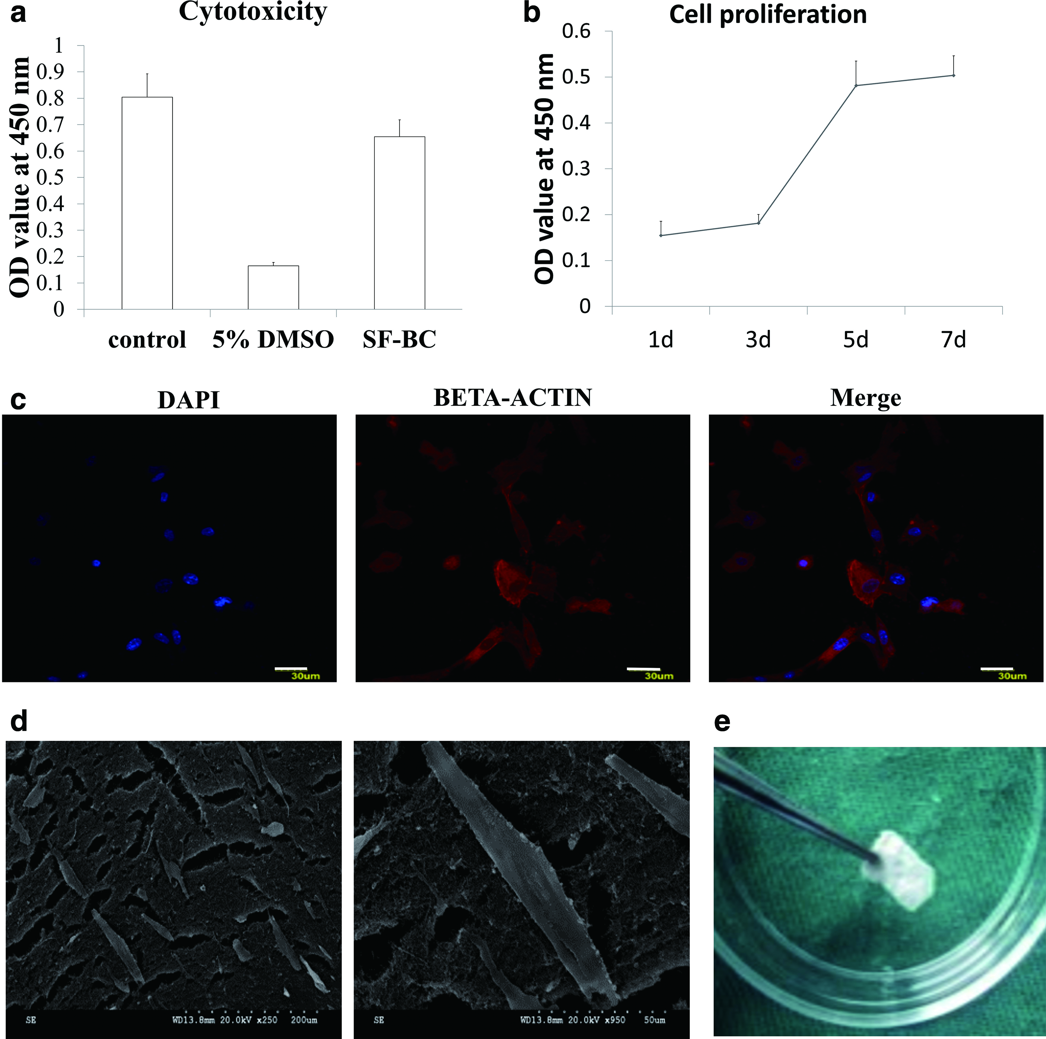
The cytotoxicity of SF-BC scaffold was assessed according to the Biological Evaluation of Medical Devices protocol (GB/T 16886.5-2003/ISO 10993-5:1999). Briefly, SF-BC was sterilized by autoclaving; a 2 × 1 cm SF-BC membrane section was immersed in 0.67 mL of high-glucose Dulbecco's Modified Eagle Medium (DMEM) (DMEM/H) containing 10% (v/v) fetal bovine serum (FBS) and 1% (w/v) penicillin–streptomycin, within a 5% CO2 incubator set at 37°C for 48 h. And 100 μL of DMEM/H containing 1000 L929 cells (Chinese Academy of Sciences, Beijing, China) was seeded in each well of a 96-well plate. After 24 h, the culture medium was replaced by media containing 5% (v/v) of extracts from the membrane, fresh DMEM/H containing 10% (v/v) FBS with 1% (w/v) penicillin–streptomycin, 5% (v/v) dimethyl sulfoxide (DMSO), and cultured for 3 days without changing the medium. The cytotoxicity was then assessed by comparing the optical density (OD) value at 450 nm with the Cell Counting KIT-8 (CCK-8; Dojindo, Kumamoto, Japan). The OD value is positively correlated with cell number. Results were presented as mean ± standard deviation (SD).
Cell proliferation assay on the SF-BC membrane
Human endometrial cells were isolated from the normal portions of the uterus of leiomyoma patients from the First Affiliated Hospital, School of Medicine, Zhejiang University. Approval for utilizing the patient samples in this study was obtained from Ethics Committee of the First Affiliated Hospital, School of Medicine, Zhejiang University. Rat uterine cells were prepared from postnatal rat uterus purchased from the Zhejiang Academy of Medical Science. All experimental procedures involving animals were approved by the Ethics Review Board for Animal Studies of Zhejiang University. Human and rat uterine cells were prepared by mincing the tissues for 5–7 min using a pair of small surgical scissors followed by digestion with type I collagenase (Gibco) in DMEM/F12 overnight at 4°C. The digested tissues were triturated into individual cell suspensions by a 1 mL pipette. Human endometrial cells were seeded on SF-BC membrane at a density of 3000 cells/well, and the OD value at 450 nm was measured with CCK8 after 1, 3, 5, and 7 days of culture. Increase in absorbance values at 450 nm corresponded to the proliferation of human uterine cells. Results were presented as mean ± SD.
Cell morphology on the SF-BC membrane
Four thousand human endometrial cells were seeded on the SF-BC membrane in a 96-well plate and were cultured in a 5% CO2 incubator at 37°C with regular changes in culture medium every 2–3 days. After 7 days, the culture medium was removed, and cells were washed thrice with PBS, followed by fixation in 4% (w/v) paraformaldehyde for 30 min, and subsequent rinsing with PBS for another three times. Next, we observed the cytoskeleton and nuclei by immunofluorescence staining of F-actin, which was detected using tetramethylrhodamine-conjugated Phalloidin (1:1500; Millipore) and 4′,6-diamidino-2-phenylindole (DAPI) (1:1200; Beyotime Institute of Biotechnology, China) separately. The cell morphology was imaged under confocal microscopy (Olympus, BX61W1-FV1000, Japan).
Migration of rat uterine cells in vitro under the influence of SDF-1α
The effects of rhSDF-1α on the migration of rat uterine cells were assessed by transwell assay (membrane pore size 8 mm, Costar 3422) as described previously. 14 Rat uterine cells (P0) were trypsinized and seeded onto the upper chamber of transwell inserts at a density of 1 × 10⁴/well within 100 μL of serum-free DMEM/F12 per well of a 24-well plate. Then 600 μL of serum-free DMEM/F12 or 600 μL of serum-free DMEM/F12 containing 100 ng/mL rhSDF-1α (R&D Systems, Minneapolis, MN) was added into the lower chamber to induce migration of rat uterine cells. 15 The cells were cultured in a 5% CO2 incubator at a temperature of 37°C for 24 h.
To confirm the effects of rhSDF-1α, rat uterine cells were pretreated with 200 ng/mL of ADM3100 (Sigma), which is an antagonist of SDF-1α, for 30 min in a 5% CO2 incubator at 37°C. Then, 100 μL of serum-free DMEM/F12 containing 1 × 10⁴ pretreated cells was seeded on the upper chamber of the transwell (Corning), and 600 μL of serum-free DMEM/F12 with 100 ng/mL rhSDF-1α was added to the lower chamber of the transwell. In another group, the cells seeded on the upper chamber of transwell were cultured in 100 μL of serum-free DMEM/F12 with 100 ng/mL rhSDF-1α, and 600 μL of serum-free DMEM/F12 with 100 ng/mL rhSDF-1α was added to the lower chamber as well.
After 24 h, the transwell inserts were taken out from 24-well plates and rinsed thrice with PBS and were fixed with 4% (w/v) paraformaldehyde for 20 min, washed another three times with PBS, and subjected to nuclear staining with DAPI (1:2000; Beyotime Institute of Biotechnology, Inc., China) for 10 min. The cells on the upper chamber were scraped to remove adherent cells. The number of cells that migrated to the lower chamber of the transwell was counted within five fields ( × 100) under microscopy.
Rat uterine horn injury/regeneration model
Twenty-nine female Sprague Dawley rats (purchased from the Zhejiang Academy of Medical Science) weighing between 200 and 250 g were selected, and the 58 uterine horns of the rats were divided into three groups, injury alone group (n = 16), SF-BC group (n = 21), and (SF-BC/rhSDF-1α) group (n = 21). All the rats were kept in a specific pathogen-free air-conditioned room and were allowed free access to food and water at the Animal Center of Zhejiang University of Medicine. All experiments were approved by the Ethics Review Board for Animal Studies of Zhejiang University. To deliver rhSDF1-α, the SF-BC membrane was soaked in a solution of rhSDF-1α at a concentration of 100 ng/mL within an incubator set at 37°C with 5% CO2 for 12 h according to previous method. 15
The procedure was partly based on the full-thickness injury model of rat uterus that was described previously.16–18 After the rats were anesthetized by intraperitoneal injection of chloral dehydrate (0.4 g/kg), a midline incision in the abdomen was made, and the uterus was exposed. In the injury alone group, a full-thickness defect was made by excising a section that was about 1.0 cm in length and 0.5 cm in width in each uterine horn (Fig. 3a), with the mesenterium being retained. The four margins of the injury were marked by 6-0 nonabsorbable silk suture. In the other two groups, the SF-BC membrane or SF-BC/rhSDF-1α membrane that was equal to the size of the uterine defect was sutured at the injury site using 7-0 absorbable proline suture through interrupted sutures. SF-BC membranes were either immersed in high-glucose DMEM (DMEM/H) alone or DMEM/H with SDF (100 ng/mL) at 37°C overnight before operation. After operation of the uterus, the abdominal cavity was washed with 0.9% (w/v) normal saline. Then the rectus abdominis was sutured by 6-0 nonabsorbable silk suture with continuous suture, and the skin and fascia were sutured by 4-0 nonabsorbable silk suture through interrupted suture. During the operation procedure, a piece of gauze, which was soaked in 0.9% (w/v) normal saline, was used to cover the uterus and viscera to avoid drying. All the operated rats were treated with intramuscular injection of penicillin (80,000 U/rat) continuously for 3 days.
Histological analysis
Hematoxylin and eosin staining
The rats were sacrificed at 30 and 90 days after surgery. The surgery site of each uterine horn (injury alone group: n = 4, SF-BC group: n = 6, and SF-BC/rhSDF-1α group: n = 6) was fixed in 4% paraformaldehyde, dehydrated with an ethanol gradient, followed by paraffin embedment and sectioning at 5 μm thickness. Then the paraffin sections were stained with hematoxylin and eosin, and the thickness of the endometrium which starts from the luminal epithelium to the muscle layer at the injury site was measured by Image-Pro Plus software (version 6.0) (Fig. 3h).
Immunohistochemical staining
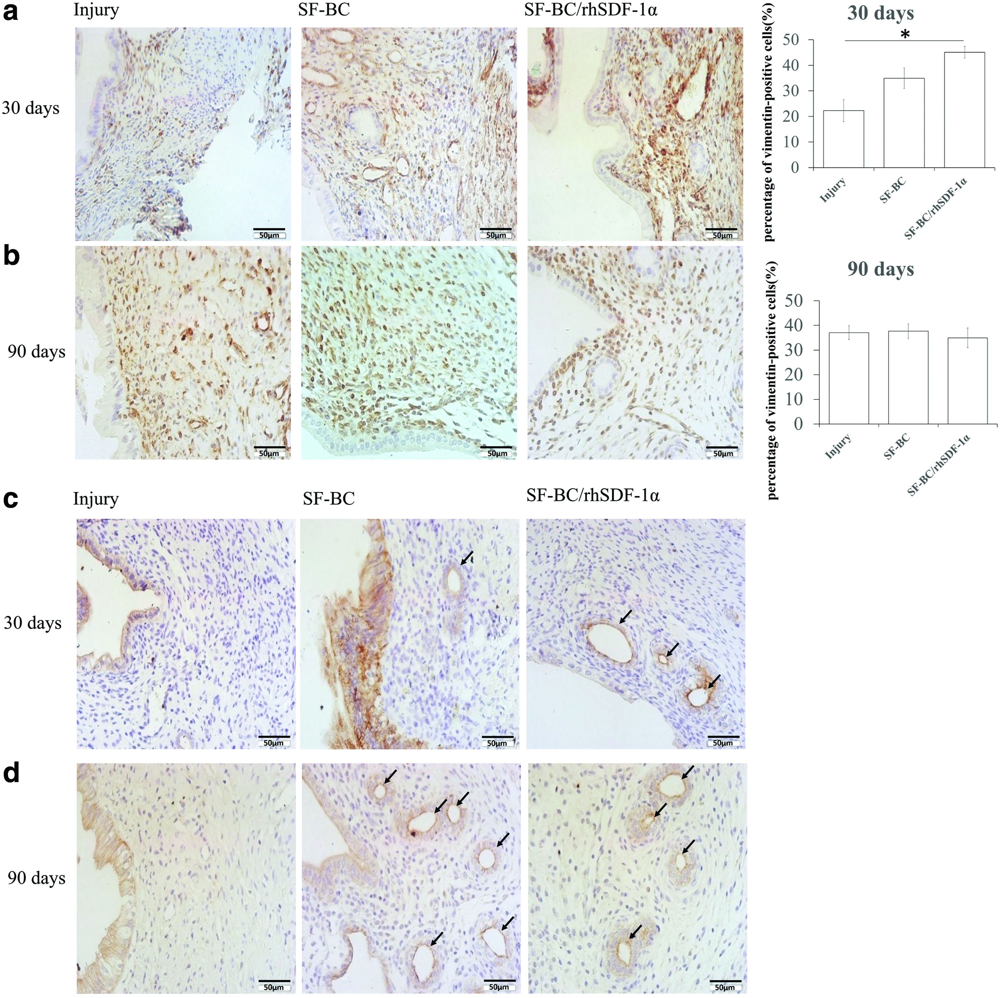
Sections were dewaxed, rehydrated, and subjected to antigen retrieval treatment with citric acid buffer (pH 6.0) or pepsin, followed by washing thrice with PBS. These were then incubated in 3% (v/v) H2O2 in methanol for 10 min to inactivate endogenous peroxidase. After rinsing with PBS, the samples were treated with blocking solution (1% w/v bovine serum albumin [BSA]) for 30 min and then incubated with primary antibodies at 4°C overnight. For indirect staining, the slides were further incubated in horseradish peroxidase-conjugated goat anti-mouse IgG in 1% (w/v) BSA for 2 h at room temperature and then washed thrice with PBS. The samples were then visualized by the 3,3′-diaminobenzidine method and counterstained with hematoxylin solution. The primary antibodies utilized were as follows: monoclonal anti-vimentin (1:50; Dakocytomation), mouse monoclonal anti-pan-cytokeratin (1:200; Abcam), and the appropriate secondary antibodies (1:50; Invitrogen, China). All the procedures were carried out according to the manufacturer's instructions. The percentage of vimentin positive stroma cells was calculated using the software IPP 6.0; the percentage was calculated as: number of vimentin positive stroma cells divided by the number of hematoxylin positive stroma cells in the regenerated region of the endometria.
Immunofluorescence staining
Sections were dewaxed, rehydrated, and subjected to antigen retrieval treatment with citric acid buffer (pH 6.0), followed by washing three times with PBS. Then the samples were treated with blocking solution (1% w/v BSA) for 30 min, and then incubated with anti-CD31 antibody (ab22783, Abcam) at 4°C overnight. After washed 3 times with PBS, the samples were then incubated with Goat anti-Rabbit IgG (H + L) Cross-Adsorbed Secondary Antibody, Alexa Fluor 488 (A-11008, Invitrogen) and DAPI (C1002, Beyotime Institute of Biotechnology). All the procedures were carried out according to the manufacturer's instructions. Immunofluorescence images were acquired with Olympus FV3000 Laser Scanning Confocal Microscope (OLYMPUS IX83-FV3000) and the number of arteries with CD31 positive signal were calculated in each high power field (HPF) of the microscopy view.
Functional test of the regenerated endometrium
To determine whether the regenerated endometrium at the surgery site is receptive to the implantation of embryos and can support subsequent embryo development,16,17 13 rats (no. of uterine horns in each group: injury alone group, n = 8; SF-BC group, n = 9; and SF-BC/rhSDF-1α group, n = 9) were mated with males, and the conception was confirmed if a large number of sperm were visible in the vagina the next day after mating. Then the rats were euthanized at late gestation (19–21 days), and pregnancy rate at the surgery site together with the number of fetuses in each uterine horn were recorded and compared.
Statistical analysis
The thickness of fibrous tissues around the implanted materials and the distance of cells migrated into the materials were measured and compared using paired T test in Prism 5.0 software. The number of cells that have migrated to the lower chamber of the transwell inserts, endometrium thickness, and number of arteries and number of fetuses in each uterine horn were compared using one-way ANOVA with Bonferroni's Multiple Comparison Test in Prism 5.0 software. The percentage of endometrial stromal cells, pregnancy rate, and pregnancy rate at the surgery site were analyzed by the X2 test and Fisher exact test for categorized variables in SPSS software (Version 19.0; SPSS, Inc., Chicago, IL), and all p values less than 0.05 were considered statistically significant.
Results
Physical and chemical properties of the SF-BC membrane
The fabricated SF-BC membrane was imaged under digital camera and SEM. Digital camera images showed that the SF-BC membrane was thin and white in color (Fig. 2e). SEM imaging revealed that SF-BC had a more porous structure compared with that of BC, with the fiber diameter at nanoscale (Fig. 1b). FTIR results showed a specific band from SF-BC at 1538.99 cm−1 that was absent in the BC membrane, which might be characteristic of the amide II group of the SF (Fig. 1c). XRD analysis of both the BC and SF-BC membrane showed three peaks (BC: 14.29°, 16.83°, 22.51°; SF-BC:14.28°, 16.38°, 22.45°) that were characteristic of the BC crystal (Fig. 1d). Table 1 presents the mechanical properties of the BC and SF-BC membranes, and the results indicated that the addition of SF (1%) could significantly increase the mechanical properties (maximum load, stiffness, and Young's modulus) of the SF-BC membranes compared with that of the BC membrane. These results showed that the SF-BC membrane has a porous structure with favorable mechanical properties. And protein release assay showed that about 56% HRP labeled goat anti-mouse IgG could be gradually released from SF-BC composite in the first 24 h, while the rest of the IgG (44%) remained in the composite after the first 24–72 h, which suggested that SF-BC membrane could be an effective system to control the release of rhSDF-1α (Fig. 1e).
Biological properties of the SF-BC membrane.
Mechanical Properties of Silk Fibroin, Bacterial Cellulose, Silk Fibroin-Bacterial Cellulose Composite Membranes, and Rodent Uterus Tissue
Data were presented as mean ± SE (standard error of the mean), difference in the letter (a and b) indicating the significant difference between two groups (p < 0.05).
BC, bacterial cellulose; SF, silk fibroin; SF-BC, silk fibroin-bacterial cellulose.
Biological properties of the SF-BC membrane
In addition, the cytotoxicity test (Fig. 2a) and cell proliferation assay (Fig. 2b) showed that the SF-BC membrane has good biocompatibility. To determine whether the SF-BC membrane affected cell morphology, human endometrial cells were seeded on it for 7 days. The confocal microscopy images showed that the cells adopted various shapes, with the cytoskeleton being stretched out on the membrane (Fig. 2c). This was also supported by the SEM images showing the cells being spread out on the membrane surface (Fig. 2d). These results thus suggest that the SF-BC membrane was biocompatible and conducive for uterine cells, which are of utmost importance for tissue regeneration at the injury site.
Regeneration of rat uterus treated with rhSDF-1α loaded SF-BC membrane after surgery
Thirty days after surgery, all of the uterine horns kept patency. The uterine horns implanted with SF-BC membrane showed only slight adhesion at the injury site. However, certain uterine horns in the injury alone group exhibited severe adhesion to the viscera, particularly to the small intestine. Moreover, there were fewer blood vessels in the injury site alone group. Although the SF-BC membrane degraded slowly, it interacted well with the adjacent tissues (Fig. 3b). Uterine horns implanted with the SF-BC membrane retained their original shape at 90 days postoperation, while uterine horns in the injury alone group exhibited stenosis at the injury site (Fig. 3c), which may have an adverse effect on the pregnancy outcomes.

Regeneration of rat uterus treated with the SF-BC membrane loaded with rhSDF-1α.
Histological analysis showed that the regenerated endometrium at the surgery site had intact luminal epithelium at 30 days postoperation (Fig. 3d), with secretory glands appearing at the injury site in both the SF-BC and SF-BC/rhSDF-1α groups, whereas only a few glands could be observed in the injury alone group (Fig. 5c). Furthermore, the endometrium in the SF-BC/rhSDF-1α group was thicker compared with both the SF-BC group and the injury alone group (259.29 ± 15.94 μm vs. 191.55 ± 4.79 μm, 259.29 ± 15.94 μm vs. 146.75 ± 13.92 μm, respectively), but there was no statistically significant difference in thickness of the endometrium between the SF-BC group and the injury alone group (Fig. 3f).
Ninety days after surgery, more secretory glands had regenerated at the injury site in both the SF-BC and the SF-BC/rhSDF-1α groups, whereas there were only a few glands present in the injury alone group (Fig. 5d). Although the endometrium thickened in all groups (Fig. 3e), the endometrium in the SF-BC/rhSDF-1α group was thicker compared with both the SF-BC group and the injury alone group (704.00 ± 29.52 μm vs. 465.40 ± 65.14 μm, 704.00 ± 29.52 μm vs. 266.08 ± 28.56 μm, respectively). The endometrium in the SF-BC group was thicker compared with the injury alone group (465.40 ± 65.14 μm vs. 266.08 ± 28.56 μm) (Fig. 3g). In conclusion, the results showed that the SF-BC/SDF composite could promote regeneration of the endometrium.
The effects of rhSDF-1α on rat uterine cells
Next, in order to investigate the potential mechanisms of the SDF1 on the promotion of regeneration of the endometria, the chemotactic effects of rhSDF-1α on rat uterine cells were tested by transwell assay in vitro, and the results showed that the number of cells that migrated through the upper chamber (in which rhSDF-1α was added) into the lower chamber was significantly more compared with the blank (18.27 ± 1.87 vs. 9.09 ± 1.38, p < 0.05). This effect was blocked by adding rhSDF-1α to the upper chamber or pretreating all of the cells with AMD3100 (an antagonist of SDF-1) (7.85 ± 1.21 vs. 9.09 ± 1.38, 8.06 ± 0.94 vs. 9.09 ± 1.38, respectively, both p > 0.05) (Fig. 3h). These results thus demonstrate that rhSDF-1α could promote the migration of rat uterine cells in vitro, which implied that the thickness of the regenerated in the composite in vivo was due to the chemotactic effects of rhSDF-1α on the rat uterine cells in vivo.
Pregnancy outcomes of the regenerated endometrium
One of the most important functions of the uterus is to receive implantation of embryos and support subsequent development of the fetus. In this study, we found that some regenerated endometrium received implantation of embryos in all three groups at 30 days postinjury (Fig. 4). In the injury alone group, 4/8 of them had fetuses in the uterine horns, which were lower than that in the SF-BC group (7/9) and the SF-BC/rhSDF-1α group (8/9). However, there was no statistically significant difference between them. Moreover, 2/8 uterine horns had an embryo implanted at the surgery site of the injury alone group, which was less than that in the SF-BC group (5/9) and the SF-BC/rhSDF-1α group (5/9), but the observed differences were not statistically significant, probably due to the small sample size (Table 2).

Pregnancy outcomes of injured uterus at 30 days postsurgery.
Pregnancy Rates, Numbers of Fetuses/Uterine Horn, and Pregnancy Rates at the Injury Site After 19–20 Days Postmating with Male Rats
No. of fetuses per uterine horn: data were presented as mean ± SE (standard error of the mean), difference in the letter (a and b) indicating the significant difference between two groups (p < 0.05).
rhSDF-1α, recombinant human stromal cell-derived factor-1α.
In addition, there was variation in the number of fetuses developed in each uterine horn. In the SF-BC/rhSDF-1α group, the number of fetuses developed per uterine horn (7.00 ± 0.99) was more than that in the injury alone group (3.00 ± 1.16). However, there was no statistically significant difference between the SF-BC group (4.67 ± 1.08) and the SF-BC/rhSDF-1α group, as well as between the injury alone group and the SF-BC group (Table 2). These data (Fig. 4g) suggest that SF-BC/rhSDF-1α improved pregnancy outcomes at 30 days after surgery.
Mechanisms of the effects of SF-BC/rhSDF-1α on the functional regeneration of the uterus
Recruitment of endometrial stromal cells at the injury site
To determine the proportion of endometrial stromal cells within the regenerated endometrium, sections of the endometria were stained with an anti-vimentin antibody. The percentage of endometrial stromal cells in the SF-BC/rhSDF-1α group was higher than that in the injury alone group at 30 days postsurgery (45.11% ± 2.27% vs. 22.25% ± 4.25%) (Fig. 5a). However, there were no statistically significant differences between the SF-BC group and the injury alone group, as well as between the SF-BC/rhSDF-1α group and the SF-BC group (34.95% ± 3.98% vs. 22.25% ± 4.25%, 45.11% ± 2.27% vs. 34.95% ± 3.98%). Figure 5b showed that there were no statistically significant differences in the percentages of endometrial stromal cells among the three groups (SF-BC/rhSDF-1α group: 34.95% ± 3.98%, SF-BC group: 37.64% ± 3.02%, and injury alone group: 37.04% ± 2.82%) at 90 days postinjury (Fig. 5b). This may be due to the proliferation of stromal cells at the injury site and migration of stromal cells from the adjacent normal site. These data suggest that SF-BC/rhSDF-1α plays a role in the migration of endometrial stromal cells at the surgery site within a short duration.
Characterization of regenerated endometria of the rat uterus.
Formation of endometrial epithelial cells at the injury site
Endometrial epithelial cells can be classified into two distinct types as follows: luminal epithelial cells and glandular epithelial cells. The latter plays an important role in decidualization, implantation, and embryo development. After the sections were labeled with an anti-pan-cytokeratin antibody, luminal epithelial cells could be detected in the injury site among the three groups at 30 days postsurgery. However, significant numbers of glandular epithelial cells were present only in the SF-BC group and the SF-BC/rhSDF-1α group, while few of these were present in the injury alone group (Fig. 5c).
Ninety days after injury, more glandular epithelial cells were detected at the surgery site of the SF-BC group and the SF-BC/rhSDF-1α group. Nevertheless, the number of glandular epithelial cells in the injury alone group at 90 days postsurgery was similar to that at 30 days postsurgery (Fig. 5d). These data suggest that the SF-BC membrane facilitated the formation of endometrial secretory glands, and this may contribute to differences in pregnancy outcomes among the three groups. The lack of secretory glands in the regenerated endometrium of the injury alone group would result in endometrial dysfunction, which may affect subsequent pregnancy outcomes.
Arteriogenesis in the regenerative endometrium
Arteriogenesis is crucial in tissue repair as the rich blood supply not only provides nutrients but also various types of growth factors, which can modulate cell migration, proliferation, and differentiation. To investigate whether rhSDF-1α could enhance arteriogenesis, the sections were stained with an anti-CD31 antibody. The number of arteries was estimated by counting CD31 positive blood vessels in nine HPF.
At 30 days postsurgery, the number of arteries in the SF-BC/rhSDF-1α group was more than that in the injury alone group and the SF-BC group (7.45 ± 0.38 vs. 1.13 ± 0.31, 7.45 ± 0.38 vs. 2.20 ± 0.20, separately) (Fig. 6a, c). There was no significant difference between the SF-BC group and the injury alone group (2.20 ± 0.20 vs. 1.13 ± 0.31) (Fig. 6a, c). At 90 days postsurgery, the number of arteries in the injury alone group was less than that in the SF-BC group and SF-BC/rhSDF-1α groups (3.03 ± 0.41 vs. 6.65 ± 0.40, 3.03 ± 0.41 vs. 5.50 ± 0.23, respectively) (Fig. 6b, d). No significant difference in the number of arteries was found between the SF-BC group and the SF-BC/rhSDF-1α group (Fig. 6b, d). The data thus demonstrated that SF-BC/rhSDF-1α promoted arteriogenesis at the injury site within a short time duration and that the SF-BC membrane was beneficial for the formation of arteries at the injury site over a longer duration.

Immunofluorescence staining of arteries at the injury site.
Discussion
Curettage, infections, and cesarean section may lead to amenorrhea, infertility, and pregnancy complications. 1 In recent years, tissue engineering has been applied to address this clinical challenge. Some studies attempted to utilize SIS to reconstruct the injured uterus; however, small intestine submucosa (SIS) collapsed or became twisted when it is longer than 1 cm. 16 Miyazaki and Maruyama found that decellularized uterine matrix demonstrated some efficacy in promoting uterine regeneration. 18 Nevertheless, human uterine tissue is not readily available due to ethical and legal issues, which limit the application of the aforementioned scaffold. Because the uterine cavity in humans needs to expand during late gestation, scaffolds suitable for uterine repair should have good biocompatibility and excellent mechanical properties, besides being readily and easily accessible. In this study, we used a composite scaffold of SF and BC, which can fulfill these requirements and could act as a good carrier for the delivery of biological factors.
SF and BC are two biological materials widely utilized in scaffold fabrication and tissue engineering.4,6 We fabricated the composite scaffold of SF and BC to synergize the strength of SF with the water absorption properties of BC. 7 In the process of fabricating the composite membrane scaffold (Fig. 1), the BC and SF were fermented in a strongly acidic environment (pH <3). This resulted in intermolecular dehydration, in situ polymerization between cellulosic residues (–OH) and SF residues (–COOH and –NH2), and the binding of SF molecules to the BC, which resulted in pore formation within the composite membrane scaffold (Fig. 1b).
The SF-BC showed slow degradation rate. As previous study showed that the total resorption time of SF would be more than 2 years, 19 our results also showed that the SF-BC would not fully degrade even at 90-day postoperation (Fig. 3e), but the cells began to migrate into the scaffold as early as 30-day postoperation (Fig. 3d), which was consistent with previous results that SF-BC would not be completely degraded at 8-week postoperation when used to promote bone regeneration. 19 The dense network of BC would display pore sizes not enough to allow migration and proliferation, supplementation of SF increased as the space between the fibers increased, and the porous interior of the composite membrane scaffold made it easier for somatic cells to get access to the internal space and grow, which could further facilitate the growth and differentiation of cells. And 90% of the amino acid composition of SF are glycine, alanine, and serine that would facilitate cell proliferation and tissue regeneration when gradually degraded in vivo.5,20 These results could explain the increase in proportion of vimentin-positive stromal cells detected in the SF-BC composite membrane group in vivo (Fig. 5). The SF-BC composite membrane fabricated in this study was safe and biocompatible with negligible cytotoxicity and could support the proliferation of uterine cells (Fig. 2b). Because the plasticity and tensile strength of the composite membrane scaffold were similar to that of the rat uterus, this could promote regeneration of the uterus after injury and physically expand to support the growth of implanted embryos. The structure of the composite SF-BC membrane was also favorable to the loading of exogenous factors, which could further promote repair and regeneration of injured tissues.
Stromal cell-derived factor-1α (SDF-1α), also known as CXCL12, is a homing factor for certain stem/progenitor cells. In this study, we found that rhSDF-1α could promote the migration of uterine cells in vitro, as well as enhance regeneration of the injured endometrium in vivo, leading to improved pregnancy outcome after uterine injury at 30 days postsurgery. In contrast, uterine repair with other factors, for example, fibroblast growth factor-basic and vascular endothelial growth factor (VEGF) could only result in blastocyst implantation at about 90 days postsurgery, as reported by previous studies.2,21 The data thus suggest that rhSDF-1α was more efficacious and could hasten regeneration and pregnancy outcomes 60 days earlier, compared to previous studies. In addition, the number of fetuses per uterine horn (1.3 ± 0.5) in postinjury uterus that was partially reconstructed by seeding primary uterine cells and MSCs on decellularized uterine matrix 18 was less than the group implanted with SF-BC loaded with rhSDF-1α (7.00 ± 0.99) in this study (Table 2). Differences in the pregnancy outcomes between the injury group and the SF-BC/rhSDF1 composite group illustrated that SF-BC loaded with rhSDF-1α could initiate functional uterine regeneration that led to an improved pregnancy rate.
It has been reported that SDF-1α can promote the regeneration of various tissues by recruiting endogenous cells to the injury site and facilitating the formation of blood vessels through recruitment of endothelial cells.9,11,22–25 In this study, SDF-1 released from SF-BC may promote the migration of adjacent uterine cells to the injury site to take part in repair and regeneration of the uterus. Hence, the better pregnancy outcomes observed in this study may be due to the following mechanisms: (1) SDF-1 promotes vascularization (2) and more mature endometrium formation.
SDF-1 promotes vascularization: In humans, uterine spiral arteries perfuse the intervillous space, enabling the exchange of nutrients and oxygen between mother and fetus. 26 With decrease in vasodilation of uterine arteries in mice, fetal growth would be restricted, implying that uterine arteries are crucial to fetal growth by providing enough blood supply to the fetus. 27 In our study, the number of arteries at the injury site of the uterus implanted with SF-BC loaded with rhSDF-1α was more than that in the uterus of the injury alone group at 90 days postsurgery. This may contribute to better pregnancy outcomes in the uterus implanted with SF-BC loaded with rhSDF-1α.
SDF-1 promotes more mature endometrium formation: uterine glands are essential for fetus development in early pregnancy, which provide nutrients, growth factors, immune-regulatory proteins, 28 and some transport carriers. 29 In our study, there were more uterine glands in the regenerated endometrium of the SF-BC loaded with rhSDF-1α group. More uterine glands at the injury site of the uterus in the SF-BC loaded with rhSDF-1α group may lead to better pregnancy outcomes than the injury alone group, by providing more nutrients, such as carbohydrates, glycoproteins, and lipids to the embryo before its implantation into the endometrium, 28 more growth factors, such as leukemia inhibitory factor, VEGF, and tumor necrosis factor-α that promote the formation of uteroplacental arteries and villous tree, more immune-regulatory proteins, like glycodelin A and MUC1 to facilitate embryo implantation, 28 and more transport carriers, such as tripeptidyl peptidase and lactoferrin to boost the immune tolerance of the feto-placenta and endometrium. 29
Conclusion
In this study, we demonstrated that the SF-BC membrane possessed good physical, chemical, and biocompatibility properties in vitro. The in vivo study showed that the incorporation of rhSDF-1α within the SF-BC membrane promoted regeneration of full-thickness uterine injury and also improved the pregnancy outcome of the damaged uterus. These results thus suggest that SF-BC loaded with rhSDF-1α has good potential in future clinical applications for the repair of uterine injury.
Footnotes
Acknowledgments
This work was supported by the National High Technology Research and Development Program of China (2018YFC1105100), the National Natural Science Foundation of China (CN) (81871127, 31870973), Zhejiang Medical and Health Science and Technology plan project (2013KYB080), and the Zhejiang Provincial Natural Science Foundation of China (LY14C100003).
Authors' Contributions
H.C., cell culture, animal experiments, acquisition of data, data analysis and interpretation, and article writing; B.W., cell culture, animal experiments, acquisition of data, and article writing; Y. Liu, animal experiments and article writing; Y. Li, acquisition of data; L.S., animal experiments; L.G., acquisition of data; Y.X., sampling of clinical samples; B.C.H., article writing; H.W., sampling of clinical samples; H.O., conception and design; Z.Z., preparation of biomaterials and article writing; and X.Z., conception and design and article writing.
Disclosure Statement
No competing financial interests exist.