
Abstract
Scaffold-guided gene transfer offers strong systems to develop noninvasive convenient therapeutic options for the treatment of articular cartilage defects, especially when targeting bone marrow aspirates from patients containing chondroregenerative mesenchymal stromal cells in a native microenvironment. In this study, we examined the feasibility of delivering reporter (red fluorescent protein [RFP], lacZ) recombinant adeno-associated virus (rAAV) vectors over time to such samples through biocompatible mechanically stable poly(ɛ-caprolactone) (PCL) films grafted with poly(sodium styrene sulfonate) (pNaSS) for improved biological responses as clinically adapted tools for cartilage repair. Effective transgene expression (RFP, lacZ) was noted over time in human bone marrow aspirates using pNaSS-grafted films (up to 90% efficiency for at least 21 days) versus control conditions (ungrafted films, absence of vector coating on the films, free or no vector treatment), without displaying cytotoxic nor detrimental effects on the osteochondrogenic or hypertrophic potential of the samples. These findings demonstrate the potential of directly modifying therapeutic bone marrow from patients by controlled delivery of rAAV using biomaterial-guided procedures as a future noninvasive strategy for clinical cartilage repair.
Impact statement
Injured articular cartilage does not fully regenerate on itself and none of the currently available clinical and experimental therapeutic procedures are capable of restoring an original hyaline cartilage in sites of injury. Biomaterial-guided gene delivery has a strong potential to enhance the processes of cartilage repair. The system presented here based on the FDA-approved biocompatible poly(ɛ-caprolactone) material provides a functional scaffold for the controlled delivery of clinically adapted recombinant adeno-associated virus vectors as an off-the-shelf compound that could be applicable in a minimally invasive manner in patients.
Introduction
The incidence of focal defects in the articular cartilage is a critical issue in orthopedic surgery as this tissue essential for the smooth frictionless weightbearing properties of the articulating joints has an inadequate capacity to heal due to the lack of access of regenerative cells in the absence of vascularization.1–3 Despite the availability of a number of therapeutic interventions (pridie drilling, microfracture, and cell transplantation),1–6 none can lead to the production of a native hyaline cartilage (proteoglycans, type-II collagen) that does not progress toward osteoarthritis and is capable of withstanding mechanical forces over time,1–6 strongly encouraging innovative research for more effective treatments.
Administration of bone marrow-derived mesenchymal stromal cells (MSCs) in sites of cartilage injury is a valuable option to enhance the local processes of cartilage repair7–10 due to the chondroreparative activities of these cells,11–13 especially when provided as marrow concentrates in their natural clinically relevant microenvironment using off-the-shelf minimally invasive procedures.14,15 Still, even with such convenient techniques, the long-term quality of repair tissue in the treated lesions remains unsatisfactory, with production of a poor fibrocartilaginous repair tissue (type-I collagen) unable to bear prolonged mechanical stress.14,15
Polymeric gene delivery 16 recently gained increased attention as a promising biomaterial-guided gene therapy strategy to enhance the processes of cartilage repair through controlled long-term delivery of gene vectors from biocompatible materials in sites of cartilage damage.17–19 Although work thus far emphasized on delivering short-lived nonviral20–30 and potentially oncogenic lentiviral vectors,31–34 there is still little information on transferring the more effective clinically preferred recombinant adeno-associated virus (rAAV) vectors using biomaterials.17,19,35–38
Interestingly, most studies focused on the value of hydrogel systems for rAAV-based cartilage regenerative medicine (fibrin, alginate, poloxamers, poloxamines, self-assembling peptides, and polypseudorotaxanes),39–46 whereas no evidence described the potential of mechanically stable solid scaffolds to guide rAAV application in sites of cartilage injury.
Among the large variety of solid scaffolds available in cartilage research, 47 those based on the biocompatible FDA-approved aliphatic polyester poly(ɛ-caprolactone) (PCL)48,49 present significant advantages as this low immunogenic biodegradable compound can mimic the anisotropic and viscoelastic biomechanical features of the articular cartilage. 50 In this study, we manipulated PCL films to further graft their surface with poly(sodium styrene sulfonate) (pNaSS), a bioactive polymer that facilitates protein adsorption and stimulates reparative cellular responses (adhesion and proliferation), 51 as potential materials to genetically modify clinical marrow samples through controlled delivery of rAAV vectors over time.
Our data demonstrate that pNaSS-grafted PCL films provide functional systems capable of supporting the effective, durable, and not cytotoxic transfer of reporter rAAV vectors in human bone marrow aspirates relative to control (vector-free) conditions, reaching levels similar to or higher than those noted using ungrafted films or upon free vector treatment. Equally important, these systems had no deleterious effects on the chondroreparative potential of the aspirates, showing the value of solid scaffold-guided rAAV gene therapy for future therapeutic approaches to treat cartilage defects in patients.
Materials and Methods
Study design
rAAV vectors (40 μL, i.e., 8 × 10⁵ transgene copies) were immobilized on PCL films that were grafted with pNaSS (low grafting: 1.11 × 10⁻⁶ mol/g; high grafting: 1.30 × 10⁻⁶ mol/g) or left ungrafted. The rAAV-coated films were placed in contact with human bone marrow aspirates (150 μL, i.e., 6 × 10⁷ cells; multiplicity of infection [MOI] = 75) for up to 21 days and processed to evaluate the efficacy of vector immobilization and release (Cy3 vector labeling, AAV Titration enzyme-linked immunosorbent assay [ELISA]) and to monitor transgene expression (live fluorescence, X-Gal staining, and immunohistochemical analysis), cell viability (WST-1 assay), and expression of osteochondrogenic factors (histological, immunohistochemical, histomorphometric, and real-time reverse-transcription polymerase chain reaction [RT-PCR] analyses).
Reagents
All reagents were purchased at Sigma (Munich, Germany) unless indicated. 4-Styrenesulfonic acid sodium salt hydrate (NaSS) was from Sigma-Aldrich (cat. no. 434574). The anti-β-galactosidase (β-gal) (GAL-13) and antitype-X collagen (COL-10) antibodies were purchased at Sigma, the anti-SOX9 (C-20) antibody was purchased at Santa Cruz Biotechnology (Heidelberg, Germany), the antitype-II collagen (AF-5710) and antitype-I collagen (AF-5610) antibodies were purchased at Acris (Hiddenhausen, Germany), the biotinylated secondary antibodies was purchased at Vector Laboratories (Alexis Deutschland GmbH, Grünberg, Germany) as well as the ABC reagent. The Cy3 Ab Labeling Kit was purchased at Amersham/GE Healthcare (Munich, Germany). The AAVanced Concentration Reagent was from System Bioscience (Heidelberg, Germany), the AAV Titration ELISA from Progen (Heidelberg, Germany), and the β-gal staining kit and Cell Proliferation Reagent WST-1 from Roche Applied Science (Mannheim, Germany).
Bone marrow aspirates
The study was approved by the ethics committee of the Saarland Physicians Council (Ärztekammer des Saarlandes, application with reference number Ha06/08) and performed in accordance with the Helsinki Declaration. Bone marrow aspirates (∼15 mL; 0.4–1.2 × 10⁹ cells/mL) were collected from the distal femurs of patients undergoing total knee arthroplasty (n = 12, age 72 ± 5 years) with informed consent given by the patients before inclusion in the study.
Preparation of the PCL films
The PCL films were prepared by the spin-coating method. 51 A PCL solution in dichloromethane (60% w/v) was dropped on a glass slide and spun for 30 s at 250 g using an SPIN150-v3 SPS. The films were air-dried for 2 h, vacuum-dried for 24 h, and cut into 4-mm disks. For pNaSS grafting, the films were ozonated for 10 min at 30°C and rinsed with distilled water to test the following conditions: no grafting, low grafting (1.11 × 10⁻⁶ mol/g pNaSS), and high grafting (1.30 × 10⁻⁵ mol/g pNaSS). The films were transferred in a degassed NaSS solution in distilled water (15% w/v) and maintained at 45°C for 3 h for graft polymerization. The samples were washed with distilled water, NaCl 0.15 M, and PBS and rinsed for vacuum drying.
Preparation of the rAAV vectors
The constructs derived from pSSV9, a parental AAV-2 genomic clone.52,53 rAAV-RFP carries the Discosoma sp. red fluorescent protein (RFP) sequence and rAAV-lacZ the lacZ gene encoding β-galactosidase (β-gal), both controlled by the cytomegalovirus immediate–early promoter.54,55 Conventional vector packaging (not self-complementary) was performed through helper-free (two-plasmid) transfection system using 293 cells with the packaging plasmid pXX2 and adenovirus helper plasmid pXX6. 54 The preparations were purified using the AAVanced Concentration Reagent and titered by real-time PCR54,55 (titers averaging 1010 transgene copies/mL, i.e., ∼1/500 functional recombinant viral particles).
rAAV vector labeling
Cy3 labeling of the rAAV vectors was performed with the Cy3 Ab Labeling Kit 43 by mixing rAAV (1 mL) with sodium carbonate/sodium bicarbonate buffer (pH 9.3) for 30 min at room temperature, followed by Cy3 labeling and dialysis purification against 20 mM HEPES (pH 7.5)/150 mL NaCl. Labeling was tested by live fluorescent microscopy using a rhodamine filter set (Olympus CKX41, Hamburg, Germany).
rAAV vector immobilization on PCL films and release
The rAAV vectors (40 μL, i.e., 8 × 10⁵ transgene copies) were incubated overnight with 0.002% poly-
rAAV-mediated gene transfer
Aliquots of bone marrow aspirates (150 μL, i.e., 6 × 10⁷ cells) with MSCs54,55 were added to the various rAAV-coated pNaSS-grafted PCL films (MOI = 75) in the presence of fibrinogen/thrombin (17 mg/mL/5 U/mL) (Baxter, Volketswil, Switzerland) in 96-well plates and maintained either in defined chondrogenic differentiation medium (DMEM high glucose 4.5 g/L, 100 U/mL penicillin, 100 μg/mL streptomycin, 0.1 μM dexamethasone, 50 μg/mL ascorbic acid, 40 μg/mL proline, 110 μg/mL pyruvate, 6.25 μg/mL of insulin, 6.25 μg/mL transferrin, 6.25 μg/mL selenious acid, 1.25 μg/mL bovine serum albumin, 5.55 μg/mL linoleic acid, and 10 ng/mL transforming growth factor beta [TGF-β]3)34,54,55 or osteogenic differentiation medium (StemPro Osteogenesis Differentiation kit with 100 U/mL penicillin and 100 μg/mL streptomycin)34,54,55 (Life Technologies GmbH, Darmstadt, Germany) at 37°C in a humidified atmosphere with 5% CO2 for up to 21 days for subsequent analyses. Control conditions included uncoated films and film-free vector treatments.
Transgene expression
RFP expression was monitored by live fluorescence using a fluorescent microscopy with a 568-nm filter (Olympus CKX41).54,55 lacZ expression was tested by X-Gal staining using a β-gal staining kit and through immunohistochemistry (specific primary antibody, biotinylated secondary antibody, ABC method with diaminobenzidine [DAB]) and visualization under light microscopy (Olympus BX45).54,55
Viability assay
Cell viability was monitored with the Cell Proliferation Reagent WST-1 (OD450nm proportional to number of cells) 54 using a GENios spectrophotometer/fluorometer (Tecan, Crailsheim, Germany).
Histology and immunohistochemistry
The samples were collected, fixed (4% formalin), dehydrated (graded alcohols), embedded (paraffin), sectioned (3 μm), and stained with hematoxylin and eosin (H&E, cellularity), safranin O (matrix proteoglycans), and alizarin red (matrix mineralization).54,55 Immunohistochemical analyses were also performed to detect the expression of the cartilage-specific sex-determining region Y-type high mobility box 9 (SOX9) transcription factor and of type-II, -I, and -X collagen using specific primary antibodies, biotinylated secondary antibodies, and the ABC method with DAB.54,55 Control conditions (absence of primary antibody) were also tested to check for secondary immunoglobulins. All sections were examined under light microscopy (Olympus BX45).
Histomorphometry
The transduction efficiencies (cells positive for β-gal immunoreactivity to total number of cells) and cell densities (number of cells per standardized area on H&E-stained sections) were examined on histological sections.54,55 Immunohistochemical/histological grading scores were performed with four sections per condition using the SIS AnalySIS program. Safranin O- and alizarin red-stained and SOX9- and type-II/-I/-X collagen-immunostained sections were scored (uniformity, intensity) using a modified Bern Score grading system 55 (0, no staining; 1, heterogeneous and/or weak staining; 2, homogeneous and/or moderate staining; 3, homogeneous and/or intense staining; 4, very intense staining). Sections were scored blind by two individuals with regard to the conditions.
Real-time RT-PCR analysis
Total cellular RNA was extracted with the RNeasy Protect Mini Kit and on-column RNase-free DNase treatment (Qiagen, Hilden, Germany). RNA was eluted in 30 μL RNase-free water and reverse transcription was performed using 8 μL of eluate and the first strand cDNA Synthesis kit for RT-PCR (AMV) (Roche Applied Science). Real-time PCR amplification was performed using 3 μL of cDNA product with Brilliant SYBR Green QPCR Master Mix (Stratagene; Agilent Technologies, Waldbronn, Germany) 55 on an Mx3000P QPCR system (Stratagene). The following conditions were used: (10 min at 95°C), 55 cycles of amplification (30 s denaturation at 95°C, 1 min annealing at 55°C, 30 s extension at 72°C), denaturation (1 min at 95°C), and final incubation (30 s at 55°C).
The primers (Invitrogen GmbH) employed were SOX9 (chondrogenic marker; forward 5′-ACACACAGCTCACTCGACCTTG-3′; reverse 5′-GGGAATTCTGGTTGGTCCTCT-3′), aggrecan (ACAN; chondrogenic marker; forward 5′-GAGATGGAGGGTGAGGTC-3′; reverse 5′-ACGCTGCCTCGGGCTTC-3′), type-II collagen (COL2A1; chondrogenic marker; forward 5′-GGACTTTTCTCCCCTCTCT-3′; reverse 5′-GACCCGAAGGTCTTACAGGA-3′), type-I collagen (COL1A1; osteogenic marker; forward 5′-ACGTCCTGGTGAAGTTGGTC-3′; reverse 5′-ACCAGGGAAGCCTCTCTCTC-3′), type-X collagen (COL10A1; marker of hypertrophy; forward 5′-CCCTCTTGTTAGTGCCAACC-3′; reverse 5′-AGATTCCAGTCCTTGGGTCA-3′), and glyceraldehyde-3-phosphate dehydrogenase (GAPDH; housekeeping gene and internal control; forward, 5′-GAAGGTGAAGGTCGGAGTC-3′; reverse, 5′-GAAGATGGTGATGGGATTTC-3′) (all 150 nM final concentration). 55
Control conditions included reactions with water and nonreverse-transcribed mRNA, and product specificity was confirmed by melting curve analysis and agarose gel electrophoresis. The threshold cycle (Ct) value for each gene was obtained for each amplification with the MxPro QPCR software (Stratagene). Values were normalized to GAPDH expression with the 2−ΔΔCt method.54,55
Statistical analysis
Data are provided as mean ± standard deviation of separate experiments. Each condition was performed in triplicate in three independent experiments per patient. Data were obtained by two individuals blinded with respect to the groups. The t test and the Mann–Whitney rank sum test were used where appropriate. A p value of <0.05 was considered statistically significant.
Results
Immobilization and release of rAAV vectors coated on pNaSS-grafted PCL films
The rAAV vectors were first coated on pNaSS-grafted versus ungrafted PCL films to evaluate the ability of these systems to support the adapted release of these constructs as an effective tool for the genetic modification of human bone marrow aspirates.
Immobilization of rAAV on the pNaSS-grafted versus ungrafted PCL films was successfully achieved as revealed by the strong fluorescent signal detected when coating Cy3-labeled vectors relative to a control condition where the films were placed in contact with unlabeled vectors or when applying Cy3 in the absence of vector coating, without notable difference between the three types of films (ungrafted PCL films, PCL films with low pNaSS grafting, i.e., 1.11 × 10⁻⁶ mol/g, PCL films with high pNaSS grafting, i.e., 1.30 × 10⁻⁶ mol/g) (Fig. 1A).
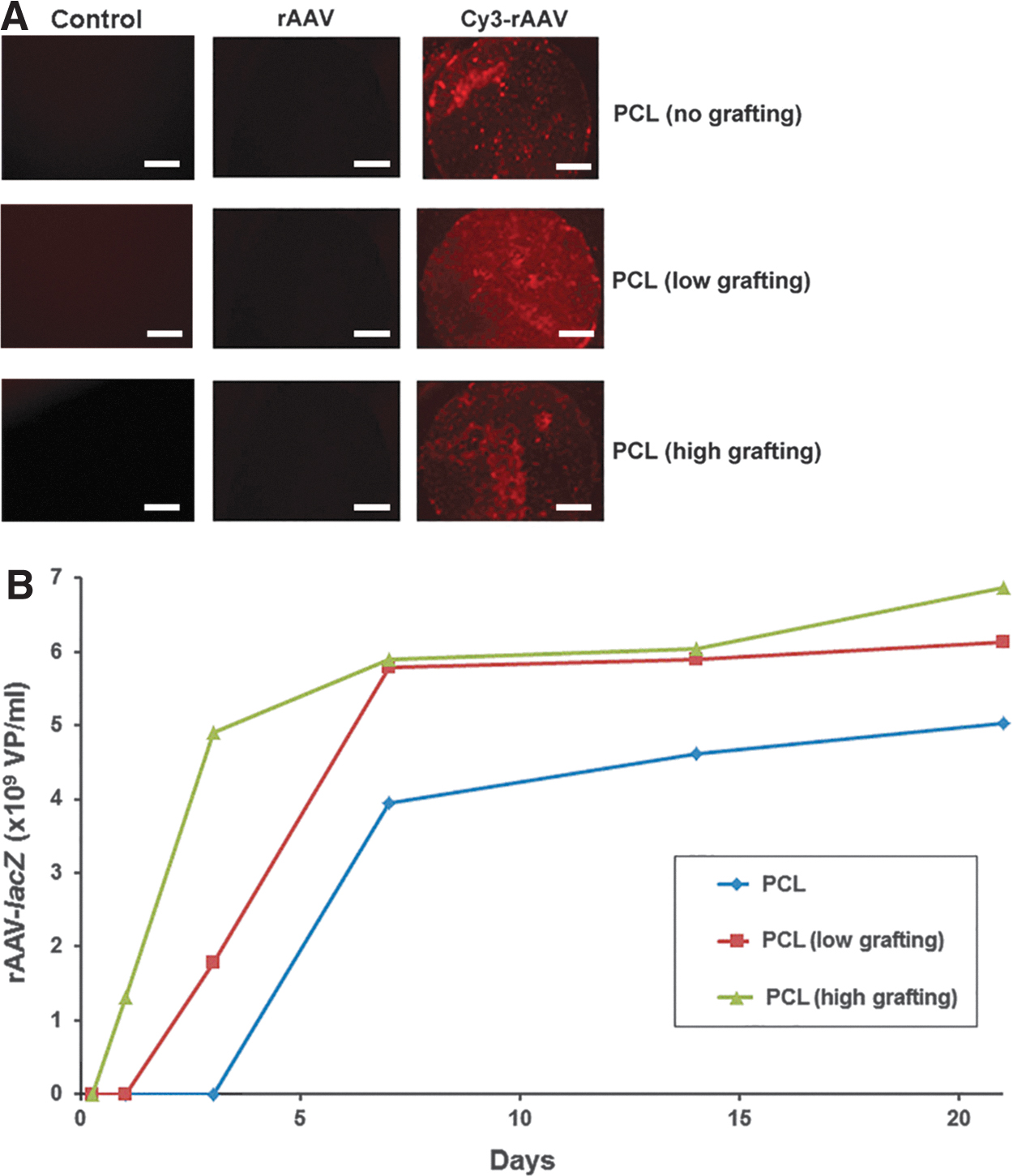
rAAV vector immobilization on and release from the pNaSS-grafted PCL films. rAAV vectors (rAAV-lacZ; 40 μL, i.e., 8 × 10⁵ transgene copies) were labeled with Cy3, immobilized on PCL films grafted with pNaSS (low grafting: 1.11 × 10⁻⁶ mol/g; high grafting: 1.30 × 10⁻⁶ mol/g) or let ungrafted, and placed in culture medium as described in the Materials and Methods section.
After immobilization, the various rAAV-coated pNaSS-grafted and ungrafted PCL films were capable of properly releasing the vectors in the culture medium over an extended period of time (up to 21 days), especially those at high and low grafting levels (1.4- and 1.2-fold difference relative to ungrafted films on day 21, respectively, p ≤ 0.001) that released rAAV starting on day 1 (high grafting) and day 3 (low grafting) compared with ∼day 5 (no grafting) (Fig. 1B).
Effective and not cytotoxic rAAV-mediated genetic modification of human bone marrow aspirates through vector delivery using pNaSS-grafted PCL films
The rAAV-coated pNaSS-grafted and ungrafted PCL films were then employed to test their ability to allow for the not cytotoxic overexpression of rAAV-delivered reporter (RFP, lacZ) genes upon direct contact with human bone marrow aspirates over extended periods of time.
Effective pNaSS-grafted or ungrafted PCL film-mediated rAAV gene transfer was achieved in human bone marrow aspirates over time as evidenced by strong fluorescent signal upon rAAV-RFP delivery versus uncoated films where no signal was detectable (Fig. 2A). RFP expression was already observed 2 days after contact between the constructs and the samples and for at least 21 days (the longest time point evaluated), reaching levels of expression that were higher than or similar to those noted upon film-free vector treatment (Fig. 2A).

Transgene expression in human bone marrow aspirates incubated with the rAAV-coated pNaSS-grafted PCL films. The rAAV-RFP
Similar results were reported when using a lacZ reporter gene instead of RFP, showing sustained more intense lacZ expression in human bone marrow aspirates treated with rAAV-lacZ-coated pNaSS-grafted or ungrafted PCL films relative to uncoated films where no signal was detectable, with X-Gal staining already noted after 2 days and for up to 21 days and with expression levels higher than or similar to those seen upon film-free vector treatment (Fig. 2B).
An estimation of the transduction efficiencies in the human bone marrow aspirates after 21 days revealed always higher values in samples treated with rAAV-lacZ-coated pNaSS-grafted or ungrafted PCL films relative to their counterparts without vector coating or using free or no vectors (up to 39-fold, p ≤ 0.003) (Fig. 2B and Table 1). Most notably, the transduction efficiencies reached rAAV-lacZ-coated pNaSS-grafted films were higher than those with ungrafted films (1.2-fold difference with low- and high-grafted films, p ≤ 0.001), yet without significant difference between grafted films (p = 0.394).
Histomorphometric Analyses in Human Bone Marrow Aspirates Incubated with the Recombinant Adeno-associated Virus-Coated Poly(Sodium Styrene Sulfonate)-Grafted Poly(ɛ-Caprolactone) Films (Day 21)
The β-gal+ cells and the cell densities are in percentage. Stained (safranin O, alizarin red) and immunostained (SOX9, type-II, -I, and -X collagen) sections were scored for uniformity and intensity according to a modified Bern Score grading system 55 as follows: 0 (no staining), 1 (heterogeneous and/or weak staining), 2 (homogeneous and/or moderate staining), 3 (homogeneous and/or intense staining), and 4 (very intense staining). Data are given as mean ± SD.
Statistically significant relative to corresponding condition without coated vector or lack of free vector treatment.
Statistically significant relative to ungrafted film.
Statistically significant relative to free rAAV-lacZ treatment.
PCL, poly(ɛ-caprolactone); pNaSS, poly(sodium styrene sulfonate); SD, standard deviation.
Administration of rAAV-lacZ to human bone marrow aspirates through pNaSS-grafted and ungrafted PCL films occurred in a not cytotoxic manner, as noted when evaluating the indices of cell viability in the samples compared with the control conditions (uncoated films, film-free vector treatment) (always ≥75%; p ≥ 0.057) (Fig. 3). These observations were corroborated by an evaluation of the cell densities on H&E-stained histological sections from samples (p ≥ 0.233) (Fig. 4 and Table 1).
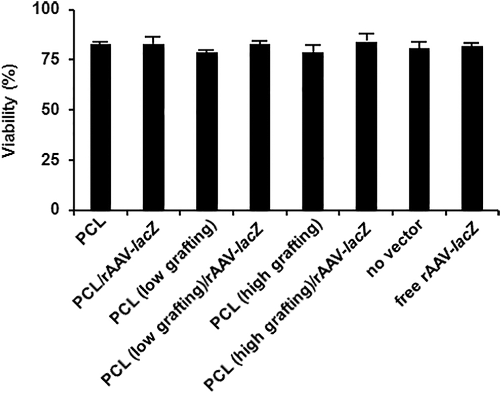
Cell viability in human bone marrow aspirates incubated with the rAAV-coated pNaSS-grafted PCL films. The rAAV-lacZ vector was immobilized on pNaSS-grafted PCL films and the rAAV-coated pNaSS-grafted PCL films were then placed in contact with aliquots of bone marrow aspirates as described in Figure 2 and in the Materials and Methods section. Control conditions included uncoated films and film-free vector treatment. Cell proliferation indices were monitored after 21 days with the Cell Proliferation Reagent WST-1 as described in the Materials and Methods section.

Biological activities and expression of chondrogenic factors in human bone marrow aspirates incubated with the rAAV-coated pNaSS-grafted PCL films. The rAAV-lacZ vector was immobilized on pNaSS-grafted PCL films and the rAAV-coated pNaSS-grafted PCL films were then placed in contact with aliquots of bone marrow aspirates as described in Figures 2 and 3 and in the Materials and Methods section. Control conditions included uncoated films and film-free vector treatment. Samples were processed after 21 days to detect cellularity (H&E staining), the deposition of matrix proteoglycans (safranin O staining) and of type-II collagen (immunohistochemistry), and the expression of SOX9 (immunohistochemistry) (magnification × 40; all representative data) as described in the Materials and Methods section (insets: uncoated films or film-free vector treatment; all representative data). H&E, hematoxylin and eosin.
Differentiation potential of human bone marrow aspirates modified by rAAV vectors released from pNaSS-grafted PCL films
The rAAV-coated pNaSS-grafted and ungrafted PCL films were further tested to evidence potential deleterious effects on the ability of human bone marrow aspirates to commit toward the chondrogenic versus osteogenic and hypertrophic phenotype over time.
Importantly, an evaluation of the biosynthetic activities and expression of chondrogenic factors in the human bone marrow aspirates assessed by estimating the intensities of safranin O staining and those of type-II collagen and SOX9 immunostaining revealed the effective deposition of proteoglycans and type-II collagen after 21 days in samples where rAAV-lacZ-coated pNaSS-grafted or ungrafted PCL films were applied, without significant difference with conditions where no vectors were coated or with free or absent vector administration (p ≥ 0.059) (Fig. 4 and Table 1).
No detrimental effects were also reported when analyzing the expression of osteogenic (alizarin red staining for matrix mineralization and type-I collagen deposition) and hypertrophic factors (type-X collagen deposition) in the human bone marrow aspirates treated with rAAV-lacZ-coated pNaSS-grafted or ungrafted PCL films relative to all other control conditions (p ≥ 0.071) (Fig. 5 and Table 1). Subsequent experiments were thus performed only using aspirates where rAAV-coated pNaSS-grafted or ungrafted PCL films were applied.

Expression of osteogenic and hypertrophic factors in human bone marrow aspirates incubated with the rAAV-coated pNaSS-grafted PCL films. The rAAV-lacZ vector was immobilized on pNaSS-grafted PCL films and the rAAV-coated pNaSS-grafted PCL films were then placed in contact with aliquots of bone marrow aspirates as described in Figures 2–4 and in the Materials and Methods section. Control conditions included uncoated films and film-free vector treatment. Samples were processed after 21 days to detect matrix mineralization (alizarin red staining) and the deposition of type-I and type-X collagen (immunohistochemistry) (magnification × 20; all representative data) as described in the Materials and Methods section (insets: uncoated films or film-free vector treatment; all representative data).
Real-time RT-PCR analyses in human bone marrow aspirates modified by rAAV vectors released from pNaSS-grafted PCL films
These results were substantiated by a real-time RT-PCR analysis performed in human bone marrow aspirates placed in contact with the rAAV-coated pNaSS-grafted and ungrafted PCL films over time.
Effective expression of chondrogenic factors was observed in samples where any of the PCL films were applied as revealed by an estimation of the SOX9, ACAN, and COL2A gene expression profiles and relative to samples receiving ungrafted PCL films without rAAV, without deleterious effects of rAAV application (always p ≥ 0.066) (Fig. 6).
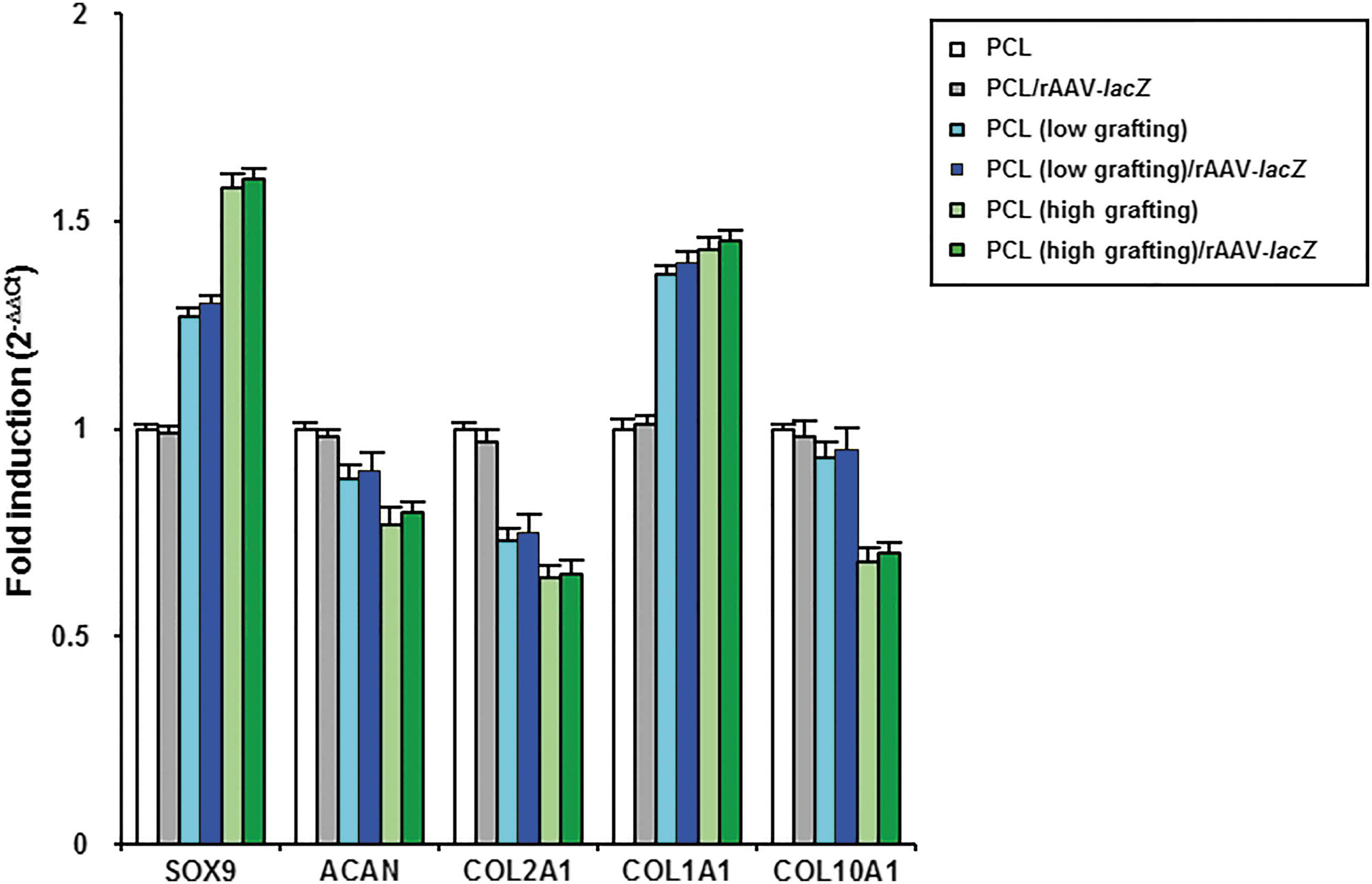
Real-time RT-PCR analyses in human bone marrow aspirates incubated with the rAAV-coated pNaSS-grafted PCL films. The rAAV-lacZ vector was immobilized on pNaSS-grafted PCL films and the rAAV-coated pNaSS-grafted PCL films were then placed in contact with aliquots of bone marrow aspirates as described in Figures 2–5 and in the Materials and Methods section. Control conditions included uncoated films. Samples were processed after 21 days to monitor the gene expression profiles by real-time RT-PCR as described in the Materials and Methods section. The genes analyzed included the transcription factor SOX9, aggrecan (ACAN), type-II collagen (COL2A1), type-I collagen (COL1A1), and type-X collagen (COL10A1), with GAPDH serving as a housekeeping gene and internal control. Threshold cycle (Ct) values were obtained for each target and GAPDH as a control for normalization, and fold inductions (relative to samples receiving ungrafted PCL films without rAAV) were measured using the 2−ΔΔCt method. GAPDH, glyceraldehyde-3-phosphate dehydrogenase; RT-PCR, reverse transcriptase polymerase chain reaction.
There was also no detrimental effects on the expression of osteogenic and hypertophic factors in samples where any of the PCL films were applied as noted by an evaluation of the COL1A1 and COL10A1 gene expression profiles versus samples receiving ungrafted PCL films without rAAV, again without detrimental effects of rAAV application (always p ≥ 0.065) (Fig. 6).
Discussion
Biomaterial-guided gene transfer is a promising strategy to develop effective tools for cartilage regenerative medicine17–19 especially for the controlled delivery of clinically adapted rAAV vectors.35–37,55 In this study, and for the first time to our best knowledge, we examined the ability of solid scaffolds based on PCL, a biocompatible FDA-approved compound that provides a mechanical environment suited for cartilage research, to effectively deliver reporter rAAV vectors to human bone marrow aspirates after pNaSS grafting of the PCL films. Such an approach may provide novel, more effective, and less invasive therapeutic options to treat focal cartilage lesions relative to direct scaffold-free rAAV administration 54 and to the indirect implantation of rAAV-treated aspirates seeded on such a material. 55
Our results first reveal that PCL films can successfully immobilize and subsequently release rAAV vectors over extended periods of time (21 days) especially upon grafting of the films with pNaSS. As a result, the released rAAV constructs (reporter RFP and lacZ vectors) were capable of promoting the effective, durable, and not cytotoxic genetic modification of human bone marrow aspirates, most particularly when providing the vectors through pNaSS-grafted PCL films (up to 90% transduction efficiencies with at least 75% viability for up to 21 days, the longest time point evaluated) relative to various control conditions (ungrafted films, lack of vector coating on the films, and free or absence of vector treatment), as observed in direct scaffold-free rAAV delivery and in indirect (PCL-assisted) implantation approaches. 55
The data next indicate that effective prolonged rAAV-mediated (reporter) gene transfer through pNaSS-grafted PCL films had no detrimental effects on the expression of chondrogenic factors of human bone marrow aspirates (matrix proteoglycan and type-II collagen deposition) over a period of at least 21 days (a time point adapted to evaluate chondrogenesis in the marrow environment) 54 compared with the control treatments, concordant with previous work using direct or indirect rAAV gene transfer in such samples.54,55
Also remarkably, delivery of rAAV through pNaSS-grafted PCL films did not activate undesirable expression of osteogenic or hypertrophic factors over time in the aspirates relative to control treatments (matrix mineralization, deposition of type-I and type-X collagen), again consistent with work through direct (film-free) rAAV transduction 54 or using rAAV-modified aspirates seeded in PCL scaffolds. 55
In conclusion, this study demonstrates the benefits of applying rAAV vectors to human bone marrow aspirates through pNaSS-grafted PCL films as a novel, highly effective, and convenient method to generate off-the-shelf mechanically adapted therapeutic platforms for cartilage repair relative to hydrogel systems. 37 Experimental work is currently ongoing to translate the findings in animal bone marrow aspirates for application in clinically relevant (orthotopic) cartilage defects in vivo21,23,24,26,56–59 that may direct the choice and future delivery of chondrotherapeutic candidates like for instance the TGF-β,21,24,59 insulin-like growth factor I (IGF-I), 58 basic fibroblast growth factor (FGF-2), 56 or the SOX family of transcription factors (SOX5, SOX6, and SOX9).23,26,57 Overall, this study provides evidence showing the potential of solid scaffold-guided delivery of rAAV vectors in chondrocompetent human bone marrow aspirates as a translational strategy to conveniently treat articular cartilage lesions in patients in the future.
Footnotes
Acknowledgments
We thank R.J. Samulski (The Gene Therapy Center, University of North Carolina, Chapel Hill, NC), X. Xiao (The Gene Therapy Center, University of Pittsburgh, Pittsburgh, PA), and E.F. Terwilliger (Division of Experimental Medicine, Harvard Institutes of Medicine and Beth Israel Deaconess Medical Center, Boston, MA) for providing the genomic AAV-2 plasmid clones and the 293 cell line.
Disclosure Statement
No competing financial interests exist.
Funding Information
This research was funded by a grant from the Deutsche Forschungsgemeinschaft (DFG VE 1099/1-1 to J.K.V. and M.C.) and University Paris 13, Sorbonne Paris Cité.