
Abstract
Erectile dysfunction caused by damage to the cavernous nerve is a common complication of radical prostatectomy for patients with localized prostate cancer. Various studies have investigated repair of damaged tissue and prevention of fibrosis in the corpus cavernosum using stem cell therapy. However, stem cell therapy has limitations, including insufficient nutrient and oxygen supply to transplanted stem cells. This study investigated whether stem cell/oxygen-releasing hollow microparticles (HPs) had therapeutic effect on erectile dysfunction in a rat model of bilateral cavernous nerve injury (BCNI). Therapeutic effects were observed in the BCNI model at 1, 2, and 4 weeks postcavernous nerve injury. Erectile function further improved after treatment with stem cell/oxygen-releasing HP system compared with treatment with only stem cells at 4 weeks. Stem cell/oxygen-releasing HP system increased cyclic guanosine monophosphate (cGMP) level and neuronal nitric oxide synthase (nNOS), endothelial nitric oxide synthase (eNOS), α-smooth muscle actin (α-SMA), and muscarinic acetylcholine receptor 3 (M3) expression while decreasing fibrosis and apoptosis in the corpus cavernosum. Our results clearly show that stem cell survival increases around transplanted stem cell/oxygen-releasing hybrid system site. Taken together, an oxygen-releasing HP system supported prolonged stem cell survival, sustaining the paracrine effect of the stem cells, and consequently enhancing erectile function. These findings show promise with regard to prolonged stem cell survival in stem cell applications for various diseases and types of tissue damage.
Impact statement
In this study, we used an oxygen-releasing hollow microparticles (HPs) system with stem cells to attempt to overcome certain limitations of stem cell therapy, including insufficient nutrient and oxygen supplies for transplanted stem cells. Our results demonstrated that a stem cell/oxygen-releasing HP hybrid system could further improve erectile function, cyclic guanosine monophosphate (cGMP) level, and NOS level in a bilateral cavernous nerve injury rat model through prolonged stem cell survival. Our data suggest that a stem cell/oxygen-releasing HP system is a promising clinical treatment option for postprostatectomy erectile dysfunction. Furthermore, this system may be relevant in different disease therapies and regenerative medicine.
Introduction
Radical prostatectomy is a treatment option for patients with localized prostate cancer. Despite technical advances, this surgery may still cause some complications, such as erectile dysfunction and urinary incontinence. 1 Post radical prostatectomy erectile dysfunction (pRP-ED) is caused by damage and neurapraxia of the cavernous nerve (CN), which can cause vascular atrophy and fibrosis in the penis.1,2 Although there are a variety of treatments available for pRP-ED, including vacuum erection devices and phosphodiesterase-5 inhibitors, these cannot completely restore erectile function.1,2 For that reason, many researchers have investigated regeneration of the CN and prevention of apoptosis and fibrosis in the corpus cavernosum using tissue engineering techniques, including stem cells and biomaterials. 3
Stem cells have been extensively studied for regenerating tissue defects of diverse organ and have showed therapeutic effects. 4 Numerous studies have approached stem cell therapy as a way to restore the erectile function of rats with CN and have demonstrated prevention of fibrosis and improved erectile function. 5 Current stem cell regenerative medicine is advancing toward using combinations of stem cell and diverse scaffolds to increase survival and maintain function of stem cells. 6 There is particular focus on adequate oxygen diffusion from three-dimensional (3D) scaffolds, which is important for survival, growth, and function of stem cells. Preclinical evidence has shown that supplying oxygen from an oxygen carrier was successful to prolong the survival of transplanted stem cells and further restore damaged tissue.7–11 Various researchers have developed oxygen carriers using hemoglobin, perfluorocarbon (PFC), and peroxides to increase oxygen delivery.8,12 Above all, PFC-based oxygen carriers have the advantage of biocompatibility and continuous oxygen release compared to peroxide-based oxygen carriers. 12
In a previous study, we developed hollow microparticles (HPs) contiating perfluorrooctane (PFO) emmulsion (oxygen carrier:PFO-HPs). The PFO-HPs act as a 3D construct and carries oxygen to cells adhered on the HPs. 13
In this study, we injected stem cells seeded on PFO-HPs at the site of CN injury. We identify whether oxygen continuously released from PFO-HPs affected survival of transplanted stem cells. Furthermore, our study demostrated that stem cells seeded on PFO-HPs more enhanced erecitle function in a bilateral cavernous nerve injury (BCNI) rat model (Fig. 1).
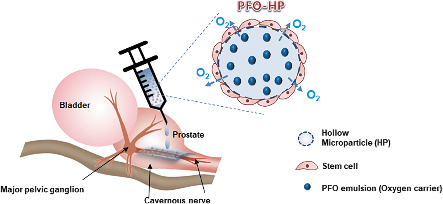
Schematic diagram showing application of stem cells attached to PFO-HPs (oxygen supply) on injured CN. CN, cavernous nerve; PFO, perfluorooctane.
Experimental Section
Fabrication of perfluorooctane-hollow microparticles
Oxygen carrier (PFO emulsion)-loaded hollow microparticles (PFO-HPs) were produced by water-in-oil-in-water emulsion solvent evaporation method (for Poly-ɛ-caprolactone [PCL] HP). Then infiltration of PFO emulsion fabricated by ultrasonication was infiltrated into the PCL HP, as described elsewhere.13,14 To prepare PCL HPs, PCL pellets were dissolved in ethyl acetate (EA) to 5% (w/w; 4 mL) at room temperature, and 1% (w/w) Pluronic F127 aqueous solution (1 mL) was added to the PCL solution with stirring (400 rpm) to emulsify the mixture solution. The emulsified mixture was directly poured into 0.5% (w/v) poly(vinyl alcohol) (PVA; Sigma-Aldrich) aqueous solution (1 L) with gentle stirring (400 rpm), and the PCL-based droplets were solidified for 10 min. Solidified PCL HPs were washed with excess water to remove EA, Pluronic F127, and PVA. The PCL HPs were separated in size ranges of 100–500 μm by wet microsieving using standard testing sieves (Chunggye Industrial Co.) and were freeze-dried. 15 To produce PFO emulsion, PFO, 20% (w/w) Pluronic F68 aqueous solution, and 4% (w/w) 1,2-Diacyl-sn-glycero-3-phosphocholine (egg yolk phospholipid; EYP; Sigma-Aldrich) aqueous solution (PFO/Pluronic F68 solution/EYP solution, 6/2/2 [v/v/v]) were vigorously agitated using a prove type ultrasonic wave homogenizer (Branson sonifier model 185). Ultrasonication at frequency of 18,000 Hz (30 s ON and 20 s OFF; 10 cycles) was directly applied to the mixture solution placed in a warm water bath (60°C). To load the PFO emulsion into the PCL HPs, the prepared PFO emulsion (15 mL) was poured in a needle tip-stopped 20-mL syringe filled with PCL HPs (5 mL). The PFO emulsion was infiltrated into PCL HPs through a shell membrane with micro-sized pores under positive pressure when the syringe piston was pushed in. Then PFO emulsion-loaded PCL HPs (PFO-HPs) were carefully washed with phosphate-buffered saline (PBS) to remove unloaded PFO emulsion. As a control group, the PBS-loaded PCL HPs (PBS-HPs) were also prepared using the same procedures described above. 15
Cell culture and differentiation
Human bone marrow-derived mesenchymal stem cells (Catholic MASTER Cells, hBMSCs) were obtained from Catholic Institute of Cell Therapy (Seoul, Korea). The cells were cultivated in Dulbecco's modified Eagle's medium (Life Technologies) containing 20% fetal bovine serum and 1% penicillin–streptomycin at 37°C in a humidified atmosphere of 5% CO2.
In our previous study, hBMSCs were induced using neural induction media in a hypoxic environment; a similar procedure was used here to stimulate neural cell differentiation in hypoxic environment using PBS0HP and PFO-HP. 16 Briefly, before cell seeding on HP, HPs (5 mL) placed in a 20-mL sample tube (SPL) were stored in an aseptic oxygen chamber for 5 min to achieve oxygen saturation. The oxygen-saturated HPs were immediately transferred to nontreated 24-well polystyrene (PS) dishes. One milliliter of cell suspension in hBMSC growth media (cell density: 1 × 10⁶ cells/mL) was seeded on HPs and shaken mildly under 50 rpm for 24 h for uniform cell adhesion on the HPs in a normal incubator (21% O2, 5% CO2, 37°C). Then, cells seeded on HPs were carefully transferred to new nontreated 24-well PS dishes. Neural induction media was added into each well (2 mL), and samples were incubated in an anaerobic chamber (0% O2, 5% CO2, 37°C) for 3 days.
Immunocytochemistry
After neural induction, cells were washed with PBS and fixed for 20 min in 4% paraformaldehyde in PBS at room temperature, washed with PBS again, and then permeabilized in 0.5% Triton X-100 in PBS for 10 min. These cells were washed with PBS twice and then blocked by incubation with 5% bovine serum albumin for 1 h at room temperature. Cells were stained with anti-neuron-specific β-III tubulin (Tuj-1, diluted 1:200, ab78078; Abcam). After washing, incubation with secondary antibody (Alexa 488-conjugated goat anti-mouse) was followed for 2 h at room temperature. Samples were stained with DAPI (Vector Laboratories, Burlingame, CA) to visualize cell nuclei. Fluorescent images were obtained using a confocal microscope (LSM 800 w/Airyscan; Carl Zeiss).
Animal experiments
Sprague-Dawley rats (rats; 250–300 g) were purchased from Orient Bio (Gyeonggi, Korea), and all protocols were performed in accordance with guidelines and regulations pertinent to animal experiments, as detailed by the Institutional Animal Care and Use Committee (IACUC, CUMC-2016-0031-02) at the Catholic University of Korea (Seoul, Korea). Rats were randomly divided into four groups (n = 7 per group): an age-matched normal control group (Normal), a BCNI group (BCNI), hBMSC seeded on the PBS-HP treatment group (PBS-HP), and hBMSC seeded on the PFO-HP treatment group (PFO-HP). Rats were anesthetized with a subcutaneous injection of Zoletil 50 (Virbac Laboratories, Carros, France) at a dose of 15 mg/kg and xylazine hydrochloride (Bayer, Seoul, Korea) at a dose of 5 mg/kg. The BCNI rat model has been previously described.17–19 Briefly, a low midline incision was used to open the abdominal wall and expose the urinary bladder and prostate. Bilateral CN located below the major pelvic ganglion (MPG) was compressed with a hemostat clamp for 2 min. To track the implanted hBMSC, cells were labeled with green fluorescent dye PKH-67 (PKH-67 fluorescent cell linker kits; Sigma-Aldrich) according to the manufacturer's protocol. In PBS-HP and PFO-HP groups, 200 μL of hBMSC seeded on PBS-HP and hBMSC seeded on PFO-HP were injected around each injured CN.
Erectile function measurement
After 1, 2, and 4 weeks, erectile function was assessed. The CN and carotid artery were exposed for measurement of intracavernosal pressure (ICP) and mean arterial pressure (MAP). A 23-gauge butterfly needle, filled with 250 U/mL heparin solution, was inserted into the proximal corpus cavernosum and then connected to a pressure transducer (Grass Instrument; Astro-Med, Inc., West Warwick, RI) for ICP measurement. A bipolar stainless-steel electrical stimulator was then used to stimulate the MPG at 10 V for 50 s and 2.4 mA with a 3.5-ms pulse. During nerve electrostimulation, the ICP peak was calculated from an isometric force transducer and recorded on a computer with a commercial data acquisition system (PowerLab; AD Instruments, Dunedin, New Zealand). To measure MAP, PE-50 tubing (BD INTRAMEDIC, Franklin Lakes, NJ) was inserted into the carotid artery. The ratio of ICP to MAP was used to determine erectile function. After ICP and MAP measurement, rats were euthanized, and the penises and prostates were immediately removed for histologic and molecular analysis. Each rat's penis was cut in half. One piece was harvested, fixed in 4% paraformaldehyde, and embedded in paraffin wax. Another piece was stored at −70°C for western blotting and other further assays. Each rat's prostate was harvested, fixed in 4% paraformaldehyde, and embedded in paraffin wax.
Immunohistochemistry
For immunohistochemical analyses, paraffin-embedded CN sections were immune stained with the following primary antibodies: anti-Tuj-1 and cell nuclei (DAPI; Vector Laboratories). Paraffin-embedded penile tissue sections were immunostained with anti α-smooth muscle actin (α-SMA, diluted 1: 250, ab5694; Abcam), anti-neuronal nitric oxide synthase (nNOS, diluted 1: 100; ab76067; Abcam), and anti Tuj-1 and stained with Masson's trichrome staining. Fluorescent images were obtained using a Zeiss LSM 510 Meta Confocal microscope (Zeiss), and the mean fluorescent intensity was calculated using ZEN 2012 (Zeiss). Digital images were obtained using an Olympus BX50 optical microscope.
TUNEL assay
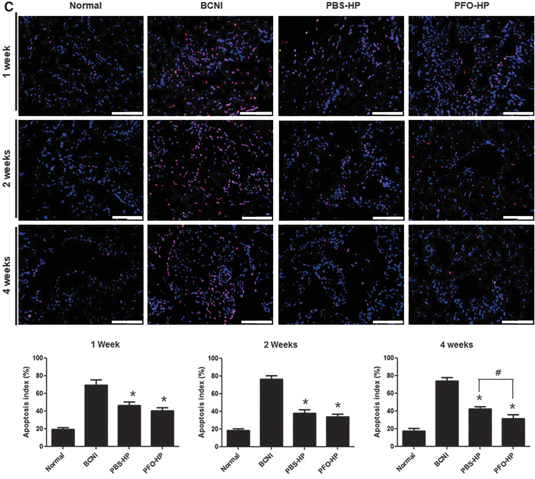
Paraffin-embedded penile tissue sections were stained using in situ Cell Death Detection Kit, TMR red (Roche, Mannheim, Germany), and TUNEL staining was performed according to the manufacturer's guidelines. Nuclei were stained with DAPI. Fluorescent images were obtained using an Olympus BX50 optical microscope. The apoptotic index (TUNEL-positive cell number/total number of nuclear × 100%) was calculated.
Western blot analyses
After harvesting penises, tissues were frozen in liquid nitrogen for western blot analysis and enzyme-linked immunosorbent assay. Tissue was homogenized in RIPA buffer (Cell Signaling Technology, Danvers, MA). The homogenized sample was then centrifuged at 12,000 g for 10 min at 4°C, and the supernatant was transferred to a new tube. The protein was electrophoresed on NuPAGE 4–12% Bis-Tris gel (Invitrogen, Carlsbad, CA) and then transferred onto nitrocellulose membrane. After the transfer, the membrane was blocked and then incubated with the following primary antibodies at 4°C overnight: rabbit endothelial nitric oxide synthase (eNOS, diluted 1: 1000, ab5589; Abcam), M3 (muscarinic acetylcholine receptor 3; diluted 1: 1000, ab28364; Abcam), and b-actin (1:2500; Santa). After incubation, the membranes were washed and then incubated with a secondary antibody conjugated to HRP for 1 h at room temperature. The chemiluminescence method (Amersham, Arlington Heights, IL) was used to develop protein bands.
Cyclic guanosine monophosphate level measurement
Fifty milligrams of penile tissue was added to 300 μL of 0.1 M HCL with silica beads (BioSpec Products, Inc., Guelph, Canada). Each sample was homogenized with a homogenizer (Precellys 24; Bertin Technologies, Montigny-le-Bretonneux, France) and then centrifuged at 12,000 g for 10 min at 4°C, and the supernatant was collected. Cyclic guanosine monophosphate (cGMP) levels were measured by cGMP Direct Immunoassay Kit (K372-100; BioVision, Edmonton, Canada) according to the manufacturer's guidelines.
Statistical analyses
The GraphPad Prism Software v5 (GraphPad Prism Software, Inc., San Diego, CA) was used for statistical analyses. All data were expressed as mean ± standard deviation. The differences between groups were examined by one-way analysis of variance test followed by Tukey post hoc test; p-value <0.05 was considered statistically significant.
Results
Characterization of PFO-HPs
As already demonstrated in our previous studies,13,14 produced PCL HPs were 100 ∼ 500 μm hollow structures with a porous shell membrane (thickness ∼10 μm and pore size, ∼2 μm), and the average size of emulsified PFO droplets was ∼300 nm. PFO emulsion easily infiltrated into hollow spaces under positive pressure and stably remained in PCL HPs. PFO-HPs could provide an appropriate environment for cell survival even in hypoxic environments by providing a sufficient oxygen delivery.
Cell differentiation of human bone marrow-derived mesenchymal stem cells seeded on PFO-HP
In recent studies, hBMSC had decreased differentiation capacity and induced apoptosis in hypoxic culture conditions. 20 To investigate whether oxygen from PFO-HPs affect cell differentiation capacity in hypoxic environments, hBMSCs were seeded onto PFO-HP or PBS-HP and induced into neural cells for 3 days in a hypoxic environment. In Figure 2, the neural differentiation of hBMSC seeded on HPs (PBS-HP and PFO-HP) was evaluated with Tuj-1 (neuronal lineage marker) immunocytochemistry. When observing PBS-HP or PFO-HP by light microscopy, PFO-HP was full of PFO emulsion, but PBS-HP was empty in PCL-HP. Three days after inducing differentiation, the difference in morphology and Tuj-1 expression was confirmed for PBS-HP and PFO-HP groups. Generally, hBMSC displayed fibroblast-like morphology. hBMSC seeded on PFO-HP retained fibroblast-like morphology, whereas hBMSC seeded on PBS-HP showed loss of fibroblast-like features. The fluorescent microscope image in Figure 2 shows that expression of Tuj-1 of cells seeded on PFO-HP is higher than in the PBS-HP group. Our results suggest that oxygen from PFO-HP affects the stabilization and differentiation capacity of hBMSC in hypoxic conditions.
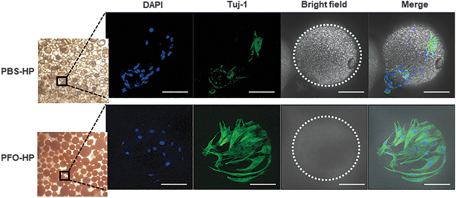
Confocal laser scanning microscope images of hBMSC neuronal differentiation on PBS-HPs and PFO-HPs. hBMSCs attached to PBS-HPs or PFO-HPs induced neuronal differentiation in induction media in hypoxic environments. Nuclei were counterstained with DAPI (blue), neuronal specific marker was stained with Tju-1 (green), and dot circles are HPs (scale bar: 100 μm). hBMSC, Human bone marrow-derived mesenchymal stem cell; HPs, hollow microparticles.
hBMSC seeding on PFO-HP improves erectile function in bilateral cavernous nerve injury model
To investigate the therapeutic potential of hBMSC seeded on PFO-HP in a rat model of BCNI (Fig. 1), maximal ICP and MAP were measured in all groups 1, 2, and 4 weeks after operation. A representative ICP curve 4 weeks after operation is shown in Figure 3A.
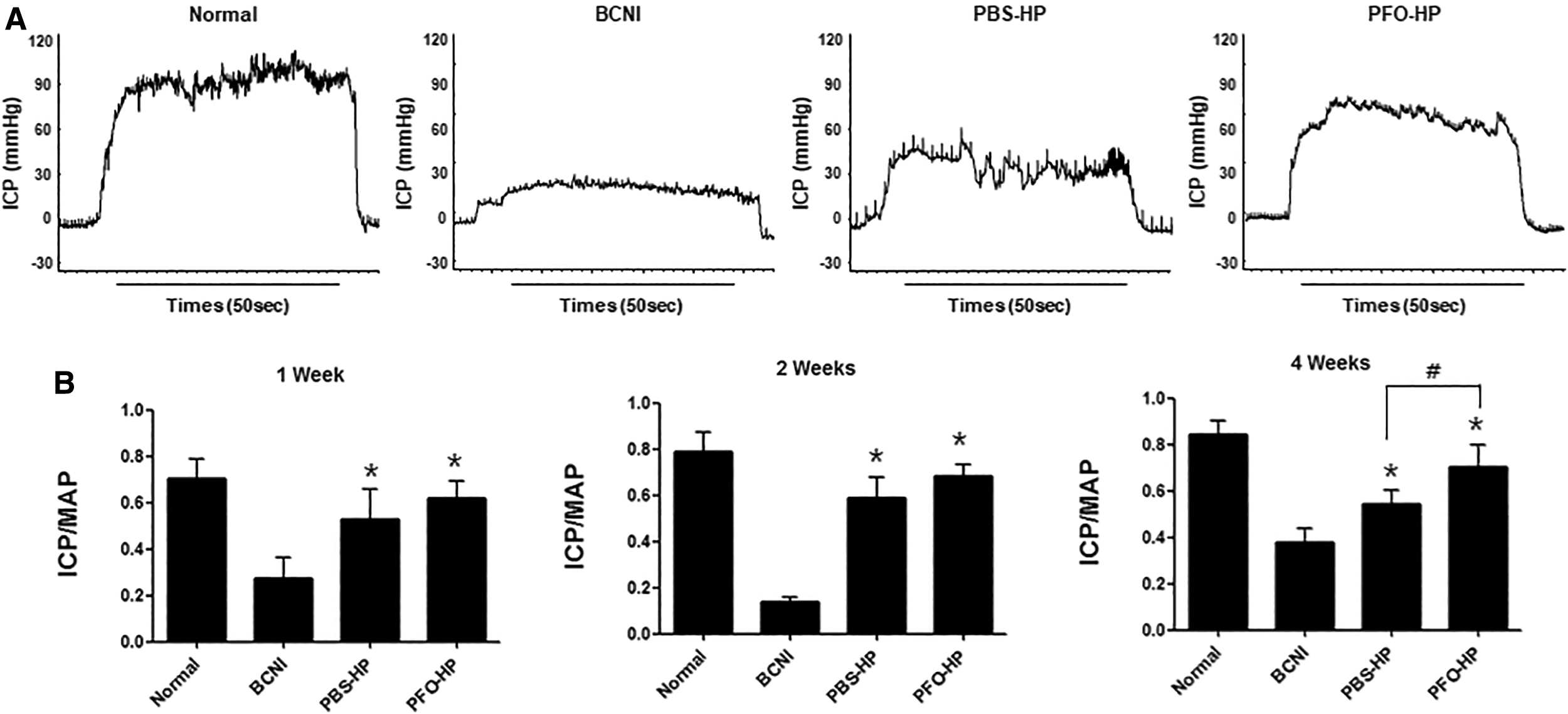
Erectile function assessment.
Figure 3B shows ICP/MAP ratio in all groups 1, 2, and 4 weeks after operation. The ICP/MAP ratio was significantly higher in the hBMSC-treated group (PFO-HP and PBS-HP group) than in the BCNI group for the duration of all experiments (p < 0.05). Interestingly, the difference in ICP/MAP ratio between the PFO-HP and PBS-HP group was not statistically significant at 1 and 2 weeks. However, the ICP/MAP ratio showed significant differences at 4 weeks (p < 0.05). We speculate that the difference in ICP/MAP ratio between PBS-HPs and PFO-HP groups at 4 weeks may be associated with prolonged hBMSC survival or hBMSC differentiation capacity through oxygen release. To confirm this, PKH-67 (green color) labeled hBMSCs around the CN (red color) were investigated. Figure 4 is a representative image of all groups 4 weeks after operation. The hBMSCs adhered on HPs were located around the CN in the hBMSC-treated groups (arrow), and Tuj-1 expression of CN fibers in hBMSC-treated groups was increased compared with the BCNI group. This result showed that implanting hBMSCs prevents CN damage. More importantly, PKH-67 expression was stronger on HP particles in the PFO-HP group than in the PBS-HP group, indicating increased survival of injected hBMSCs. A few green-fluorescent hBMSCs co-stained with red-fluorescent Tuj-1 showed a yellow color (thin arrow), which may be evidence for hBMSC differentiation toward nerve in vivo.
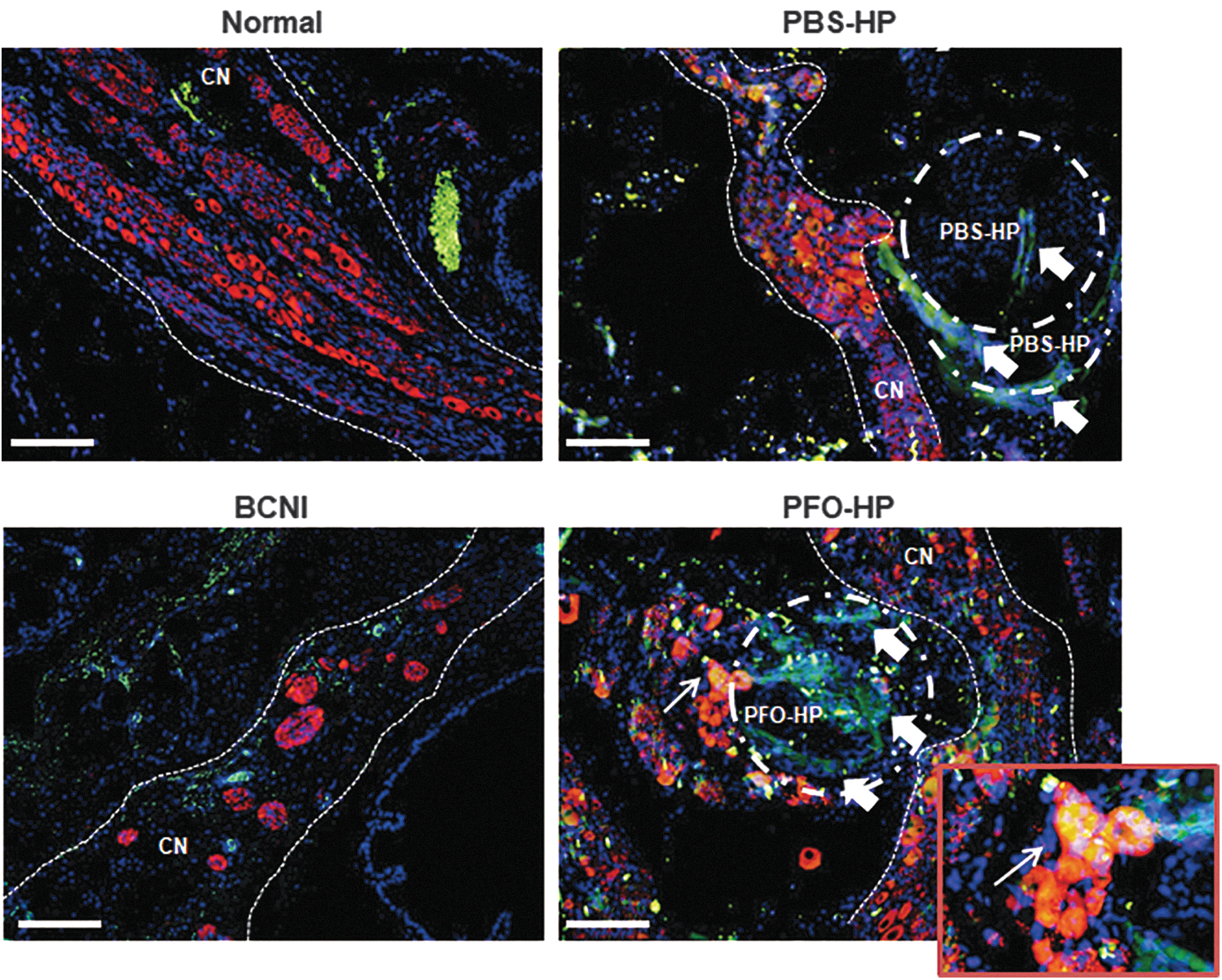
In vivo tracking of PKH67-labeled hBMSCs seeded onto PFO-HPs or PBS-HPs (dot circle; HP) in BCNI model. Immunostaining using Tuj1 (red) and PKH-67 (green) obtained around CN 4 weeks after surgery. PKH67-labeled hBMSCs seeded onto PFO-HP and PBS-HP groups are shown around CN. Scale bar is 100 μm. Merged hBMSCs (yellow, thin arrow) in PFO-HP group showed colocalization of PKH-67 labeled hBMSCs and CN (small image, bottom). CN, thin dotted line.
hBMSC seeding on PFO-HP increases nitric oxide synthase expression in the dorsal nerve
The nNOS expression in dorsal penile nerves evaluated with immunohistochemistry and sections was stained with green-fluorescent Tuj-1 and red-fluorescent nNOS to discriminate nNOS content in the dorsal nerve bundle. Figure 5 shows a representative image and nNOS positive area in all groups at 1, 2, and 4 weeks after operation. The nNOS positive area increased in hBMSC-treated groups compared with the BCNI group at 1, 2, and 4 weeks. In particular, 4 weeks after operation, expression of nNOS in the PFO-HP group was significantly increased compared with that of the PBS-HP group (p < 0.05).

Expression of nNOS in dorsal penile nerve. Representative images showed dorsal penile nerve for each group 1, 2, and 4 weeks after surgery. Tuj-1 (dorsal penile nerve) was stained green, and nNOS was stained red. Scale bar is 50 μm. Quantification of nNOS expressed as percentage of positive expression area in surrounding dorsal penile nerve 1, 2, and 4 weeks after surgery. Each bar shows mean values (±SD). N = 7 rats per group; *p < 0.05 compared with BCNI, #p < 0.05 compared with PBS-HP. nNOS, neuronal nitric oxide synthase.
hBMSC seeded on PFO-HP prevents fibrosis and apoptosis in corpus cavernosum tissue
Masson's trichrome staining was used to evaluate fibrosis in the corpus cavernosum. Representative histologic images for all groups at 1, 2, and 4 weeks are shown in Figure 6A. Staining verified that smooth muscle/collagen ratio in the BCNI group was reduced in the corpus cavernosum compared to hBMSC-treated groups. In other words, hBMSC-treated groups had prevented fibrosis in the corpus cavernosum. When PBS-HP and PFO-HP groups were compared, the SMA/collagen ratio in the PBS-HP group was significantly higher than in the PFO-HP group at 4 weeks.
Histologic analyses of corpus cavernosum.
Immunohistochemistry was performed to evaluate inhibition of smooth muscle atrophy in the corpus cavernosum (Fig. 6B, C). In the BCNI group, α-SMA expression was reduced (Fig. 6B) and TUNEL-positive cells increased (Fig. 6C) in the corpus cavernosum compared with hBMSC-treated groups (p < 0.05). These results indicated that hBMSC treatments prevent apoptosis of smooth muscle in the penis by CN damage regardless of oxygen supply. Furthermore, 4 weeks after operation, both expressions of α-SMA and TUNEL were significantly different between the PBS-HP group and PFO-HP group (p < 0.05). The above results suggest that the PFO-HP group prevented fibrosis and smooth muscle apoptosis and in the penis by CN damage through prolonged hBMSC survivals.
hBMSC seeded on PFO-HP increases expression of p-eNOS and M3 in the corpus cavernosum
To confirm eNOS and M3 levels in the corpus cavernosum, protein expression was examined by western blotting. Figure 7 shows representative protein bands and relative density in all groups. Expression of eNOS and M3 was increased in all hBMSC-treated groups compared to the BCNI group. In addition, 4 weeks after operation, both phosphorylated eNOS and M3 expression were significantly increased in the PFO-HP group compared with the PBS-HP group (p < 0.05).

eNOS and M3 expression analyses and cGMP concentration in corpus cavernosum.
hBMSC seeded on PFO-HP restores cyclic guanosine monophosphate concentration in the corpus cavernosum
Next, cGMP level was evaluated to confirm NO/cGMP pathway (Fig. 7). cGMP concentration in BCNI group was reduced after CN injury; however, cGMP concentration in hBMSC-treated groups was restored. In particular, the cGMP concentration of the PFO-HP group was significantly higher at 4 weeks after operation (p < 0.05). Collectively, the aforementioned findings reveal that hBMSCs maintained survival through oxygen release and restored NO/cGMP pathway, further improving erectile function in the BCNI model.
Discussion
Biomaterials incorporating stem cells are being developed for use in regeneration of tissue defects. 21 The mechanisms of stem cell therapy have been characterized as transdifferentiation into target cells and paracrine effect releasing soluble factors from stem cells for tissue regeneration. 22 Diverse researchers have investigated if biomaterials and stem cells can improve erectile function. 3 Our previous study demonstrated that combined application of stem cells and diverse biomaterials can improve erectile function in a CN injury model by promoting nerve regeneration.19,23–25 However, inadequate oxygen supply to transplanted cells is the main concern of biomaterials incorporating stem cells due to poor cell survival. 8 In recent studies, BMSCs had decreased differentiation capacity, stabilization, and survival in hypoxic conditions.20,26 To overcome this crucial limitation, our previous study demonstrated that PFO-HP prevented transplanted cell necrosis through sufficient oxygen delivery to transplanted cells. Based on Henry's law, in which oxygen solubility is proportional to partial pressure, oxygen readily permeates through HP membranes to attach to stem cells in low oxygen environments. 15 Our previous results showed that PFO-HP can prolong cell survival both in vitro and in vivo by supplying oxygen for at least 10 days. 13 Consistent with this study, our results show that PFO-HPs can supply oxygen to stem cells, which affects stem cell stabilization, survival, and differentiation capacity in vitro. Based on these in vitro results, we examined whether oxygen from PFO-HPs influences the therapeutic effect of implanted stem cells on a rat model of BCNI.
In agreement with other studies, 27 our results showed that hBMSC-treated groups had significantly improved erectile function compared with the BCNI group at 1, 2, and 4 weeks. Interestingly, erectile function in the PFO-HP group was significantly improved compared to the PBS-HP group at 4 weeks postsurgery. To investigate the difference in erectile function at 4 weeks in the PBS-HP group and the PFO-HP group, immunohistochemistry was performed around the CN injury site. Results clearly showed that hBMSC survival around the CN in the PFO-HP group was increased compared to the PBS-HP group. Colocalization of hBMSCs and CN was confirmed only in the PFO-HP group. This histologic phenotype could be evidence for capacity of hBMSC differentiation into neural cells in vivo. However, there was less colocalization of CN and hBMSCs than expected. We speculate that improved erectile function in the PFO-HP group compared to the PBS-HP group results from increased and prolonged cell survival rather than neuronal differentiation. Many studies have described that MSC transplantation in experimental models might heal injured tissues by secreting soluble factors such as growth factors, cytokines, and hormones from survival stem cell rather than transdifferentiation. This hypothesis is supported by recent studies, in which MSC-derived conditioned medium or exosome treatment enhanced organ function similar to MSC treatment.22,28,29 Yiou et al. also demonstrated that hMSCs improved erectile function not by transdifferentiation but by paracrine effect through soluble factors released by hMSCs. 30 Therefore, prolonged survival of transplanted stem cells around the injury site is important for paracrine secretion of soluble factor. Our development of PFO-HPs can be a promising prolonged survival of transplanted stem cells around the injury site.
Neurodegeneration induced by CN injury can lead to damage such as fibrosis and smooth muscle atrophy in the corpus cavernosum, resulting in erectile dysfunction.31,32 You et al. reported that periprostatic implantation of hBMSCs prevented apoptosis and fibrosis in the corpus cavernosum through CNI regeneration in a BCNI model. 33 The present study confirmed that hBMSC-treated groups significantly prevented antiapoptotic and antifibrotic potencies in the corpus cavernosum at 1, 2, and 4 weeks (Fig. 6). We determined that the PFO-HP group more effectively prevented fibrosis and smooth muscle atrophy in the corpus cavernosum compared with the PBS-HP group at 4 weeks. These results suggest that prolonged stem cell survival could conserve corpus cavernosum architecture by preventing smooth muscle cell apoptosis.
Erectile function is regulated by parasympathetic nerve terminals and induced by smooth muscle relaxation and contraction. Smooth muscle relaxation in the corpus cavernosum is caused by neuroregulatory interaction and the NO/cGMP pathway. NO is mediated by calcium-calmodulin controlled isoenzymes eNOS and nNOS, respectively. Activated NOS produces NO, which stimulates guanylate cyclase to convert 5′GTP into cGMP; this pathway induces penile erection. 34 To confirm changes in the NO/cGMP pathway, nNOS, eNOS, and M3 (located on corpus cavernosum endothelium to stimulate NOS) expression and cGMP concentration were examined. As shown in Figures 5 and 7, expression of nNOS, eNOS, and M3 and cGMP concentration in the hBMSC-treated group are higher than in the BCNI group, consistent with previous studies. More importantly, the PFO-HP group showed incrementally increased expression of nNOS, eNOS, and M3 and cGMP concentration compared with the PBS-HP group at 4 weeks. This result also proved that compared with stem cell only treatment, stem cells adhered onto PFO-HP protect from deregulation of the NO/cGMP signaling pathway induced by CNI.
In summary, we showed that transplantation of hBMSC seeded on PFO-HPs into damaged CN enhanced erectile function more than hBMSC seeded on PBS-HPs. More importantly, this study identified that oxygen released by PFO-HPs prolongs stem cell survival by the sustained paracrine effect of hBMSCs. To the best of our knowledge, this is the first study to elucidate the role of a PFO-HP for sustained oxygen release system to prolong stem cell survival and enhance erectile function. Our results offer an important theoretical basis for stem cell therapy in ED. These data demonstrate the promise of this material for overcoming stem cell therapy limitations, which could ultimately facilitate tissue engineering and regeneration for various types of damaged tissue.
Conclusions
We demonstrated that a stem cell/oxygen-releasing HP system enhanced erectile function more than a stem cell/HP system. More importantly, stem cell survival after transplantation was increased, and colocalization between stem cells and CN was confirmed in the stem cell/oxygen-releasing HP system. Taken together, these results suggest that an oxygen-releasing HP system supported prolonged stem cell survival, sustaining the paracrine effect of stem cells, and consequently enhancing the erectile function compared to treatment with only stem cells. This material has potential for stem cell applications in various diseases and in different types of tissue damage through prolonged stem cell survival.
Footnotes
Data Availability Statement
The data that support the findings of this study are available from the corresponding author upon reasonable request.
Disclosure Statement
The authors declare no competing financial interests.
Funding Information
This work was partially supported by the Bio & Medical Technology Development Program of the National Research Foundation of Korea (NRF) funded by the Korean government (MSIT; No. NRF-2017M3A9B8069577). Research was also partially supported by the National Research Foundation of Korea (NRF) grant funded by the Korean government (No. NRF-2019R1A2C2086549).