
Abstract
The recent advent of 3D bioprinting of biopolymers provides a novel method for fabrication of tissue-engineered scaffolds and also offers a potentially promising avenue in cartilage regeneration. Silk fibroin (SF) is one of the most popular biopolymers used for 3D bioprinting, but further application of SF is hindered by its limited biological activities. Incorporation of growth factors (GFs) has been identified as a solution to improve biological function. Platelet-rich plasma (PRP) is an autologous resource of GFs, which has been widely used in clinic. In this study, we have developed SF-based bioinks incorporated with different concentrations of PRP (12.5%, 25%, and 50%; vol/vol). Release kinetic studies show that SF-PRP bioinks could achieve controlled release of GFs. Subsequently, SF-PRP bioinks were successfully fabricated into scaffolds by bioprinting. Our results revealed that SF-PRP scaffolds possessed proper internal pore structure, good biomechanical properties, and a suitable degradation rate for cartilage regeneration. Live/dead staining showed that 3D, printed SF-PRP scaffolds were biocompatible. Moreover, in vitro studies revealed that tissue-engineered cartilage from the SF-PRP group exhibited improved qualities compared with the pure SF controls, according to histological and immunohistochemical findings. Biochemical evaluations confirmed that SF-PRP (50% PRP, v/v) scaffolds allowed the largest increases in collagen and glycosaminoglycan concentrations, when compared with the pure SF group. These findings suggest that 3D, printed SF-PRP scaffolds could be potential candidates for cartilage tissue engineering.
Impact statement
Three-dimensional bioprinting of silk fibroin (SF) hydrogel as bioinks is a promising strategy for cartilage tissue engineering, but it lacks biological activities, which favors proliferation of seeded cells and secretion of the extracellular matrix. In this study, we have successfully added platelet-rich plasma (PRP) into SF-based bioinks as an autologous source of growth factors. The 3D, printed SF-PRP scaffold showed an enhanced biological property, thus aiding in potential future development of novel cartilage tissue engineering applications.
Introduction
Cartilage repair and regeneration remain a great challenge in the regenerative medical field and tissue-engineered cartilage is a promising solution. Three-dimensional bioprinting has made remarkable progress in versatile fields, which also provides a novel approach in cartilage tissue engineering. 1 The 3D printed scaffold has its specific advantages, including highly porous structures beneficial for nutrients and metabolic waste transport and custom structures in different shapes and sizes. Bioinks are known as printable biomaterials, which carry seeded cells and play significant parts in the bioprinting process.2–4 Both synthetic (Pluronic F1275 and polyethylene glycol [PEG]) 6 and natural materials (alginate, 7 chitosan, 8 and gelatin 9 ) have been applied to bioprinting and cartilage tissue engineering. Hydrogels made from these biomaterials are the most common forms of bioinks; researchers have attempted to adjust their properties, such as printability, mechanical properties, and degradation rate.
Silk fibroin (SF), derived from degummed natural silk fibers, has been identified as a promising natural polymer with abundant resource, good biocompatibility, and adjustable biodegradability.10,11 Previous studies have reported the applications of the SF hydrogel-derived sponge together with rabbit chondrocytes for in vitro cartilage tissue engineering.12,13 Based on our previous study, SF could be used as a structural component in a lyophilized extracellular matrix (ECM) scaffold, thus confirming its excellent mechanical property. 14 SF solutions could expediently turn into the gel state through β-sheet structure formation by physical,15,16 chemical,17,18 or photochemical reactions. 19 As safe and bioinert polymers, PEGs have been widely used as drug excipients in the pharmaceutical industry. The sol–gel transition of SF solution induced by PEG could ensure smooth extrusion and structure maintenance during the printing process.20–23 In addition, the SF/PEG hydrogel showed better mechanical properties than other forms of hydrogels by adjusting the number of β-sheet structures. 24 Recently, low-molecular-weight PEGs have been reported as effective chemical cross-linking agents in formation of the self-standing SF/PEG hydrogel for 3D printing. 25 However, owing to its relatively poor biological function, the pure SF/PEG hydrogel is unfavorable for attachment and proliferation of seeded cells. 18
The addition of growth factors (GFs) is identified as a useful method to improve biological function in tissue engineering and regenerative medicine. However, animal-derived GFs can result in disease transmission or inflammation, while recombinant GFs generated by gene engineering are too expensive to be widely used in the clinic. 26 Platelet-rich plasma (PRP) provides an autologous reservoir of various GFs, including transforming growth factor-β (TGF-β), platelet-derived growth factor (PDGF), insulin-like growth factor-1, epidermal growth factor, and vascular endothelial growth factor (VEGF), all of which are important for cell differentiation and proliferation, specifically during regeneration.27–30 However, the effectiveness of PRP is transient owing to the burst release of GFs, 28 which results in repeated and painful injections for patients. PRP combined with hydrogel or even fabricated into scaffolds could be a possible solution, 31 which may achieve not only localized delivery and sustained release of GFs but also improved cartilage regenerative ability.
Considering the above-mentioned evidences, we composed SF-based hydrogels with PRP as a resource of GFs. The novel bioinks were successfully fabricated into biodegradable scaffolds through 3D bioprinting. The characteristics of both bioinks and scaffolds were systematically evaluated. The cell-laden, 3D printed scaffolds were further cultured in vitro, providing a new strategy for cartilage tissue engineering.
Materials and Methods
Materials
Sterile, soluble lyophilized SF was provided by Simatech, Inc. (Suzhou, China). Pharmaceutical-grade polyethylene glycol (Kollisolv PEG400, average molecular weight 380–420 g mol−1) and other chemical reagents were all purchased from Sigma-Aldrich (St. Louis, MO). Cell culture medium and other reagents were purchased from Thermo Fisher Scientific. TGF-β1 and PDGF-AB enzyme-linked immunosorbent assay (ELISA) kits were purchased from Cusabio Biotech (Wuhan, China). New Zealand white rabbits were purchased from Qinglong Mountain Experimental Animal Center (Nanjing, China). All animal subjects received care in accordance with the Guide for Care of Laboratory Animals, as detailed by the National Institutes of Health.
Preparation of silk fibroin-platelet-rich plasma bioinks
PRP preparation
Extraction of autologous PRP was performed using a two-step centrifugation technique, as previously described. 32 Briefly, 40 mL of peripheral blood was collected from central auricular arteries of New Zealand white rabbits weighing 3.0–3.2 kg. Citric acid (4 mL) was added into the sterile tube as the anticoagulant. An additional aliquot of the blood sample was taken to determine the platelet count. Then, the samples were centrifuged at 1500 rpm for 10 min and red blood cells were separated from plasma. The upper platelet-containing plasma layer was transferred to another sterile tube for a second centrifugation at 3000 rpm for 10 min to pellet the platelets. The supernatant plasma was discarded, leaving ∼4.0 mL of PRP. The platelet counts of PRP and whole blood were analyzed using an automatic hematology analyzer (Sysmex XE-2100, Sysmex Corporation, Kobe, Japan).
The concentration of platelets in PRP was 8.1 times higher [(1629 ± 165) × 103/μL] than that of baseline whole blood [(201 ± 43) × 103/μL] (p < 0.05). The mean concentration of leukocytes in PRP (0.08 × 10³/μL) was significantly lower than that (9.50 × 10³/μL) of whole blood. To obtain different concentrations of PRP solutions, the pristine PRP (100%) was diluted with Dulbecco's modified Eagle's medium (DMEM). Platelet concentrations that were two (25%) and four times (50%) higher compared with the normal venous blood counts were achieved.
Isolation and expansion of rabbit chondrocytes
Rabbit chondrocytes were harvested from the trochlear region of 2-month-old New Zealand white rabbits. Briefly, the rabbit knee joint was opened to cut down superficial articular cartilage slices after euthanasia. Furthermore, the tissue was minced and washed three times with phosphate-buffered saline (PBS); the cartilage specimens were then treated with 0.2% collagenase (Sigma-Aldrich) for 6 h at 37°C. Digested chondrocytes were filtered through a sterile, 70-μm nylon mesh for purification and cells were then resuspended in culture medium. The medium was changed every 3 days following confluent growth of chondrocytes. Cells were passaged at 80% confluence.
Preparation of SF and SF-PRP solutions
SF solutions were prepared by dissolving sterile, soluble lyophilized SF in PRP solutions (25%, 50%, and 100%, v/v) to the final concentration of 10% (w/v). SF solution dissolved in pure culture medium was used as the control (without PRP, 0). Then, SF-based solutions were mixed with rabbit chondrocytes (2.5 × 10⁶ cells/mL).
Formation of bioinks: preparation of SF/PEG and SF-PRP/PEG hydrogels
PEG solution (80% [w/w]) was prepared by mixing liquid-state PEG (PEG E400) with deionized water. Then, PEG solution was filtered through a 0.22-μm filter. Hydrogels were prepared by mixing an equal volume of 10% SF solution (or 10% SF-PRP solution) with 80% PEG solution. The final concentrations of SF in all bioinks were consistently at 5% and the final proportions of PRP in bioinks were 12.5%, 25%, and 50% (v/v). Finally, the mixtures were incubated at 37°C until solidification, according to the previous study. 18
Fourier transform infrared spectroscopy
Infrared spectra of SF and SF-PRP (50% PRP; v/v) bioinks were analyzed using the Continuum FT-IR microscope (Thermo NICOLET 6700). The bioinks were washed with PBS and lyophilized overnight to prepare them for Fourier transform infrared (FTIR) analysis. Scanning was performed in the spectral range of 400–4000 cm−1.
Rheological characterization of bioinks
The rheological characteristics of bioinks with time (storage, loss modulus, and viscosity) were measured using a HAAKE MARS 40 rheometer (Thermo Scientific, Suzhou, China). The pregel solutions were prepared as described above, and the experiment was performed at 37°C and at a frequency of 1 Hz for 4000 s or until the sample had reached a plateau modulus to measure the time-dependent storage modulus (G′) and loss modulus (G′′). Dynamic viscosity of various bioinks was measured by varying the shear rate from 0.1 to 100 s−1 at 37°C.
Growth factor release kinetics
To assess the release profile of GFs from SF-PRP hydrogels (12.5%, 25%, and 50% PRP; v/v), ELISAs were performed. Briefly, 200 μL of SF-PRP hydrogel was added into a 48-well plate, followed by addition of 200 μL of PBS. The PBS was collected and refreshed every 2 days for 2 weeks. The collected PBS was stored at −80°C. Finally, the contents of TGF-β1 and PDGF-AB were quantified using rabbit-specific ELISA kits (Cusabio Biotech) according to the manufacturer's instructions.
Preparation of SF and SF-PRP scaffolds by 3D bioprinting
Preparation of 3D printed scaffolds
We assessed the printability of the fabricated bioink using a commercial regenHU 3D printer (Bio Excellence, Beijing, China). Bioinks with a composition of 5% SF (w/v) and different PRP concentrations (0%, 12.5%, 25%, and 50%; v/v) were prepared and loaded into the syringe of the printer. Then, the syringe was fixed onto the printhead carriage for printing. All geometries to be printed were designed by BioCAD and generated into G-code instructions for the 3D printer and printed using the 3D Discovery bioprinter. The printing diameter was set to 0.5 mm and the printing speed was adjusted to 300 mm/min. We finally fabricated four groups of scaffolds (pure SF, 12.5% PRP, 25% PRP, and 50% PRP). All tested scaffolds were 8 mm in diameter and 2 mm in height.
Morphology and porosity of scaffolds
The prepared SF and SF-PRP scaffolds (200 μL) were rinsed three times with PBS to remove PEG. The scaffolds were frozen at −80°C overnight and lyophilized for 48 h. The dried samples were mounted on sample stubs and sputter-coated with gold–palladium, and images were taken using a Hitachi scanning electron microscope (S3400II; Hitachi Medical Corporation, Tokyo, Japan) at 3.0 kV.
Mechanical tests
The mechanical properties of the 3D printed scaffolds were tested using an Instron tensile force tester (Instron Corporation). Young's modulus was evaluated by measuring the compression modulus (n = 3/group). The compressive speed was set at 0.2 mm/min and the test was terminated at breaking points of strain–stress curves. Compressive Young's modulus was calculated according to the compression strain–stress curves, as described. 33
In vitro degradation of scaffolds
In vitro degradation was assessed using the weight method. Dry weight (Wd) of the 3D printed scaffolds was initially determined (n = 3/group). All samples were immersed in sterile PBS for incubation and weighed weekly after lyophilization (Wi). The degradation ratio was defined based on the percentage of weight loss as follows: the degradation ratio (%) = (Wd–Wi)/Wd × 100%. 34
Cytocompatibility of 3D printed scaffolds
All scaffolds (n = 3/group) were cultured in culture medium (DMEM containing 10% fetal bovine serum and 1% antibiotic/antimycotic), and the medium was refreshed once every 2 days.
Cell proliferation on 3D printed SF and SF-PRP scaffolds was evaluated by the Cell Counting Kit-8 (CCK-8) (Beyotime, Nanjing, China), according to the manufacturer's protocol. First, the 3D printed scaffold was cultured for 1, 3, 5, and 7 days. Then, 180 μL of DMEM and 20 μL of CCK-8 solution were added to the culture medium and cultured for 3 h at 37°C in the dark for 1, 3, 5, and 7 days,. Finally, 100 μL of medium in each well was transferred to a 96-well plate. The absorbance of the solution was read at a wavelength of 450 nm using a microplate reader (Thermo Fisher, MA).
Cell viability was assessed by performing the live/dead assay. Briefly, cell-laden bioprinting was performed as described above. After printing, the cell-laden constructs were cultured in culture medium. After 7 days, cell viability was analyzed in the 3D scaffolds by adding the live/dead staining agent (Molecular Probes, Eugene, OR) based on the manufacturer's instructions. The constructs were incubated with a live/dead stain for 45 min at room temperature. The scaffolds were then observed using a fluoroscope (Zeiss, Nanjing, China) to discriminate live cells (Calcein AM stained green) from dead cells (propidium iodide stained red).
In vitro cartilage regeneration
The 3D printed constructs with rabbit chondrocytes were cultured in culture medium for 4 weeks for in vitro cartilage regeneration. All samples were harvested for cartilage regeneration evaluation.
Histological and immunohistochemical staining
The tissue-engineered cartilages were fixed in 4% paraformaldehyde, embedded in paraffin, and sectioned for histological and immunohistochemical analysis. Sections were stained according to previously established methods, using hematoxylin and eosin (HE), Safranin O, and Masson's trichrome staining to evaluate the histological structure and formation of collagen cartilage ECM. The presence of type II collagen was used to confirm the expression of cartilage-specific proteins in the regenerated cartilage. 35
Quantitative real-time polymerase chain reaction analysis for mRNA expression
The gene expression levels of collagen II (COL-II), collagen I (COL-I), collagen X (COL-X), and aggrecan (ACAN) were detected by quantitative real-time polymerase chain reaction (qRT-PCR). A total RNA isolation kit (Yeasen, Shanghai, China) was used to extract RNA from the constructs. The extracted total RNA was applied as the template and reverse transcribed using a reverse transcription kit (Yeasen) for cDNA preparation. Quantitative real-time PCR detection was done using a qPCR system (StepOne Real-Time PCR System; Applied Biosystems) under the following conditions: 95°C for 10 min, 95°C for 15 s, and 60°C for 1 min by using SYBR Green PCR Master Mix (Yeasen). PCR specificity was verified with the melting curve and glyceraldehyde-3-phosphate dehydrogenase (GAPDH) was used as a control in the 2−ΔΔCt method to calculate the level of expression of the gene. Each experiment was performed in triplicate.
Biochemical evaluations
The samples were collected and analyzed for sulfated glycosaminoglycan (sGAG), collagen, and DNA quantification at day 28. Constructs were incubated with papain (125 μL/mL papain, 5 mM L-cysteine, and 100 mM EDTA; Sigma) at pH 6.5 and at 60°C overnight. sGAG content was detected by the dimethylmethylene blue (DMMB; Sigma) assay. Collagen content was examined by quantifying hydroxyproline concentration using a conversion factor of 7.69 μg collagen/μg hydroxyproline. The ds-DNA content was evaluated by PicoGreen dsDNA assay (Invitrogen).
Statistical analysis
Data are expressed as mean value ± standard deviation (X ± SD). Statistical analysis was performed using IBM SPSS Statistics 20.0. Intergroup differences were analyzed by one-way analysis of variance after testing for the homogeneity of variance, and data between any two groups were compared using the Q test. Statistical significance was determined by analysis of variance with p < 0.05.
Results
Characterization of SF-PRP bioinks
In this study, we aimed to develop a novel SF-based hydrogel combined with PRP, which could be used as an appropriate bioink for 3D bioprinting. According to our preliminary screening studies, the SF-based bioink with a low concentration (2.5%) of SF had difficulty in maintaining its shape after printing, whereas the high concentration of SF (10%) was too viscous to be extruded through the nozzle. The SF-based bioink with a mid-concentration of SF (5%) was highly viscous, yet still injectable through the printer head. Hence, we chose 5% as the ideal concentration of SF required to fabricate different SF-based bioinks. The highest concentration of PRP was achieved by dissolving sterile soluble SF in extracted PRP. To form SF-PRP hydrogels, 10% (w/v) SF-PRP solution was mixed with 80% PEG400 solution in a glass container at a volume ratio of 1:1. The mixture was originally transparent and incubated at 37°C. With passage of time, the solution became more opaque and viscous. According to our results, the concentration of PRP (50%, v/v) did not significantly change the gelation time; both SF and SF-PRP hydrogels were formed at ∼30 min (Fig. 1A). The SF-PRP bioink was finally achieved with concentrations of 5% (w/v) SF and 50% (v/v) PRP.
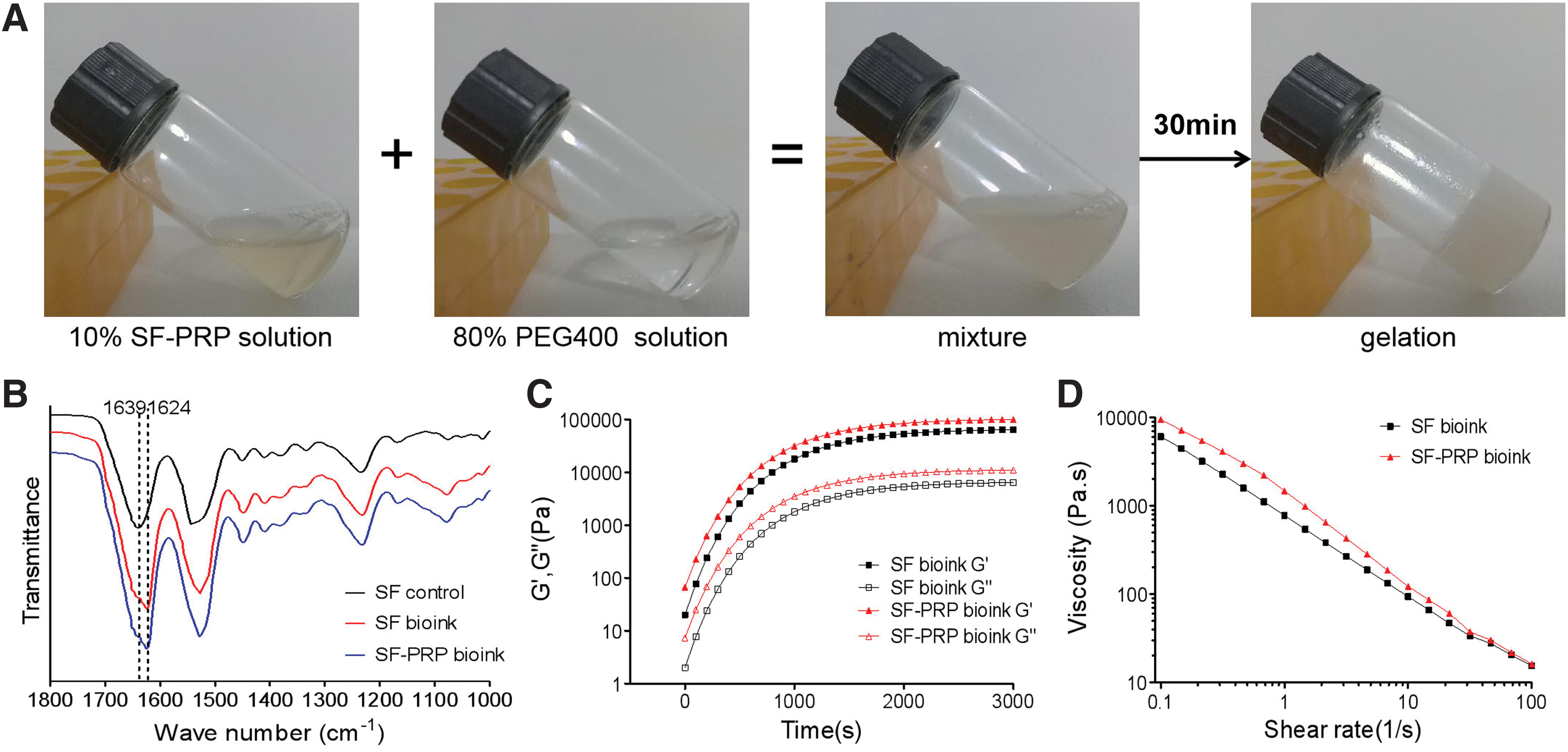
Fabrication of SF-PRP hydrogels as bioinks.
To characterize the physiochemical properties, SF solutions and SF-based bioinks were subjected to FTIR spectroscopy for crystallization measurements (Fig. 1B). The SF solution without PEG showed a major peak at 1639 cm−1 in the amide I region, indicating that the mixture was dominated by random coils. PRP in SF hydrogel did not significantly affect the major peak. After addition of PEG, SF gelation was accompanied by β-sheet structure formation, as seen by a major peak shift toward 1622–1626 cm−1 in the amide I region on the FTIR spectrum. Similar peaks were observed in the amide I region in SF and SF-PRP hydrogels.
For rheological measurements, 5% (w/v) SF-based hydrogels induced by PEG were examined with or without PRP (50%, v/v). Both G′ and G′′ were increased in response to increasing time, and the G′ values exceeded G′′ values over the whole angular frequency range (G′ > G′′), suggesting the formation of a typical gel structure. The plateau of storage modulus was around 95 kPa in the SF-PRP hydrogel (50% PRP, v/v), which was significantly higher than that (55 kPa) of pure SF hydrogel (Fig. 1C). The viscosity of both pure SF and SF-PRP hydrogels decreased in response to the linearly increasing shear rate (Fig. 1D), which confirmed that the bioinks were printable.
GF release kinetics
To evaluate the GF release kinetics in the bioinks, SF-based hydrogels with ascending PRP concentrations (12.5%, 25%, and 50%; v/v) were fabricated. ELISA was performed to test the release kinetics of GFs (TGF-β1 and PDGF-AB) from SF-PRP hydrogels. As is customary, the PRP gel (50%, v/v) activated by thrombin was used as a control. We found that in the thrombin-activated PRP gel, a burst release was observed within 2 days, after which the release of GFs (TGF-β1 and PDGF-AB) significantly slowed down. Results showed that GFs were almost linearly released from SF-PRP hydrogels for 14 days (Fig. 2), and concentrations of TGF-β1 and PDGF-AB increased with the PRP volume (p < 0.05).

Cumulative growth factors, including TGF-β1
Characterization and biocompatibility evaluation of 3D scaffolds
To construct 3D printed scaffolds with SF-PRP bioinks, a regenHU 3D printer (Bio Excellence) was used for printing (Fig. 3A1). The designed model and printed scaffold are as shown in Figure 3A2 and A3. We successfully constructed scaffolds with different diameters varying from 4 mm to 16 mm (Fig. 3B1–B4). The 3D printed scaffolds were self-standing even when attaining a height of 10 mm (Fig. 3B5). To further verify the printing resolution and shape fidelity of SF-PRP bioinks, we designed and printed a meniscus-shaped construct, which could maintain precise shape integrity (Fig. 3C).

Three-dimensional printing process with various shapes and dimensions.
Structural properties of the 3D printed scaffolds
We observed the surface morphology and pore structure of freeze-dried, 3D, printed SF-PRP (50% PRP, v/v) scaffolds through SEM and compared them with that of SF scaffolds. SF-PRP scaffolds showed a relatively looser 3D structure with larger and longer interconnected holes, possibly owing to the fibrinogen in PRP forming fibrin fibers. In enlarged images, we observed that the freeze-dried sample of SF-PRP hydrogel showed abundant platelets located in the trabecular microstructure, whereas no platelets could be observed in the SF scaffold (Fig. 4).
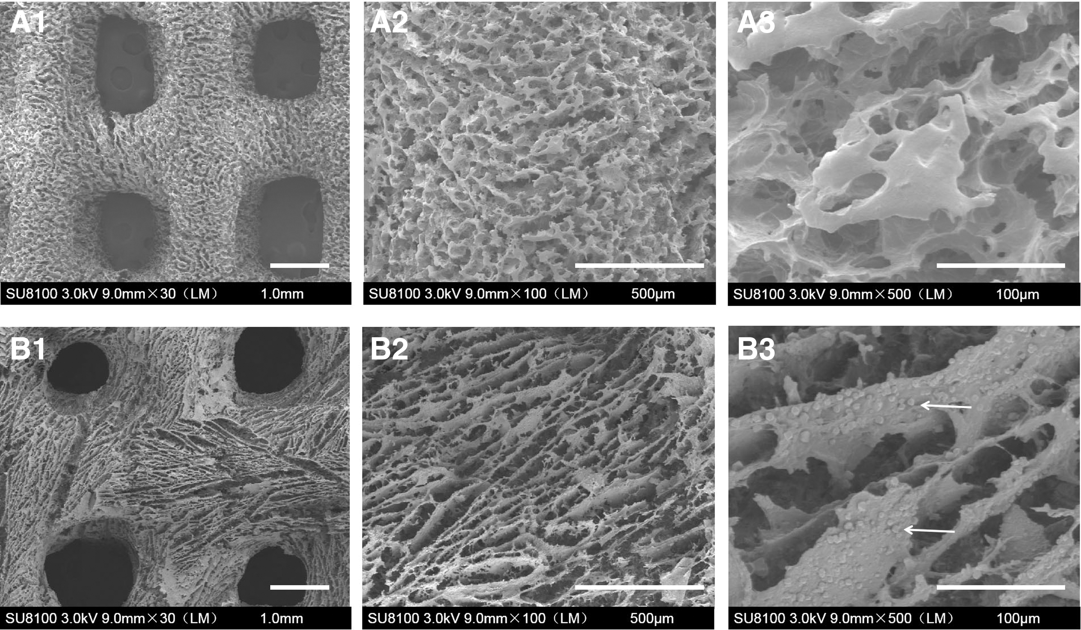
Structure properties of 3D printed pure SF
Mechanical and degradation tests of scaffolds
Mechanical tests showed that with ascending concentrations of PRP, the compressive modulus of SF-based bioinks increased. The observed compressive strength and Young's modulus were significantly higher in the 3D, printed SF-PRP (50% PRP, v/v) scaffolds (110 kPa) than that in the pure SF scaffold (70 kPa) (Fig. 5A), which was consistent with rheological findings.
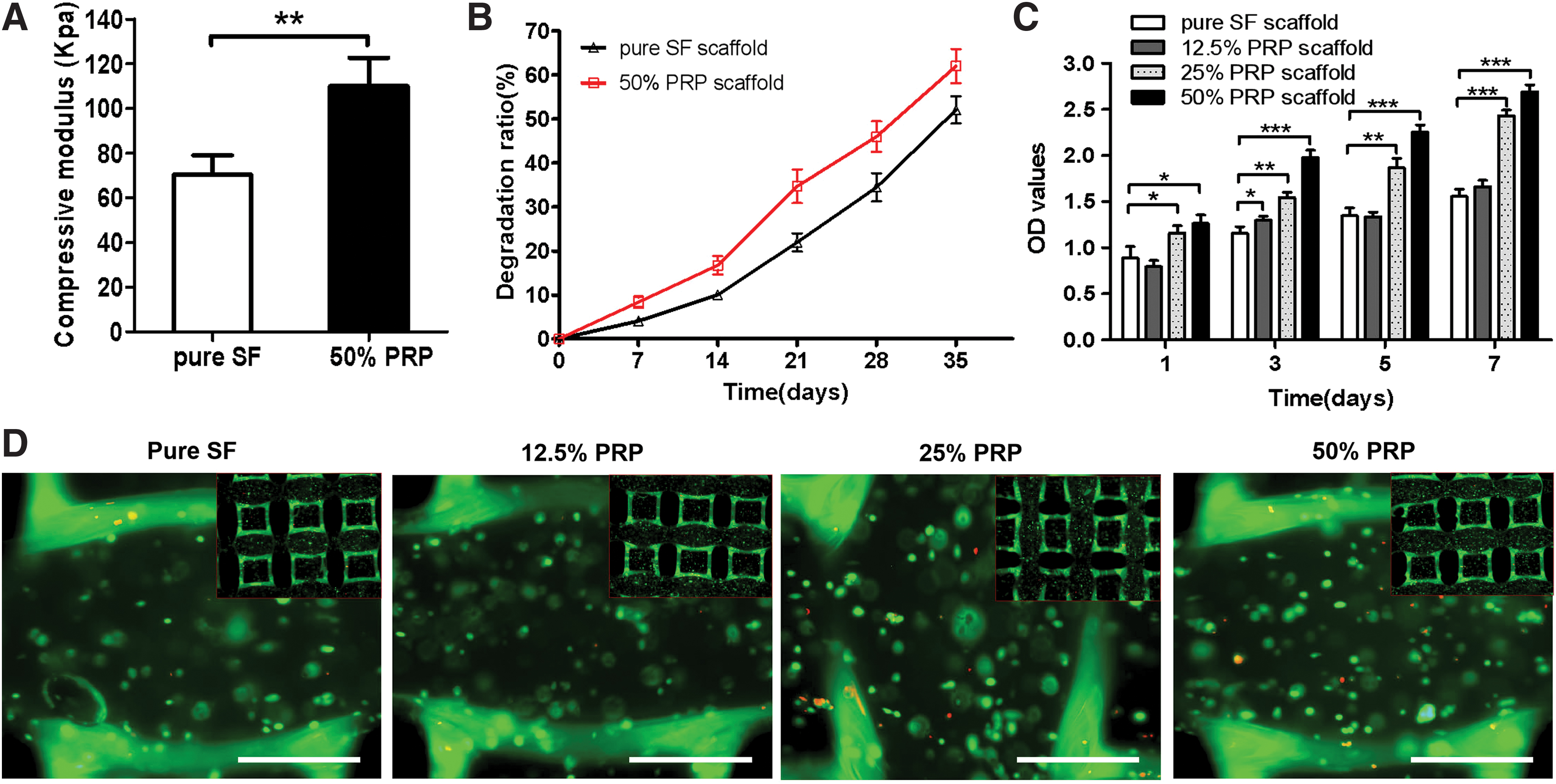
Physical characterizations and biocompatibility of 3D, printed SF scaffolds with different PRP concentrations.
The result of weight loss ratios indicated a low degradation rate of SF-based scaffolds. The in vitro degradation time of both pure SF and SF-PRP scaffolds could extend to more than 4 weeks, which could thus aid in cartilage regeneration (Fig. 5B).
Cell proliferation and viability in 3D printed constructs
The CCK8 assay was performed to assess cell proliferation and viability on days 1, 3, 5, and 7. After 7 days of incubation, constructs containing higher PRP (25% and 50%, v/v) supported more active proliferation than other constructs. In contrast, pure SF scaffolds showed relatively poor cellular proliferation and viability (Fig. 5C).
Live/dead assay staining was also used to determine the viability of chondrocytes. The cell-loaded survival rate on the 25% and 50% PRP scaffolds was over 70%. These results demonstrate that our SF-PRP bioinks exhibit low cytotoxicity and show suitability for chondrocyte proliferation (Fig. 5D).
In vitro cartilage regeneration
Gene expression
The levels of COL-II, COL-I, COL-X, and ACAN mRNA expression in different groups were quantitatively analyzed by real-time qPCR at day 28. The results showed that expression levels of COL-II and ACAN in the 50% PRP group were significantly higher than that in groups with lower PRP and the control group (p < 0.05) (Fig. 6A, D). However, no significant differences were detected in COL-I (fibrotic marker gene) and COL-X (hypertrophy marker gene) in any of the groups (Fig. 6B, C).

mRNA expression of
Histological analysis and immunohistochemical assays
To further determine whether the scaffolds were suitable for cartilage regeneration, we explored the feasibility of cartilage regeneration in vitro using 3D, printed SF-PRP scaffolds (Fig. 7). HE staining showed cells randomly distributed in the matrix and the 50% PRP group formed cartilage-like lacuna structures at 4 weeks (Fig. 7A1–A4). Safranin O and Masson's trichrome staining showed cartilage-specific ECM deposition, especially in 25% and 50% PRP groups (Fig. 7B1–B4, C1–C4). Immunohistochemistry revealed that production of collagen II was higher in the 50% PRP group than in other constructs (Fig. 7D1–D4).

Histological and immunohistochemical examination of fabricated constructs for 28 days of culture in vitro, stained with hematoxylin and eosin
Biochemical properties of regenerated cartilage
Biochemical analysis revealed that total collagen and sGAG content of in vitro-engineered cartilage-like tissue in the high concentration PRP group (50%, v/v) was significantly higher than that of low concentration PRP groups (0%, 12.5%, and 25%; v/v) (Fig. 7E, F). Addition of PRP (50%, v/v) in scaffolds resulted in significant increases in ds-DNA content (Fig. 7G). These results indicated that SF-PRP (50%, v/v) scaffolds were more suitable for in vitro cartilage regeneration.
Discussion
The present study focused on fabrication of 3D, printed SF-based scaffolds for cartilage tissue engineering. Our results confirmed that the SF-PRP bioinks could achieve sustained release of GFs, including TGF-β1 and PDGF-AB. According to previous studies, TGF-β could promote chondrocyte proliferation and ECM secretion, 36 and the combination of TGF-β and PDGF can significantly promote the synthesis of proteoglycan. 37 The biological function of SF and SF-PRP scaffolds was compared by in vitro culture for 4 weeks. The histology and biochemistry results showed that addition of PRP in SF-based scaffolds could significantly improve the quality of tissue-engineered cartilage. This might be owing to the GFs released from PRP. Reportedly, addition of TGF-β could promote GAG disposition in 3D bioprinting PCL-alginate gels. 38
In this study, we used PEG400 as a cross-linking method to fabricate SF-based bioinks. The gelation efficiency of SF is correlated with the PEG ratio. 18 To shorten the gelation time and preserve the seeded cells, we found that when equal volumes of PEG400 and SF solution were mixed, the sol–gel transition could be completed within 30 min, which was suitable for bioinks. The highest concentration of PEG400 used in drug formulations for parental injection could approach 50%. 39 In the present study, the concentration of PEG400 in SF-based hydrogels was less than 40%, which was within the limits of acceptable concentration.
We tried to optimize the concentration of PRP in 3D printed constructs. Theoretically, the concentration of GFs is in proportion to the concentration of PRP. Our results also confirmed that the SF-PRP (50%, v/v) group released the highest concentration of GFs. However, PEG should take up 50% of the volume in the present research, therefore we could add mostly 50% volumes of PRP in hydrogels. Zhang et al. reported that the SF hydrogel could achieve sustained release of VEGF. 40 We chose half concentrations of PRP gel activated by thrombin as the positive control. Our results showed that the PRP gel released most of the GFs within 2 days, whereas all the SF-PRP hydrogels achieved controlled release of GFs at 14 days. Compared with SF-PRP formation, the thrombin-activated PRP gel could only release about 80% of the total GFs.
PRP could be classified as leukocyte-rich PRP and pure PRP based on the different leukocyte concentrations. High-quality evidence supports the use of leukocyte-rich PRP for lateral epicondylitis and patellar tendinopathy.41,42 However, compared with pure PRP, leukocyte-rich PRP could result in a higher incidence of side effects in clinical osteoarthritis treatment.43,44 Leukocyte-rich PRP could deliver proinflammatory cytokines with high levels of leukocytes, including interleukin-1β and tumor necrosis factor-α, which could inhibit cartilage healing and regeneration.45,46 With this consideration, we used pure PRP with lower leukocyte concentrations in this study, which was confirmed by counting platelets and leukocytes using an automatic hematology analyzer.
Whether the existence of PRP would affect the microstructure and gelation efficiency in SF-based bioinks was a cause for concern. Our SEM results showed that the SF-PRP hydrogel has a suitable porous structure. The rheological measurements also confirmed that the addition of PRP had no significant effect on the gelling time. We thought that although PRP accounted for half of the volume, the mean plateletcrit of PRP was only 1% in this research. Interestingly, the final storage modulus of the SF-PRP hydrogel was up to 95 kPa, which was slightly higher than that of pure SF hydrogel. This might be owing to formation of a cross-linked network of platelets in PRP, which is consistent with the alginate/PRP hydrogel reported by Faramarzi et al. 26
Cell viability was confirmed by live staining using calcein AM in both SF and SF-PRP scaffolds. The toxicity of PEGs was not an issue, but the presence of initial PEG is not favorable for cell adhesion and proliferation. PEG could covalently couple to SF to modulate surface hydrophilicity and reduce cell attachment. Cytotoxicity of the SF/PEG hydrogel was assessed using rabbit chondrocytes. More than 80% of total PEG was released from SF/PEG hydrogels in 96 h. 18 Compared with the pure SF bioink, SF-PRP bioinks could promote the proliferation of rabbit chondrocytes. Once again, it indicated that PRP could improve the biological properties of SF-based hydrogels.
In summary, the present study demonstrates that the addition of PRP into SF-based bioinks could achieve sustained release of autologous growth factors. The 3D, printed SF-PRP scaffold had better biological function and could be a promising candidate for cartilage tissue engineering.
Footnotes
Acknowledgment
The authors thank International Science Editing for editing the manuscript.
Disclosure Statement
There are no competing financial interests.
Funding Information
This work was supported by the National Natural Science Foundation of China (No. 81672169, No. 81974332).