
Abstract
Mesenchymal cells derived from human umbilical cord tissue are attracting increasing attention as a source for cell therapy. However, for applying the same in tissue engineering, it has been shown that the differentiation capacity of mesenchymal stromal cells (MSCs) is influenced by the tissue from which the cells are harvested. Thus, to explore the possibility of increasing the osteogenic capacity of MSCs derived from the perivascular tissue of the human umbilical cord (human umbilical cord perivascular cells, HUCPVCs), we cultured these cells using conditioned medium (CM) derived from cultures of human bone marrow-derived mesenchymal stromal cells (hBMMSCs). However, hBM-CM contains a wide variety of growth factors, the amounts and ratios of which are considered to vary with the cell culture stage. Thus, we aimed to evaluate the effects of hBM-CM derived from different stages of hBMMSC culture on the osteogenic capacity of HUCPVCs. The stages of hBMMSC culture were defined as follows: Stage 1 (mitogenic stage) represented the period from the start of hBMMSC culture to 70% cell confluence; Stage 2 (confluent stage) represented the period from 70% confluence to the initiation of calcified nodule formation; and Stage 3 (calcification stage) represented the period following the initiation of calcified nodule formation. An analysis of growth factors contained in the CM obtained at each stage by enzyme-linked immunosorbent assay showed that insulin-like growth factor 1 (IGF-1) was significantly elevated at Stage 2, whereas vascular endothelial growth factor (VEGF) was significantly elevated at Stage 3. HUCPVCs were cultured using the CM from each of the stages for 1, 2, or 3 weeks. RUNX2 expression was the most upregulated at week 1 and then downregulated in all the groups. The expression of collagen 1 was significantly elevated in Stage 2 HUCs at week 3. Alkaline phosphatase (ALP) activity, ALP, and alizarin staining were higher in Stage 2 HUCs and Stage 3 HUCs. The calcium content was the highest in Stage 2 HUCs. The calcium content of HUCPVC obtained by the method used in this study was six times higher than that reported in the previous study. Collectively, our results show that the CM obtained at Stage 2 was most effective in driving the osteogenic differentiation of HUCPVCs.
Impact Statement
Mesenchymal stromal cells (MSCs) derived from the perivascular tissue of umbilical cords are promising candidates for regenerative medicine. Because these are able to be differentiated into bone cells, cartilage cells, and adipocytes. The number of MSCs in perivascular tissue (HUCPVCs) is ∼1/300 but the number of HUCPVCs that differentiates into osteogenic cells is quite low. In order to promote osteogenic differentiation of HUCPVCs, we cultured HUCPVCs using conditioned medium collected from human bone marrow-derived mesenchymal stromal cells. Our study suggests that the use of conditioned medium can be effective on inducing osteogenic differentiation of HUCPVCs.
Introduction
Mesenchymal cells derived from human umbilical cord tissue are attracting increasing attention as a cell therapy source. 1 Indeed, cord tissue mesenchymal stromal cells (MSCs) represent the most rapidly adopted MSC source for clinical trials in the past decade. 2 Although majority of cell therapies using MSCs rely on their paracrine signaling phenotype, the differentiation potential of cells is of predominant importance for applications in tissue engineering. However, the differentiation capacity of MSCs is influenced by the tissue from which the cells are harvested in addition to the conditions under which they are cultured. 3 At present, cell sources such as iPS cells and bone marrow-derived MSCs (BMMSCs) are most commonly used for tissue engineering. Particularly, BMMSCs have been widely applied clinically, and their effectiveness has been demonstrated.4,5 BMMSCs account for ∼0.125% of all the bone marrow cells and have been reported to differentiate into bone cells, chondrocytes, and adipocytes. 6 However, the number of MSCs in bone marrow decreases with age, 7 as does their differentiation capacity. 8
Although it is known that MSCs may be collected from various biological tissues,9–14 an advantage of collecting from perinatal tissue is that perinatal tissue is the youth of the cells contained therein. Furthermore, the umbilical cord and placenta, among these tissues, can be collected noninvasively after parturition. Numerous authors have confirmed the presence of MSCs in umbilical cord tissue, 15 and this has been reviewed. 16 Although the number of MSCs in umbilical cord blood is only 1/200,000,000, 17 this is vastly increased in the perivascular tissue to 1/300. 18 Differentiation of MSCs into bone cells, cartilage cells, and adipocytes has been shown in vitro 19 and into bone cells and cartilage in vivo.20,21 However, the number of the cells capable of differentiation is low. 21 Thus, in this study, we aimed to increase the osteogenic capacity of the perivascular tissue of the human umbilical cord (human umbilical cord perivascular cells, HUCPVCs) by culturing them in an environment modified by the addition of conditioned medium (CM) derived from bone marrow cultures (BM-CM). In fact, Osugi et al. have reported that transplantation of BM-CM in a bone defect promotes bone regeneration, 22 lending credence to the notion that BM-CM is likely to be involved in effective differentiation of MSCs. We have previously reported that osteogenic differentiation of HUCPVCs can be promoted when cultured using BM-CM. 23 However, BM-CM is known to contain >100 cytokines that are secreted by BMMSCs24–27 at different stages of culture maturation, typical examples of which are IGF-1, hepatocyte growth factor (HGF), VEGF, and transforming growth factor-β1 (TGF-β1), which may work synergistically to act as a cytokine cocktail. Indeed, the cytokine composition of the culture environment would reasonably be expected to change during the three distinct phases of osteogenic culture, which are as follows: the mitogenic, confluency, and calcification stages. Thus, we aimed to analyze the effects of BM-CM produced at each of the three culture stages on the osteogenic differentiation of HUCPVCs.
Materials and Methods
This study was approved by the Ethics Committee of the School of Dental Medicine, Tsurumi University (approval No. 1011) and was conducted in accordance with the “Guidelines on Clinical Research Using Human Stem Cells,” the notification issued by the Ministry of Health, Labour and Welfare.
Cells
HUCPVCs were harvested from umbilical cords obtained from consenting patients who had undergone full-term cesarean section after ethical approval was obtained from the University of Toronto and University Health Network, Toronto. The cords were acquired immediately upon delivery, and cell harvesting was conducted by Tissue Regeneration Therapeutics, Inc., (Toronto) as follows: Human umbilical cords (UCs) from full-term patients were obtained immediately upon delivery in a vessel containing 80% minimum essential medium Eagle, alpha modification (Gibco Burlington, Canada) and 20% antibiotics [167 U/mL penicillin G (Sigma, Oakville, Canada), 50 μg/mL gentamicin (Sigma), and 0.3 μg/mL amphotericin B (Sigma)]. UC sections 4–5 cm in length were dissected by first separating the epithelium of the UC section along its length to expose the underlying Wharton's jelly (WJ). Each vessel was pulled away from the surrounding WJ matrix and was placed into a 50 mL tube containing 1 mg/mL collagenase (Sigma) with phosphate-buffered saline (PBS) at 37°C. After 18–24 h, vessels were removed from the suspension, which was then diluted with PBS to reduce the viscosity of the suspension, and centrifuged. The HUCPVCs were transported at the temperature of liquid nitrogen to Tsurumi University, where they were cultured according to the method of Ennis et al. and displayed CD105+, CD90+, CD73+, CD45−, CD34−, and MHC-II− phenotypes by flow cytometry, as reported previously. 28 BMMSCs (Lot No.372262, human mesenchymal “stem” cells, hMSCs) were purchased from LONZA Japan (Tokyo, Japan).
HUCPVCs and BMMSCs were cultured at 37°C in the presence of 5% CO2 with MSCGM-CD Bulletkit™ medium (LONZA Japan) and MSCGM Bulletkit medium (LONZA Japan), respectively.
Collection of CM
BMMSCs were seeded in T75 flasks at 8.0 × 10⁴ cells in 10 mL culture medium. The cells at the third passages were used for experiments. BMMSCs were cultured in the osteogenic induction medium supplemented with β-glycerophosphate (10 mM/L),
Enzyme-linked immunosorbent assay analyses
Enzyme-linked immunosorbent assay (ELISA) was conducted to analyze the growth factors (VEGF, IGF-1, TGF-β1, bone morphogenetic protein-2 [BMP-2], and fibroblast growth factor [FGF]) contained in the BM-CM collected at each stage. The concentration levels of these growth factors were determined using the Human Quantikine ELISA kit (R&D Systems, Inc., Minneapolis, MN) in accordance with the manufacturer's instructions. Specifically, the individual samples were added to a 96-well microplate coated with monoclonal antibodies corresponding to the target factors and incubated for 2 h. After washing with PBS, a horseradish peroxidase-conjugated cytokine or growth factor-specific antibody was added to each well of the plate, incubated for 2 h, and washed. A substrate solution was added, and the plate was incubated for 30 min, after which the reaction was stopped. The concentration level of the cytokine or growth factor was determined by measuring the absorbance at 450 nm using an EMax® Plus Microplate Reader (Molecular Devices, CA).
Cell culture
BMMSCs or HUCPVCs were cultured in the six different groups, as described hereunder.
BM: BMMSCs were cultured in the osteogenic induction medium.
HUC: HUCPVCs were cultured in the osteogenic induction medium.
CMHUC: HUCPVCs were cultured in the CM of BMMSCs cultured simultaneously.
Stage 1 HUC: HUCPVCs were cultured in BM-CM produced in Stage 1.
Stage 2 HUC: HUCPVCs were cultured in BM-CM produced in Stage 2.
Stage 3 HUC: HUCPVCs were cultured in BM-CM produced in Stage 3.
The cells were seeded in six-well plates at 1.0 × 10⁴ cells/well in 2 mL culture medium. The culture media for the groups with CM were replaced with a 50/50 volume split of a new osteogenic induction medium and CM. PCR assays, ALP assays, ALP staining, alizarin red staining, and calcium (Ca) content determination were performed at weeks 1, 2, and 3 of cell culture.
PCR for calcification-related gene expression
In each experimental group, the cells were cultured in a six-well plate for 1, 2, or 3 weeks to verify the expression levels of type I collagen (COL I) and Runt-related transcription factor 2 (RUNX2), which are hard-tissue-related genes. Total RNA was collected using NucleoSpin RNA XS (Macherey-Nagel, GmbH & Co. KG., Duren, Germany). The collected RNA was reverse transcribed to cDNA using SuperScript VILO cDNA Synthesis Kit (Life Technologies, Inc., Carlsbad, CA). The PCR assay was performed using the FastStart Essential DNA Green Master (Roche, Ltd., Basel, Switzerland). The sequences of individual primers are given in Table 1.
Primer and Probe Sequences
Used in the real-time polymerase chain reaction.
Alkaline phosphatase activity
The differentiation status of each group was assessed by measuring ALP activity. The cells were washed with PBS 3 times after 1, 2, and 3 weeks of culturing. Sterile distilled water containing 10 mM 2-amino-2-(hydroxyl)methyl-1, 3-propanediol hydrochloride (pH 10.0), and 5 mM MgC12 was added to the plate, and the cells were collected using a cell scraper. The collected cell suspension was mixed in a vortex for 1 min and centrifuged at 3000 rpm for 10 min. The obtained medium was transferred to a 96-well plate, and 10 mM p-nitrophenylphosphate (pNPP) was added as the substrate to activate the reaction. The absorbance of the produced p-nitrophenol was determined at 405 nm using a microplate reader (Ultramark; BIO-RAD, Tokyo, Japan). The measured value was divided by the protein content in each group, and the resultant value was assumed to be the ALP assay value for the group.
Alkaline phosphatase staining
The differentiation status of each group was also assessed by ALP staining. The cells were washed with PBS 3 times after 1, 2, and 3 weeks of culturing and immobilized with 4% paraformaldehyde. NAS-MXP (naphthol AS-MX phosphate; Sigma, St. Louis, MO) was used as the enzyme substrate. A reaction was induced with 0.1 M Tris-HC1 (pH 8.5) containing 2 mM MgC12, the produced naphthol hydrochloride was subjected to reaction with Fast Blue B salt (Sigma), and the cells were stained with azo dye and precipitated. The stained cell images were observed under a phase-contrast microscope (Keyence BZ-8100).
Alizarin red staining
The calcification status of each group was assessed by alizarin red staining. After cell culture, individual plates were washed with PBS, and the cells were immobilized in 100% ethanol and washed with distilled water three times. Next, 1.0% alizarin red S dye was added, the solution was left at room temperature for 10 min, and the cells were washed three times again before observation.
Ca content determination
After each of the culture periods, the Ca content in the six-well plate was determined. After removing the CM in the wells, the wells were washed with PBS. The adherent cells were lysed in 0.5 M hydrochloric acid. A suspension was used for determining the Ca content. The Ca content was determined using the Ca E-test (Wako, Osaka, Japan) and calculated by measuring the absorbance at 570 nm using a microplate reader.
Statistical analysis
Based on the results of our previous study, 23 we calculated the number of samples in this study using the formula N = 16(SD/Δ), 2 where SD is the average standard deviation between the two groups and Δ is the difference between the average values of the two groups. It was calculated at a significance level α of 0.05 and power of 80% (β = 0.2). As the result, all experiments were performed with n = 3. All the experiments were performed at least three times. The obtained data were analyzed by the closed test to control multiple test errors. SPSS Statistics version 19 (IBM Japan, Tokyo, Japan) was used for statistical analysis. The median and interquartile range values of the measured parameters were calculated for each of the six groups. Multiple comparisons of the five groups excluding the BM group were performed using Kruskal–Wallis nonparametric test for evaluation. Values of p < 0.05 were considered statistically significant.
Results
ELISA analyses
The concentration levels of VEGF, IGF-1, TGF-β, BMP-2, and FGF in the BM-CM obtained at each stage were determined by ELISA. The VEGF level was significantly higher at Stage 3 than that at Stage 1. The IGF-1 level significantly increased at Stage 2 compared with that at Stage 1. No significant difference in the TGF-β and FGF levels was observed among the stages. The BMP-2 level significantly increased at Stage 1 compared with that at Stage 3 (Fig. 1).
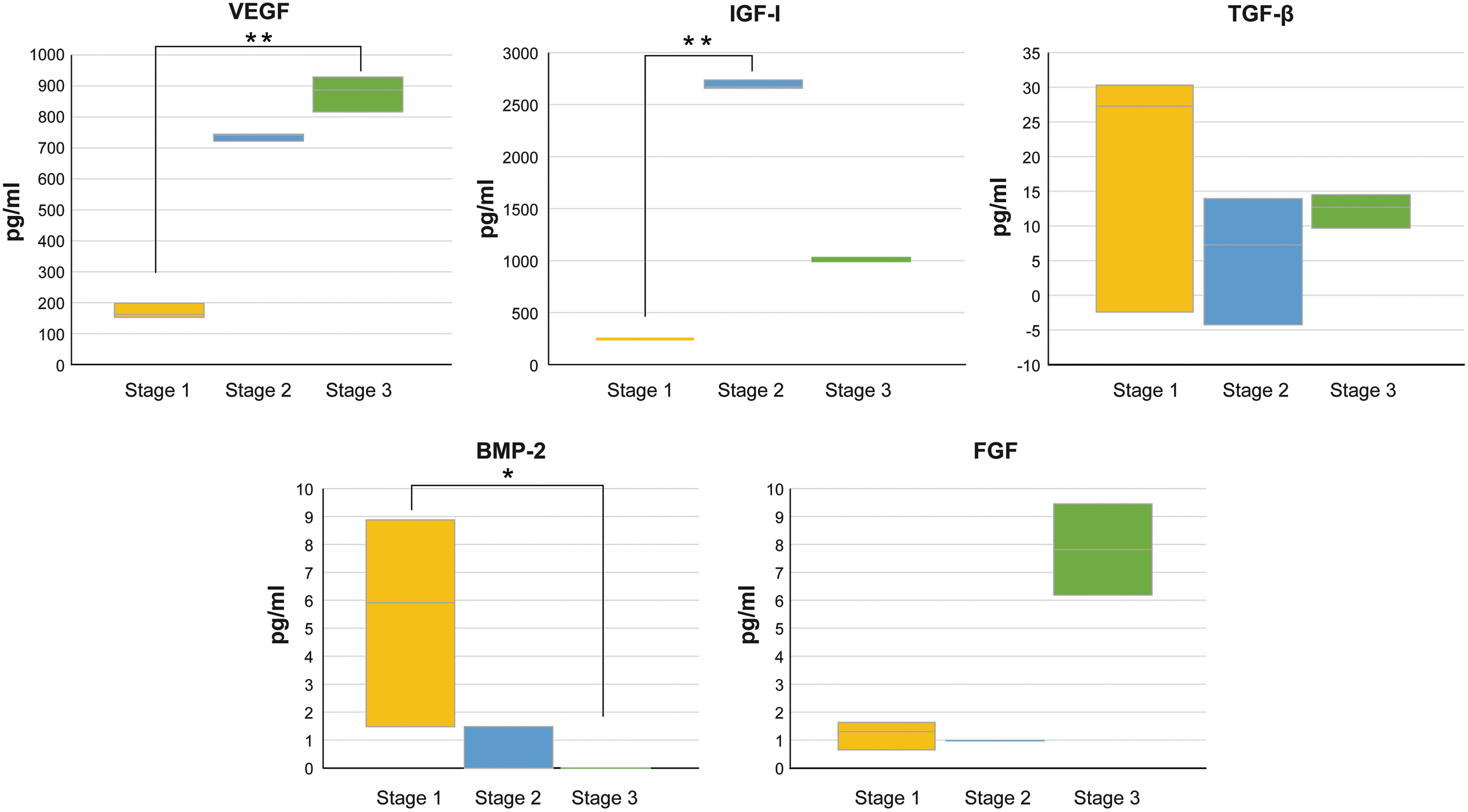
ELISA analysis of conditioned medium from BMMSCs at each stage. The concentration level of VEGF significantly increased at Stage 3 compared with that at Stage 1 (p < 0.01). The concentration level of IGF-1 significantly increased at Stage 2 compared with that at Stage 1 (p < 0.01). The BMP-2 level significantly increased at Stage 1 compared with that at Stage 3 (p < 0.05). *p < 0.05, **p < 0.01. BMMSC, bone marrow-derived mesenchymal stromal cell; VEGF, vascular endothelial growth factor; BMP-2, bone morphogenetic protein-2.
Calcification-related gene expressions
The gene expression level of RUNX2 was highest at week 1 in all the groups and then decreased subsequently. A comparison of the RUNX2 levels among stages showed that the level was significantly higher at week 2 in the Stage 2 HUC group than those in the Stage 1 HUC group (Fig. 2a).
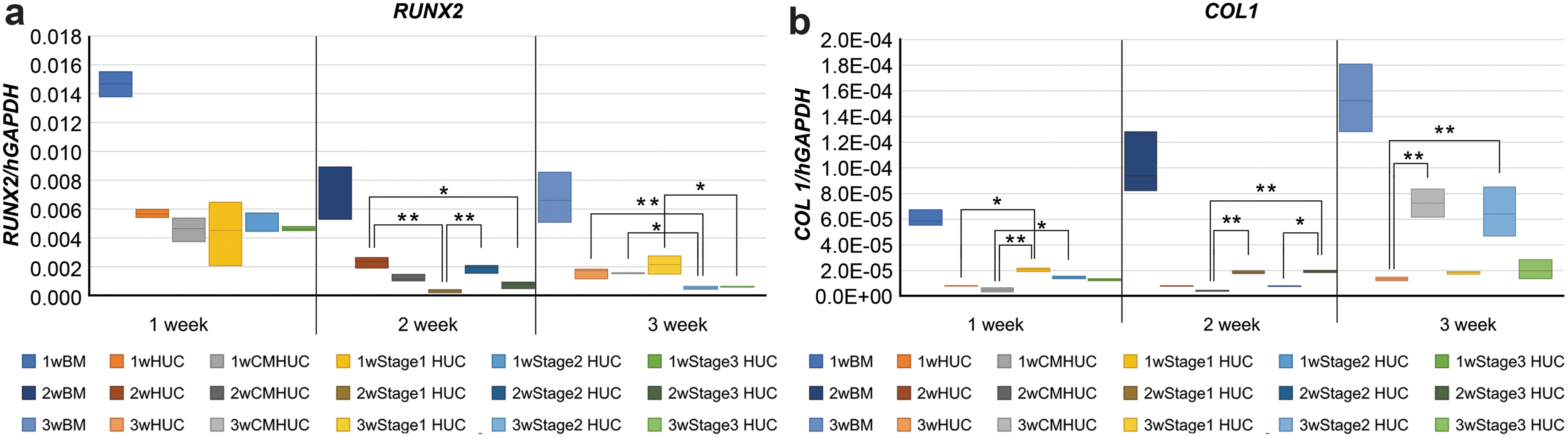
PCR analysis of calcification-related gene expression.
The gene expression level of COL 1 was upregulated in the BM group over time. In HUCPVCs, the COL 1 expression level significantly increased at week 3 in the CMHUC and Stage 2 HUC groups compared with that at HUC (Fig. 2b).
ALP assay and ALP staining
The ALP activity was high in the BM group (Fig. 3a). A comparison of ALP activity levels only among the HUCPVC experimental groups showed that its level was significantly higher at weeks 1 and 2 in the Stage 2 HUC and Stage 3 HUC groups than that in Stage 1 (p < 0.05) (Fig. 3b). The ALP activity of HUCPVCs cultured in blank CM were as follows: week 1: 17.01 ± 8.95; week 2: 16.73 ± 1.03; and week 3: 14.13 ± 1.72 μg mol/g. ALP staining revealed no ALP-positive cells at week 1 in any experimental group. No ALP-positive cells were observed at week 2 in the HUC and Stage 1 HUC groups. Some ALP-positive cells were observed at week 2 in the CMHUC group, and more ALP-positive cells were observed at week 2 in the BM, Stage 2 HUC, and Stage 3 HUC groups. By week 3, ALP-positive cells were visible in the HUC and Stage 1 HUC groups, whereas they were considerably more in the BM, CMHUC, Stage 2 HUC, and Stage 3 HUC groups (Fig. 4).
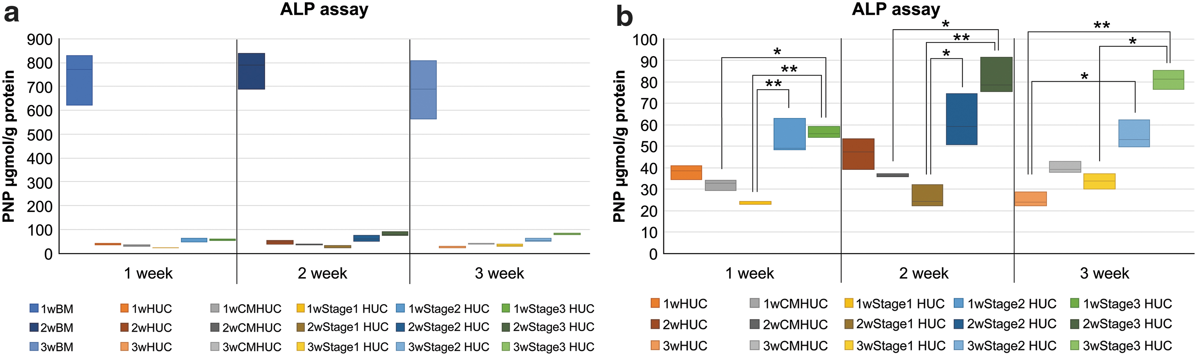
ALP activity in each group.
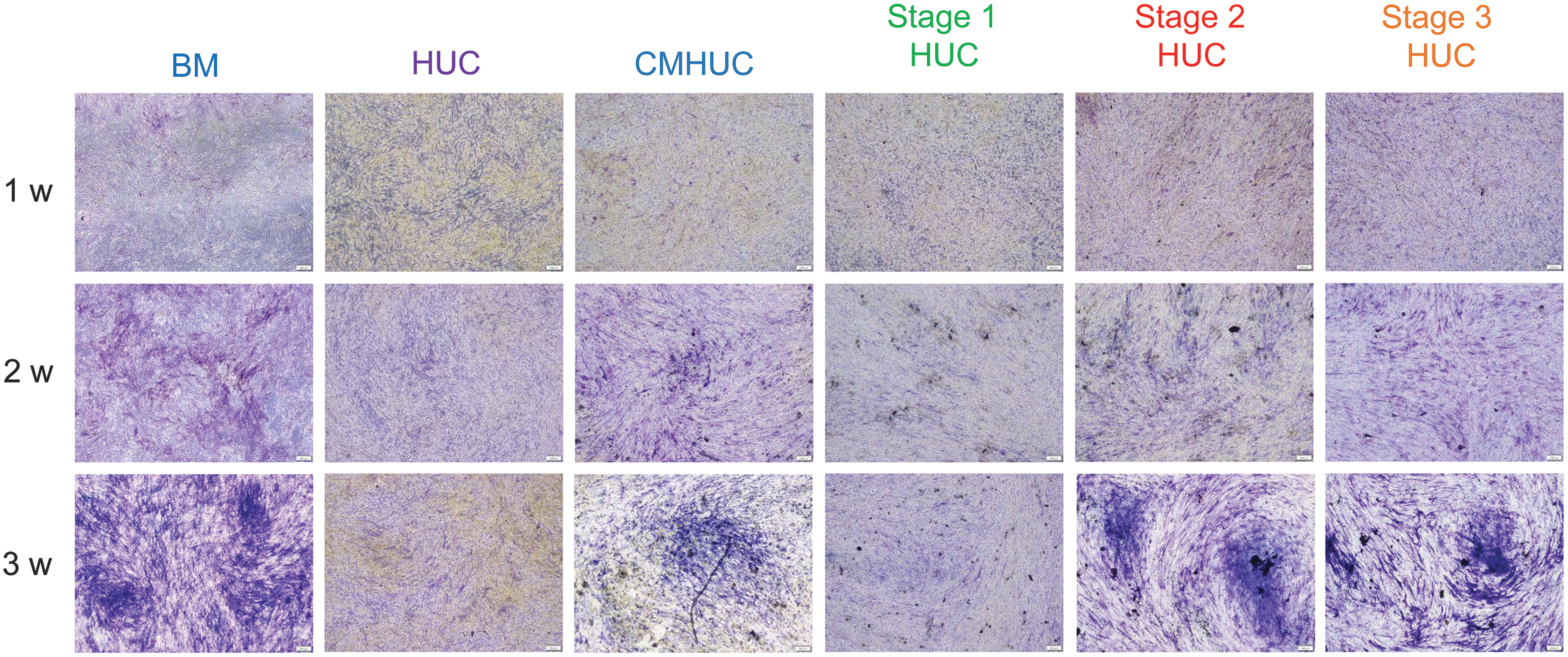
ALP staining in each group. No ALP-positive cells were observed at week 2 in the HUC and Stage 1 HUC groups. Only a few ALP-positive cells were seen in the CMHUC group. ALP-positive cells were observed in the BM, Stage 2 HUC, and Stage 3 HUC groups. At week 3, some ALP-positive cells were observed in the HUC and Stage 1 HUC groups with more in the BM, CMHUC, Stage 2 HUC and 3 HUC groups.
Alizarin red staining
Alizarin red staining revealed no evidence of calcification at week 1 in any group. At week 2, some evidence of positive staining was present in the CMHUC, BM, Stage 2 HUC, and Stage 3 HUC groups but none in the HUC and Stage 1 HUC groups. By week 3, more definitive evidence of positive staining was evident in the BM, CMHUC, Stage 2 HUC, and Stage 3 HUC groups but none in the HUC and Stage 1 HUC groups (Fig. 5).
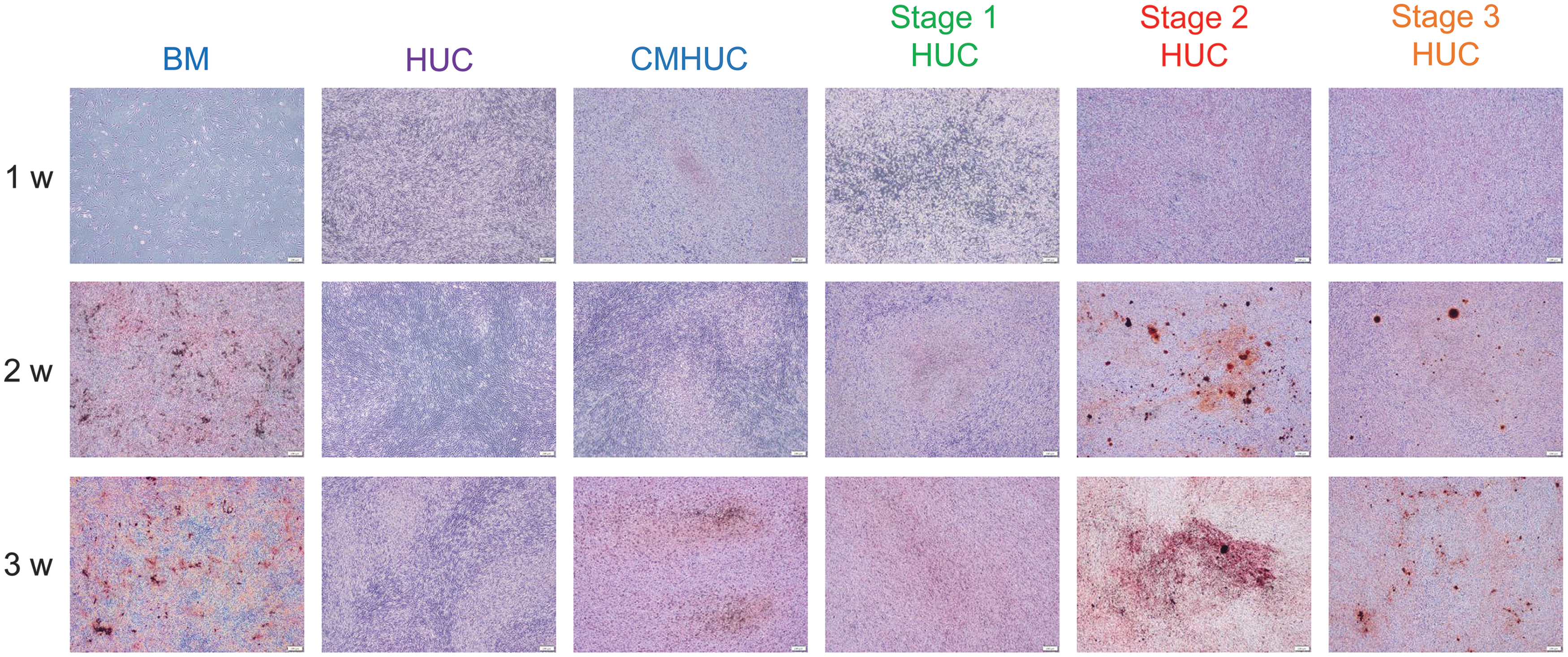
Alizarin red staining in each group. At week 2, staining was observed in a few cells of the CMHUC group. Staining was clearly noted in the BM, Stage 2 HUC, and Stage 3 HUC groups. At week 3, staining was observed in the BM, CMHUC, Stage 2 HUC, and Stage 3 HUC groups.
Ca content
The Ca content of each well was determined at weeks 1, 2, and 3. The Ca concentration increased in the BM group over time. A comparison of the concentration levels of Ca among HUCPVC groups showed that its level was significantly higher in Stage 3 HUC of week 2 and Stage 2 HUC of week 3 compared with those in the HUC, CMHUC, and Stage 1 HUC groups. In the Stage 2 HUC group, in particular, a time-course increase in calcium content was observed. The Stage 2 HUC group was six times higher than the CMHUC group (Fig. 6). The Ca contents of HUCPVCs cultured in blank CM were as follows: week 1: 0.26 ± 0.33; week 2: 0.39 ± 0.33; and week 3: 1.05 ± 0.18 μg/dL.
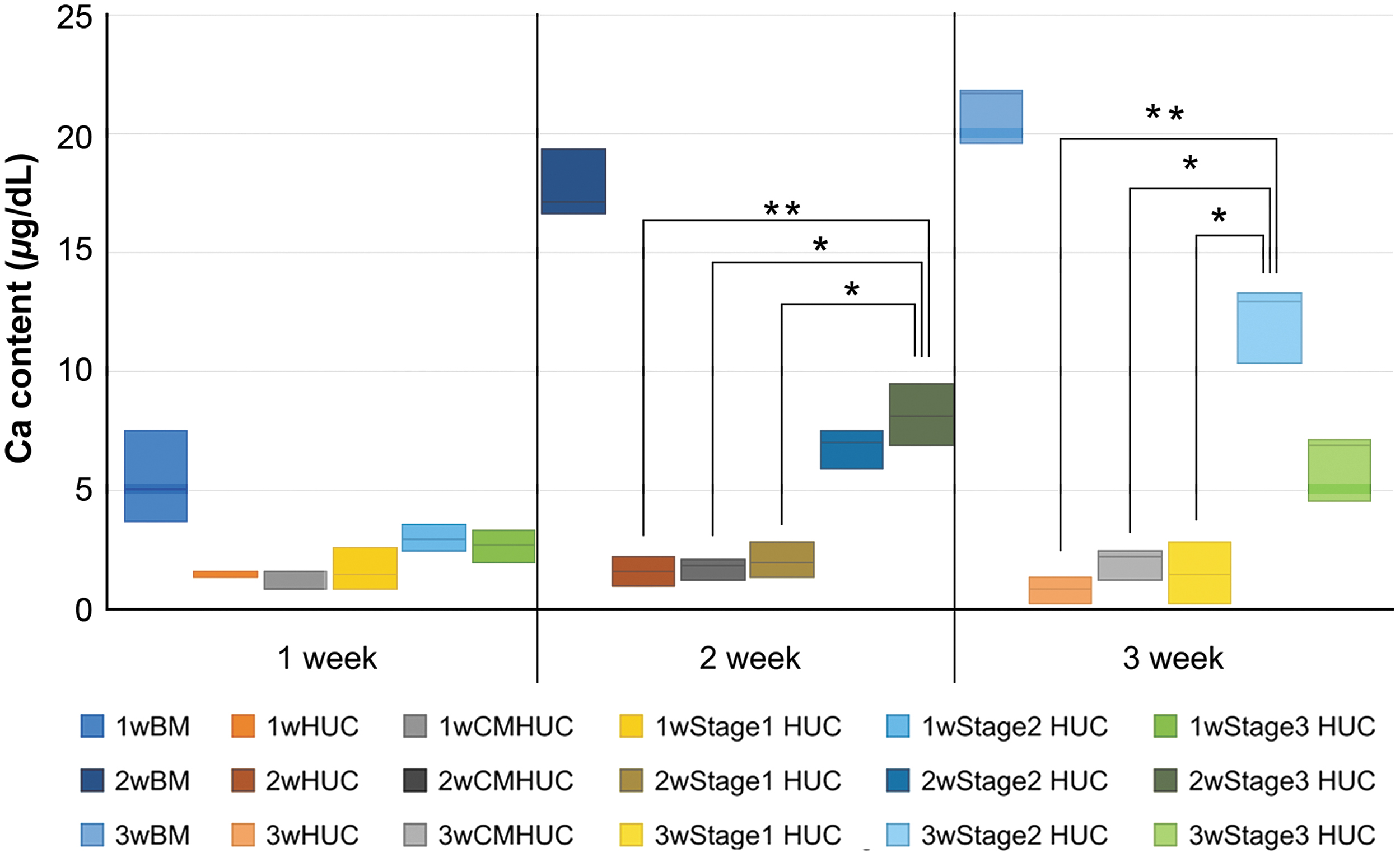
Calcium measurement in each group. Higher calcium levels were observed in the BM group. In Stage 3 HUC of week 2 and Stage 2 HUC of week 3, the calcium content was significantly higher than the HUC, CMHUC, and Stage 1 HUC groups. In the Stage 2 HUC group, in particular, a time-course increase in calcium content was observed. The Stage 2 HUC group was six times higher than the CMHUC group. *p < 0.05, **p < 0.01.
Discussion
Somatic stem cells having multipotency play an important role in regenerating damaged tissues. Much translational research has been conducted on these cells, which are contained in all tissues, including the umbilical cord, with the aim of targeting clinical applications.19,29,30 An advantage of HUCPVCs is that they are harvested from tissue commonly considered medical waste. Similar to other MSCs, they are immune evasive because they express no MHC class II antigens. HUCPVCs have anti-inflammatory effects and may be a stronger candidate than BMMSC to treat inflammatory diseases. 31 The differentiation of mesenchymal stem cells to targeted stromal cells cannot be achieved by adding single growth factors. Multiple growth factors in the CM of iPS cells promote the differentiation of mesenchymal stem cells in damaged tissues to the target stromal cells that, in turn, promote tissue regeneration. 27 We have verified in our study that the use of HUCPVCs cultured in the CM obtained from BMMSCs induces osteogenic differentiation and promotes bone formation. 23
The expression levels of VEGF, IGF-1, TGF-β, BMP-2, and FGF in the samples harvested from Stages 1, 2, and 3, classified depending on the collection time of the CM, were determined by ELISA. Their expression levels differed with the harvest stage. The BMP-2 level was high at Stage 1, and the VEGF and IGF-1 levels were high at Stage 2. The VEGF and FGF levels were high at Stage 3. High levels of VEGF are secreted by MSCs 32 ; accordingly, BMMSCs from which the CM were harvested had the characteristics of MSCs. IGF-1 is necessary for the activation and mitogenic activity of MSCs and are likely involved in the differentiation of MSCs into osteoblasts and subsequent bone formation.33,34 IGF-I induces the expression of VEGF in osteoblasts through the PI3K pathway.35,36 Cells activate signaling pathways and transcription factors by autocrine factors, including various growth factors and cytokines or paracrine factors, to secrete protein. The ratios of the secreted factors differed with the culture stage from which the medium was harvested.
RUNX2, an early gene associated with the differentiation of MSC into osteoblasts, exhibited high expression levels at week 1 in all groups. Thus, the differentiation into precursor osteoblasts was evident in the BM and HUC groups. Osteoblasts secrete type 1 collagen and deposit Ca phosphate to synthesize a calcified matrix. High expression of type 1 collagen at week 3, particularly in the CMHUC and Stage 2 HUC groups, suggests that the production of extracellular matrix was progressing toward calcification.
ALP activity increased considerably in the BM group compared with those in other groups, whereas ALP staining demonstrated that the ALP-positive cell count was higher in the Stages 2 and 3 HUC groups than that in the BM group. This result may partly be because the ALP values were normalized for total protein and also because the concentration of osteogenic supplements would have been higher in the BM cultures, which received no CM. Thus, the differentiation into osteoblasts was earlier in the BM group, whereas higher mitogenic activity would have preceded osteoblast differentiation in the HUCPVC groups.
Alizarin red staining showed the same results as those obtained from ALP staining. Dye affinity at Stage 2 HUC and Stage 3 HUC groups at week 2 was almost equivalent to that in the CMHUC group at week 3. This suggests that Stages 2 and 3 CM allow for the induction of differentiation into osteoblasts a week earlier than that by conventional use of CMHUC.
The Ca content was high with Stage 2 and Stage 3 CM cultures, especially at week 3 of Stage 2. This suggests that Stage 2 CM is effective in causing the HUCPVCs to differentiate into osteogenic cells.
Throughout the experimental period, the HUC groups showed lower levels of ALP and cell expressions than the positive control BM. In our previous report, the HUC groups showed lower levels of calcification-related gene expressions and ALP in vitro compared with the BM group. However, bone volume, histological analyses, and immunohistochemistry in vivo significantly increased in the HUC groups compared with the BM group. The MSC population can be sufficiently influenced by the local environment to cause differentiation into the functional phenotype required in the targeted connective tissue compartment. In the previous study, it was the bony compartment. We considered that the local host environment was the predominant regulator of the increased bone formation in the HUC groups. That is, HUCPVCs are more likely to differentiate into cells in the surrounding environment of transplanted compared with BMMSCs. There was a significant difference in the osteogenic differentiation of HUCPVCs by the time of BM-CM collection. The calcium content of HUCPVC obtained by the method used in this study was six times higher than that reported in the previous study. 23 In the future, the transplantation of HUCPVCs that are differentiated into osteogenic cells by confluent-stage BM-CM can effectively lead to bone formation in vivo. HUCPVCs have definite advantages over BMMSCs in terms of the amount of stem cells collected and invasiveness during collection.
CM contains multiple factors including unknown growth factors.24–26 Extracellular vesicles could mediate the paracrine effects of MSCs. They considered that exogenous protein (IGF-1) can be carried into extracellular vesicles derived from HUCPVCs and can be an alternative to bone marrow cell therapy in liver fibrosis. 37 Here, partial evaluation of these factors revealed that based on ALP and alizarin red staining and Ca measurement, the CM harvested from Stage 2 is considered the most effective in causing HUCPVC osteogenic differentiation than that harvested from two other culture stages. An increase in the level of type 1 collagen in the Stage 2 HUC group at week 3 supports this osteogenic differentiation partly owing to the cytokine cocktail containing IGF-1 and VEGF.
In conclusion, our results show that the CM from Stage 2 BM cultures was effective in inducing osteogenic differentiation of HUCPVCs. The use of such a CM may reflect the curative benefit of the in vivo environment, which cannot be reflected by the use of a single growth factor, and provides a putative approach to the induction of differentiation of somatic stem cells.
Footnotes
Disclosure Statement
No competing financial interests exist.
Funding Information
This work was supported by JSPS KAKENHI Grant-in-Aid for Young Scientists (B) 16K20547. The authors thank Crimson Interactive Pvt. Ltd. (Ulatus) for their assistance in article translation and editing.