
Abstract
Corneal endothelial dysfunction is a major cause of corneal blindness and is mainly treated by corneal transplantation. However, the global shortage of donor cornea hampers its application. Intracameral injection of cultured primary corneal endothelial cells (CECs) was recently confirmed in clinical trials. However, abnormal adhesion of the grafted CECs affects the application of this strategy. In this study, we explored if laminin 511 (LN511) improves the therapeutic function of the intracameral CEC injection for corneal endothelial dysfunction. To mimic the late stage of corneal endothelial diseases, intense scraping was developed to remove CECs and extracellular matrix of the posterior Descemet's membrane (DM) without DM removal in rabbits. Then, Dulbecco's phosphate-buffered saline (DPBS) and LN511 were intracamerally injected as the control and intervention groups, respectively. We found that the injected LN511 could settle and form a coating on the posterior surface of DM. After CEC transplantation, corneal clarity of rabbits in the LN511 group was rapidly recovered within 7 days, whereas the corneal recovery took 14 days in the DPBS group. Corneal thickness of LN511 group decreased to 413.3 ± 20.8 μm 7 days after operation, which was significantly lower than 1086.3 ± 78.6 μm of DPBS group (p < 0.01). Moreover, for the grafted CECs, LN511 promoted the rapid adhesion, tight junction formation, and expression of Na+/K+-ATPase and ZO-1. In vitro analysis revealed that the functions of LN511 on the cultured human CECs mechanistically depended on the cell density and the nuclear-cytoplasmic translocation of the Yes-associated protein. Our study demonstrated that LN511 precoating promoted the adhesion of the transplanted CECs and enhanced the functional regeneration of the corneal endothelium. Thus, our data suggested that the strategy of LN511 precoating and CECs' intracameral injection could be a potential method for the therapy of corneal endothelial dysfunction.
Impact statement
Intracameral injection of cultured corneal endothelial cells (CECs) is a potential alternative therapy for corneal endothelial dysfunction and has been proven to be effective in clinical trials. However, abnormal adhesion of the grafted CECs affects its application. In this study, intense scraping was developed to remove CECs and extracellular matrix of the posterior Descemet's membrane (DM) without DM removal for the therapy of late stage of corneal endothelial diseases. Laminin 511 was intracamerally injected to form a coating, improve the posterior DM, enhance the adhesion of the grafted CECs, and promote the functional regeneration of CEC transplantation through Yes-associated protein signaling.
Introduction
Corneal endothelium, an innermost monolayer of the cornea, maintains corneal transparency through its pump and barrier functions. 1 As human corneal endothelial cells (HCECs) have limited proliferative capacity, several pathologic conditions can cause the reduction of corneal endothelial cells (CECs)’ density, leading to corneal stromal edema known as bullous keratopathy.2–4 Nowadays, both Descemet stripping endothelial keratoplasty (DSEAK) and Descemet's membrane endothelial keratoplasty (DMEK) are commonly utilized for treating corneal endothelial disorders such as Fuchs' endothelial dystrophy and bullous keratopathy.5–9 However, there is a worldwide shortage of transplant-grade donor corneas.
Technologies of tissue engineering cornea are being investigated for treating corneal endothelial dysfunction, such as the expansion of in vitro CECs, scaffold material, and delivery systems.10–12 Two main delivery systems have been reported as follows: tissue-engineered endothelial sheet grafts and intracameral injection of CECs' suspension.13–15 Similar to the transitional DMEK and DSEAK, it is technically challenging to transplant a structurally flexible tissue-engineered endothelial sheet into the anterior chamber, and the artificial carriers for endothelial sheet increase the risk of immunological rejection.16–19
Recently, the intracameral injection of CECs' suspension was developed and confirmed in bullous keratopathy patients, although there were risks of abnormal adhesion in adjacent tissues of the anterior chamber. 20 Several strategies were developed to resolve this problem, such as magnetic cell guidance, combined Y27632-CEC transplantation, and our previous mini sheet injection.13,15,21 Besides, intracameral injection of CECs' suspension prefers the early stage of corneal endothelial diseases and has limited therapeutic efficacy for the late stage of diseases with abnormal Descemet's membrane (DM), like guttae. 14
Laminin (LN) is an extracellular matrix (ECM) glycoprotein composed of three trimeric (α, β, and γ) chains, and it is the major noncollagenous component of DM.22–24 LNs are reported to modulate several cellular functions, such as adhesion, migration, survival, proliferation, and differentiation.25,26 The spatial and temporal patterns of expression of different LN chains, especially during development, suggest that different isoforms play distinct roles.27,28 Laminin 511 (LN511) is one of the earliest ECM molecules found during embryogenesis, and its deletion causes the absence of eye. 29 In vitro, LN511 has many functional roles, including the self-renewal of human embryonic stem cells (hESCs), expansion of neural cells, and retinal differentiation.25,30–32 It is expressed in the corneal endothelial basement membrane and modulates CECs' adhesion and proliferation.33,34
In this study, we developed intense scraping to remove CECs and ECM of the posterior DM without DM removal to mimic the late stage of corneal endothelial diseases. LN511 was intracamerally injected and forms a coating on the posterior DM. LN511 precoating promoted the adhesion, facilitated the recovery of corneal clarity, thickness, and endothelial density of the transplanted CECs. Moreover, we found that LN511 promoted the translocation of Yes-associated protein (YAP) from the nucleus to the cytoplasm.
Methods and Materials
Animals
New Zealand white male rabbits were purchased from Ji'nan Xiling Kok Breeding Center (Shandong, China) and maintained in the animal facility of the Shandong Eye Institute. Animal care and procedures were conducted in accordance with the Principles of Laboratory Animal Care. All animal experiments were carried out according to the principles of the Association for Research in Vision and Ophthalmology Statement for the use of animals in ophthalmic and vision research.
Cell culture
Twenty rabbits were euthanatized by intravenous injection of pelltobarbitalum natricum (50 mg/kg; Shanghai, China), and the rabbit corneal endothelial cells (RCECs) were subsequently collected. Primary RCECs were isolated and cultured according to previously reported methods. 35 Briefly, DM was torn off from the cornea and RCECs still attached on the inner side of DM. DM tissues were first incubated in the culture media supplemented with 10 μM ROCK inhibitor Y-27632 (StemCell Technologies, Vancouver, Canada) overnight and then were incubated in 0.6 U/mL of Collagenase I (Sigma-Aldrich, St. Louis, MO) for 1 h to release the RCECs from DM. The obtained RCECs were resuspended in culture medium and plated in 12-well plate (RCECs of two corneas/one well) after 1 h incubation at 37°C.
RCECs were cultured in medium composed of Dulbecco's modified Eagle's medium (DMEM; Corning, Manassas, VA), 10% fetal bovine serum (FBS; Gibco, Grand Island, NY), 2 ng/mL human basic fibroblast growth factor (bFGF; R&D Systems, Minneapolis, MN), 1 × insulin-transferrin-selenium (ITS 100X; Gibco), 10 μM small-molecule inhibitor SB505124 (Sigma-Aldrich), 1 × penicillin streptomycin solution (PS 100 X; Corning), and 10 μM Y-27632. Cells were incubated at 37°C in 5% CO2, and the medium was changed every 2–3 days. RCECs were trypsinized by 0.25% trypsin-EDTA (Sigma-Aldrich) for 5 min at 37°C, subcultured with 6-cm cell dishes, and passaged at ratios of 1:2. Cultivated RCECs at passages 2 through 3 were used for the transplantation.
The HCEC line B4G12 was purchased from Creative Bioarray and seeded in six well plates coated with or without 20 μg/mL LN511 (BioLamina, Sundbyberg, Sweden) and was cultured in medium composed of human endothelial serum-free medium (Creative Bioarray), 2% FBS (Gibco), and 10 ng/mL bFGF. Cultures were incubated at 37°C in 5% CO2, and the medium was changed every 2 days.
HCECs were cultured in a 12-well plate by inoculating 4 × 10⁵ cells per well precoated with LN511 or Dulbecco's phosphate-buffered saline (DPBS; Solarbio). The nonadherent cells were rinsed away with phosphate-buffered saline (PBS) after 30 min, and new medium was added. HCECs coated with LN511 were further treated by 20 μM blebbistatin (Ble; Sigma-Aldrich) or 0.5 μM verteporfin (VF; Selleck Chemicals). Cells were harvested for further detection after consecutively cultured for 48 h.
Cell preparation for transplantation
Confluent RCECs were prepared into mini sheet for transplantation according to our previous study. 21 The microscopy morphology and the ZO-1 and N-cadherin staining of cultured RCECs in vitro before transplantation are shown in Supplementary Figure S1. Cells were dissociated with Accutase (StemCell Technologies) for 3 min at 37°C and gently triturated into mini sheet suspension. The mini sheets were subsequently suspended in 250 μL DMEM supplied with 100 μM Y-27632. To trace the transplanted cells, 2 μM CellTracker CM-Dil (Invitrogen, Life Technologies) was used to label cells for 5 min at 37°C and then for an additional 15 min at 4°C. After labeling, RCECs were washed by PBS and suspended for transplantation.
Surgical process and postoperative detection
Thirty six rabbits received RCEC transplant by anterior chamber injection, and six of them received the labeled cells. The rabbits were anesthetized with intraperitoneal injection of pelltobarbitalum natricum (25 mg/kg; Shanghai, China). The surgery was performed only on the right eye, and the left was untreated. The corneal endothelium of the rabbits was intensely removed with a 20-gauge soft silicone needle (Inami, Tokyo, Japan), 9 mm in diameter, which was different from our previous description 21 (Supplementary Fig. S2). The tip of the needle is made of soft silicone and was used to scrape CECs gently, while the metal part of the back end of the silicone was used to intensely remove CECs and ECM of the posterior DM. LN511 (10 μg/mL; the LN511 group) or the vehicle of DPBS (the DPBS group) was injected into the anterior chamber, and then the eye was kept down for 1 h to form a coating on the DM surface. RCECs at a density of 3.0 × 10⁵ cells were suspended in 250 μL DMEM supplemented with 100 μM Y-27632 and then injected into the anterior chamber of the right eyes according to the previous descriptions.21,36 The rabbits were maintained with the eye-down position for 3 h to promote cell attachment.
The corneal clarity was observed and photographed using slit-lamp microscopy (SL-D7; Topcon, Tokyo, Japan). Corneal thickness was measured by an Ultrasound Handy Pachymeter (Tomey, Nagoya, Japan). The intraocular pressure was examined with a tonometer (Tono-Pen AVIA). Endothelium morphology and density were detected with in vivo confocal microscopy (Heidelberg Engineering, Heidelberg, Germany), a noninvasive detector. The attached cell number and cell density were acquired by counting the cell number per unit area.
Immunofluorescence staining
The rabbit eyes were retrieved after euthanasia, and the transplanted corneas were resected and then fixed with 4% paraformaldehyde for 12 min, permeabilized with 0.5% Triton X-100 for 1–5 min, and blocked with 2.5% bovine serum albumin (BSA) for 1 h at room temperature. The samples were incubated with primary antibodies (Table 1) at 4°C overnight. After treatment with secondary antibodies (Invitrogen) or phalloidin staining for F-actin (1:150; Phalloidin; Invitrogen) for 1 h at room temperature, the nuclei were stained by 4,6-diamidino-2-phenylindole (DAPI; Beyotime, Shanghai, China). Digital images were observed and captured with a laser scanning confocal microscope (LSM 800; Zeiss, Jena, Germany).
Antibodies for Immunofluorescence Staining and Western Blot
GAPDH, glyceraldehyde-3-phosphate dehydrogenase; IF, immunofluorescence; p-MLC, phosphorylated myosin light chain; WB, Western blot; YAP, Yes-associated protein.
Real-time polymerase chain reaction
Total RNA was extracted from HCECs with the MiniBEST Universal RNA Extraction Kit (Takara, Dalian, China). Complementary DNA (cDNA) was synthesized by a Mir-X microRNA (miRNA) First-Strand Kit (Takara). Real-time polymerase chain reaction (PCR) was carried out using the ChamQTM Universal SYBR® qPCR (quantitative PCR) Master Mix (Vazyme, Nanjing, China) and the Applied Biosystems 7500 Real-Time PCR System (Applied Biosystems, Foster City, CA). The cycling conditions were 10 s at 95°C followed by 40 two-step cycles (10 s at 95°C and 30 s at 60°C). The quantification data were analyzed with the Sequence Detection System software (Applied Biosystems) using glyceraldehyde-3-phosphate dehydrogenase (GAPDH) as an internal control. The primer sequences for the connective tissue growth factor (CTGF) were 5′-CGA AGC TGA CCT GGA AGA GAA-3′ (forward) and 5′-CAT CGG CCG TCG GTA CAT AC-3′ (reverse). The primer sequences for cysteine-rich 61 (CYR61) gene were 5′-GCT CCC TGT TTT TGG AAT GGA-3′ (forward) and 5′-CGG CAC TCA GGG TTG TCA T-3′ (reverse), and the sequences for GAPDH were 5′-CAT GTT CGT CAT GGG TGT GAA-3′ (forward) and 5′- GGC ATG GAC TGT GGT CAT GAG-3′ (reverse).
Western blot analysis
Cultured HCECs were collected and lysed in the RIPA buffer (Beyotime) containing EDTA-free protease inhibitor cocktail (Roche). Samples were run on 10% sodium dodecyl sulphate polyacrylamide gel electrophoresis separating gels and then transferred to polyvinylidene fluoride membranes (Millipore, Billerica, MA). Membranes were blocked in 5% BSA and incubated overnight at 4°C with primary antibodies (Table 1). Horseradish peroxidase (HRP)-conjugated monoclonal mouse anti-GAPDH (Sanjian, Tianjin, China) was used as the internal control. After treatment with HRP secondary anti-mouse or anti-rabbit antibodies, the blots were visualized using the chemiluminescent HRP substrate (Millipore) and the ChemiDocTM Touch Imaging System (Bio-Rad).
Statistical analysis
All experiments were performed at least thrice, and the data were expressed as mean ± standard deviation. Statistical analysis was conducted with SPSS 19.0 software (SPSS, Chicago, IL). Differences between the two groups were tested with Student's t-test. The value of p < 0.05 was considered to be statistically significant.
Results
LN511 precoating improves the posterior surface of Descemet's membrane for corneal endothelial cell transplantation
The strategy of scraping the damaged corneal endothelium and injection of primary CEC suspension have been effective in corneal endothelium dysfunction in animal and clinical trials.20,21 As shown in Figure 1A, we modified this novel strategy and injected LN511 into the anterior chamber before cell transplantation. Before cell transplantation, the recipient corneal endothelium should be first removed by intense scraping to mimic the late stage of corneal endothelial diseases, which removes CECs and ECM of the posterior DM without DM removal. LAMA5 staining revealed a continuous expression in the internal surface of the normal rabbit cornea. Scraping treatment considerably removed the LAMA5, and the harder the scraping operation, the more thorough the removal of LAMA5 (Supplementary Fig. S1). After an intense scrape, no obvious LAMA5 was stained in the posterior DM with successive injections of DPBS and CECs, while LAMA5 was found with LN511 precoating and CEC injection (Fig. 1B). Therefore, the injected LN511 can settle and form a coating on the posterior DM, which would supply a suitable basement membrane for the adhesion of the transplanted CECs (Fig. 1B).

Effects of LN511 precoating after scraping on the posterior surface of DM for transplanted CECs.
LN511 precoating enhances the recovery of corneal clarity, thickness, and endothelial density in rabbits
LN511 is a component of the basement membrane, which is secreted by CECs. 35 To determine whether LN511 could enhance the functional recovery of corneal endothelium with CEC transplantation in vivo, we scraped the corneal endothelium of rabbits and transplanted the CECs' suspension 1 h after LN511 injection. The observation of slit lamp microscopy indicated that the corneal clarity rapidly recovered within 7 days after CECs' transplantation in the LN511 group, whereas the recovery took 14 days in the DPBS group (Fig. 2A). Compared with the normal corneal thickness in the rabbits (302.9 ± 10.8 μm), the thickness of LN511 precoating corneas decreased rapidly to 413.3 ± 20.8 μm and remained stable since 7 days after cell transplantation, whereas the DPBS group exhibited a protracted reduction in corneal thickness of 1086.3 ± 78.6 μm in the first 7 days after the operation and approached the normal level till 14 days (**p < 0.01) (Fig. 2B). Importantly, the intracameral injection of neither LN511 nor DPBS increased the intraocular pressure (p > 0.05) (Fig. 2C).

Effects of LN511 precoating on the recovery of corneal clarity, thickness, and endothelial density in rabbits. One hour after LN511 was intracamerally injected to form a coating, the cultured RCECs were transplanted into the anterior chamber. The corneal clarity
Confocal microscope was used to examine the corneas of rabbits at 14 and 21 days after CEC transplantation with or without precoated LN511. The results indicated that the compact and increasing arranged cells appeared in the corneal endothelium in the LN511 group 14 days later. However, the cells in the DPBS group exhibited enlarged and loose morphology 14 and 21 days after transplantation (Fig. 2D). Endothelial cell counts indicated that the LN511 group had a higher cell density than the DPBS group at 14 and 21 days (3930 ± 159 cells/mm2 vs. 2667 ± 113 cells/mm2 at 14 days, 3973 ± 221 cells/mm2 vs. 2736 ± 98 cells/mm2 at 21 days, **p < 0.01), whereas the cell density in normal rabbits was 3407 ± 89 cells/mm2 (**p < 0.01) (Fig. 2E).
LN511 precoating accelerates the adhesion and functional regeneration of the transplanted CECs
To analyze the roles of LN511 for the recovery of corneal endothelial wound, we determined the adhesion and functional regeneration of corneal endothelium after CECs' transplantation with or without LN511 precoating. F-actin staining revealed that the cells with LN511 precoating adhered rapidly and spread on the surface of the DM with the retained cytoskeleton structure 3 h later, the duration of the face-down position of rabbits, whereas the DPBS group exhibited scattered adhesion (Fig. 3A). Furthermore, the number of adhered RCECs with LN511 precoating was approximately fivefold that of the DPBS group (3265 ± 513 cells/mm2 vs. 735 ± 143 cells/mm2). The transplanted cells with LN511 injection appeared to have a continuous distribution of N-cadherin and Na+/K+-ATPase at 48 h, the functional markers of the corneal endothelium, which were close to the normal levels of the corneal endothelium. However, no N-cadherin signals were noted in the DPBS group, and the staining of Na+/K+-ATPase was weak and irregular (Fig. 3B).
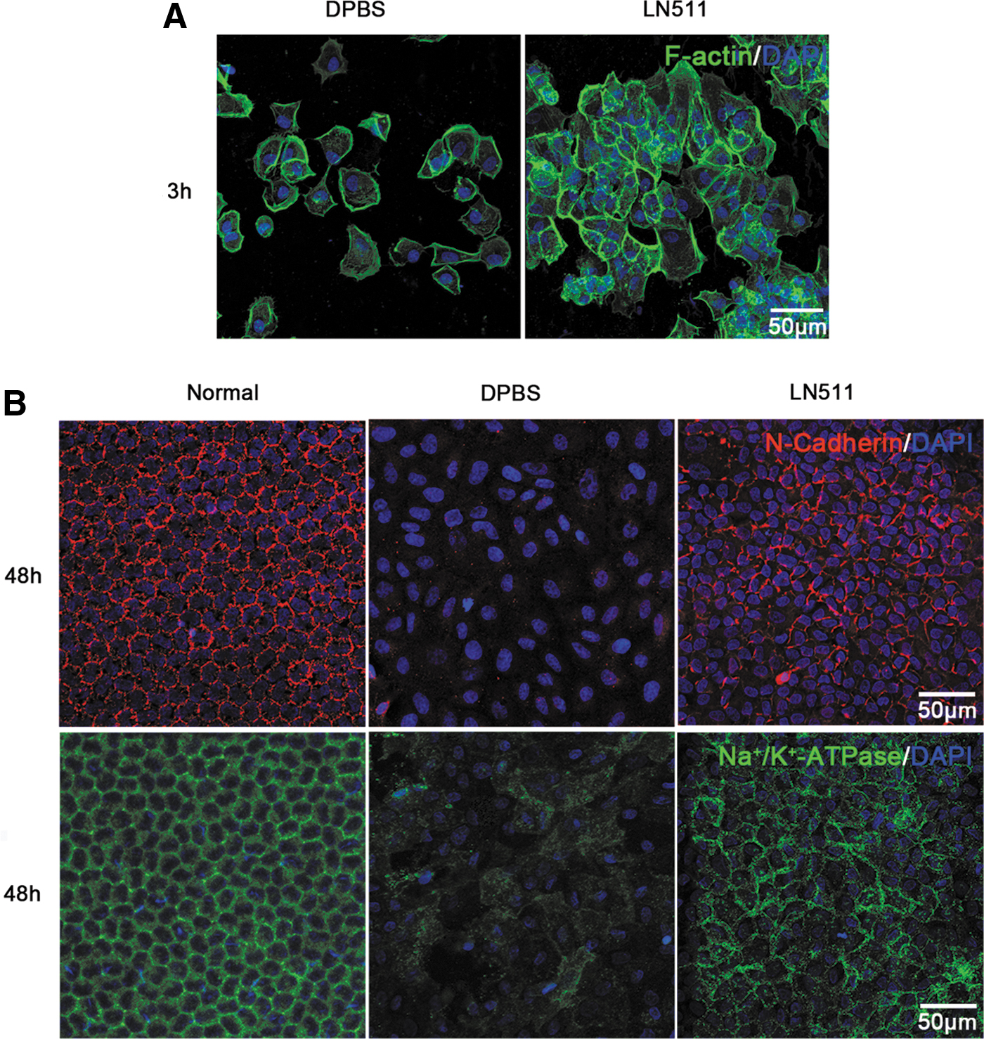
Effects of LN511 on the adhesion and functional regeneration of transplanted CECs.
LN511 precoating promotes the functional recovery of the transplanted CECs
Corneal endothelium maintains corneal transparency by its barrier and pump function. 1 We detected the expressions of the functional markers of the corneal endothelium with immunofluorescence. Similar to the normal corneal endothelium, ZO-1 and Na+/K+-ATPase were extensively distributed at the plasma membrane and displayed a regular polygonal morphology in the LN511 group. However, transplanted CECs in the DPBS group exhibited weak and sparse expression of ZO-1 and Na+/K+-ATPase expression, and F-actin staining displayed an irregular arrangement of the large cells (Fig. 4A, B). Moreover, CM-Dil was used to trace the transplanted cells. The results indicated that the cells in the central area of corneal endothelium were positive for CM-Dil 14 days after transplantation in both LN511 and DPBS groups, which suggested that the functional corneal endothelium was derived from the transplanted cells and was not regenerated by CECs of the receptor (Fig. 4C, D).
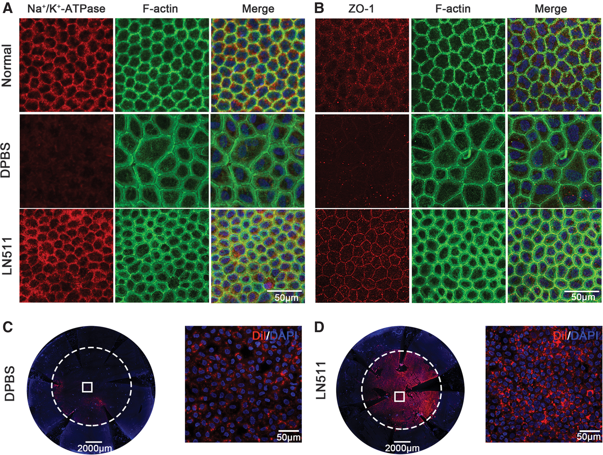
Effects of LN511 on the functional recovery of transplanted CECs. The corneas were obtained 14 days after CECs' injection.
LN511 promotes the nuclear–cytoplasmic translocation of yes-associated protein
YAP is a transcription factor that senses cell density and regulates cell function. 37 LN511 increased the density of the transplanted CECs and significantly reduced the nuclear location of YAP than that observed in the DPBS group in vivo (Figs. 3B and 5A). To assess whether the functions of LN511 related to CECs are affected through the YAP signaling, the HCEC line B4G12 was used in an in vitro study. Compared with the DPBS group, HCECs cultured in LN511-coated dishes exhibited high density and regular and continuous distributions of ZO-1 and Na+/K+-ATPase at the plasma membrane (Fig. 5B), and the YAP staining revealed positive signals distributed in the cell cytoplasm (Fig. 5C). Expressions of YAP downstream genes, CTGF and CYR61, were significantly lower in HCECs cultured on LN511 (Fig. 5D, E).
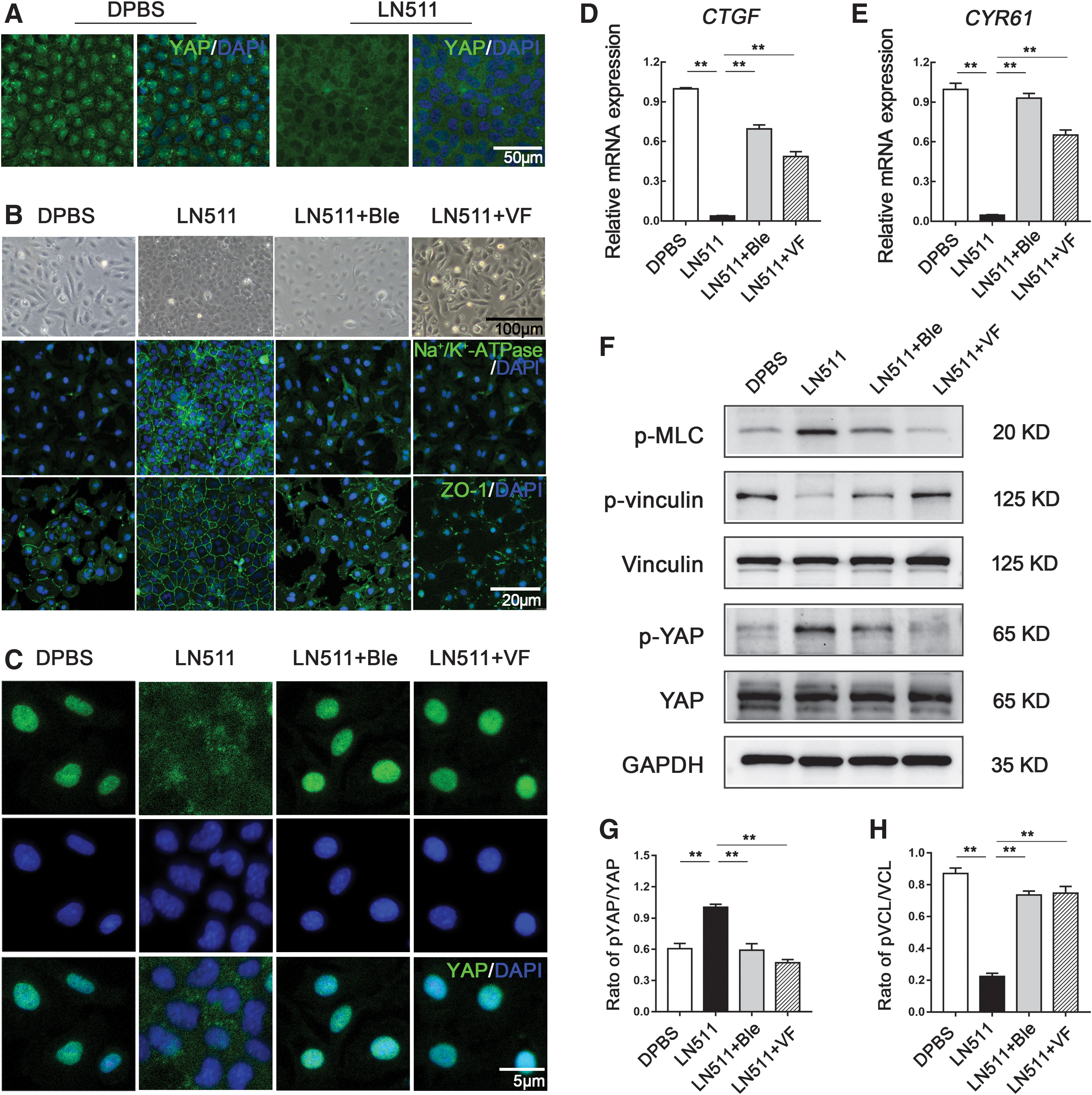
Effects of LN511 on the nuclear–cytoplasmic translocation of YAP.
Actomyosin contractile force was reported to enhance the compact morphology of cells. 38 We found that LN511 promoted the expression of phosphorylated myosin light chain (p-MLC), a contractile force marker, and reduced expression of phosphorylation of vinculin, which recognizes cell–cell but not cell–matrix adhesions (Fig. 5F). 30 Ble and VF inhibit myosin heavy chains and the YAP/TAZ signaling pathway, respectively, to abolish the compact morphology of cells.39,40 Our results indicated that 20 μM Ble or 0.5 μM VF treatment reversed the effects of LN511, including reduction of cell density, suppression of ZO-1 and Na+/K+-ATPase, promotion of the nuclear location of YAP staining, and expressions of CTGF and CYR61 (Fig. 5B, E). Ble or VF treatment also decreased the protein levels of p-MLC and p-YAP and increased p-vinculin in the cell cultured on LN511 (Fig. 5F). Compared with the control group, the ratio of phosphorylated YAP and phosphorylated MLC increased in the LN511 group, while that of phosphorylated vinculin decreased significantly (Fig. 5G, H). The complete gels of the western blots are shown in Supplementary Figure S3. These results indicated that LN511 enhanced the formation of CECs' compact morphology through the activation of actomyosin contraction force and the nuclear–cytoplasmic translocation of YAP.
Discussion
Corneal transplantation is the main therapy for corneal endothelial dysfunction. The anterior chamber injection of cultured HCECs has garnered increasing attention in recent years.20,41,42 We previously developed a “mini sheet injection” method, which promoted adhesion and tight junction formation of the grafted CECs, as well as the functional recovery of the cornea in a rabbit model. In this study, we developed intense scraping to remove CECs and ECM of the posterior DM without DM removal to mimic the late stage of corneal endothelial diseases in a rabbit model. Then, LN511, as a coating material, was injected into the anterior chamber to improve the scraped posterior DM, which enhanced the adhesion of the grafted CECs, increased corneal endothelium regeneration, and facilitated corneal recovery with CEC transplantation.
For CEC transplantation, two main methods have been reported: transplantation of corneal endothelial sheets and injection of CECs' suspension.43,44 The former one is based on the technique of DSEAK, which demands extensive surgical experience. Moreover, the construction of corneal endothelial sheet requires a suitable carrier, such as an intact DM from donor tissue, the artificial gelatin methacryloyl based hydrogels, and biosynthetic DM-like basement membranes.45,46 Compared with this strategy, anterior chamber injection of CECs' suspension is a simpler and less invasive method for CEC transplantation. 44 However, because of the flow of aqueous humor, the issue of the weak adhesion greatly affects the efficacy of the transplanted cells, and several strategies were also developed to resolve this.36,47–49 Previously, ECM coating was reported to promote cell/ECM interactions and improve the retention and survival of grafted cells in various tissues.50,51 For the first time, we used LN511 to enhance the adhesion of grafted cells in vivo. Before CEC transplantation, LN511 was first injected into the anterior chamber to form a coating on the endothelial face of DM, which improved the DM posterior surface to supply the suitable basement membrane for CEC adhesion. Moreover, LN511 precoating did not increase the intraocular pressure, suggesting that it did not block the trabecular meshwork.
According to the condition of the DM, the pathological process of corneal endothelial diseases can be divided into early and late stages. 3 In the early stage, DM is not seriously damaged, and the injection of CEC suspension can be applied after CEC scraping without DM removal. 13 The strategy of LN511 precoating reported in the present study would enhance the efficacy of the CEC injection therapy. In the late stages, DM was usually abnormal. For example, scarring of DM form guttae, and dysfunction of ECM secretion of abnormal CECs induces deficiency of DM. 3 These abnormal DM would induce CECs' degeneration and have to be removed. Conventionally, the transplantation of a corneal endothelial sheet is more suitable than the injection of CECs' suspension for this stage. 6
In this study, intense scraping could be used to remove the damaged CECs and scarring of the posterior DM without DM removal. Then LN511 was injected to improve the scraped posterior DM and facilitate corneal recovery of the grafted CECs. Besides, we also detected the efficacy of LN511 precoating with DM removal. LN511 could promote the adhesion of the grafted CECs, but the center area of the cornea was still hazy 2 weeks later (Data not shown). We think that the removal of an area of DM as large as 9 mm in diameter may be too big for the therapeutic function of LN511 precoating and CECs injection. It was reported that when DM removal is small (4 mm in diameter), it could still allow CEC injection to recover the corneal transparency, 14 which suggests that the combination with a small descemetorhexis and LN511 precoating might also be beneficial for CECs' transplantation in the therapy of later stages of corneal endothelial diseases.
ECMs are mainly fibrous forming proteins, which serve as scaffolds and regulate many cellular processes, including survival, growth, migration, and differentiation.52,53 DM is formed by CEC-secreted ECMs and is composed of collagen VIII, collagen IV, LNs, and fibronectin.54–56 LN511 promotes CEC adhesion and enhances the in vitro expansion of HCECs.33,34 LN511 was reported to enhance cell adhesion and proliferation by activating FAK signaling through α3β1 and α6β1 integrins. 57 It can also enable the proliferation of pluripotent stem cells by actomyosin contraction and YAP inactivation. 30
Consistently, LN511 precoating enhanced the expression of the barrier and pump function-related genes of the grafted CECs in vivo and further promoted the cytoplasmic localization of YAP in the grafted CECs, suggesting the upregulation of YAP phosphorylation. LN511 also enhanced the formation of compact morphology of HCECs, promoted the expressions of p-MLC and p-YAP, and reduced the expressions of p-vinculin and YAP downstream genes in the cultured HCECs. In addition, the inhibitors VF and Ble abolished the effects of LN511 on HCECs, suggesting that roles of LN511 in HCECs are affected through the activation of actomyosin contraction force and the nuclear–cytoplasmic translocation of YAP.
Cell transplantation has progressively been used for the therapy of multiple diseases or disorders, such as hematopoietic disease and neurological disorders.58,59 Microenvironment of the receptor greatly decides the survival and functions of the grafted cells. Several methods, such as ECM manipulation, engineering biomaterials, and vehicle composition, have been developed to improve the transplanted microenvironment.60–62 Natural ECM components, such as collagen, fibrin, and LN, are being used as cell vehicles to facilitate the constructive remodeling in several tissues during cell transplantation.63,64
This is the first time that ECM is used to improve the microenvironment of CEC transplantation. Beside LN, we also detected the staining changes of other DM components collagen VIII and IV after intense scraping and found that the staining of collagen VIII and IV on the posterior surface of the DM was significantly decreased, as well as LAMA5 (Supplementary Fig. S4). This suggested that other DM components or a cocktail of ECM components might be more beneficial than LN511 alone for treating corneal endothelial diseases. Moreover, optimization of the vehicle composition might be effective for improving CEC transplantation. For example, a cell therapy vehicle (CTV) was generated by retaining the clinically translatable materials, and CECs' injection in CTV efficiently promoted corneal endothelium regeneration in rabbits. 43 The addition of growth factors or small molecules, such as Y27632 and niacinamide (NIC), would also be a good approach.36,65,66 The ROCK inhibitor Y27632 effectively reduced corneal edema in some clinical trials in combination with HCEC injection. 20
Finally, to find an alternative CEC source is promising for the strategy of CECs' transplantation, and many types of stem cells were used to generate CEC-like cells, such as hESCs, induced pluripotent stem cells, and adult stem cells. 67 Overall, the approach of CECs injection is promising for treating corneal endothelial diseases, and subsequent basic and clinical studies will be required to further improve and evaluate its efficacy and safety.
Conclusions
In conclusion, we developed intense scraping to remove CECs and ECM of the posterior DM without DM removal for the therapy of late stage of corneal endothelial diseases. We also demonstrated that the LN511 precoating improved the posterior DM and enabled the adhesion and the functional recovery of the grafted CECs. These could serve as new strategies for cell-based therapy of corneal endothelial diseases.
Authors' Contributions
C.Z., Z.L., and W.S. conceived and designed the study. C.Z., H.D., X.W., Y.J., Y.G., W.L., and C.D. performed the experiments. C.Z. and Z.L. supervised the study and oversaw the data collection. C.Z., X.W., H.D., and Y.G. collected the data. Z.L. and Q.Z. guided the data analysis. C.Z. wrote the initial article draft. Q.Z., Z.L., and W.S. critically revised the article and gave valuable insight to the study concept. All authors read and approved the final article.
Footnotes
Disclosure Statement
No competing financial interests exist.
Funding Information
This work is partially supported, in part, by the National Natural Science Foundation of China (81700811, 81900834) and the Natural Science Foundation of Shandong Province (ZR2019ZD37, ZR2019PH110, ZR2018LH008). W.S. and Q.Z. are partially supported by the Taishan Scholar Program (20150215, 20161059) and the Innovation Project of Shandong Academy of Medical Science.
References
Supplementary Material
Please find the following supplemental material available below.
For Open Access articles published under a Creative Commons License, all supplemental material carries the same license as the article it is associated with.
For non-Open Access articles published, all supplemental material carries a non-exclusive license, and permission requests for re-use of supplemental material or any part of supplemental material shall be sent directly to the copyright owner as specified in the copyright notice associated with the article.