
Abstract
Hyaluronic acid (HA) is an ideal initial material for preparing hydrogels, which may be used as scaffolds in soft tissue engineering based on their advantageous physical and biological properties. In this study, two crosslinking agents, divinyl sulfone (DVS) and butanediol diglycidyl ether, were used to investigate their effect on the properties of HA hydrogels. As HA hydrogels alone do not promote cell adhesion on the scaffold, fibrin and serum from platelet-rich fibrin (SPRF) were combined with the scaffold; the aim was to create a material intended to be used as soft tissue implant that facilitates new tissue formation, and degrades over time. The chemical changes were characterized and cell attachment capacity of the protein-containing gels was examined using human mesenchymal stem cells, and viability was assessed using live–dead staining. Fourier-transform infrared measurements revealed that linking fibrin into the gel was more effective than linking SPRF. The scaffolds were found to be able to support cell adherence onto the hydrogels, and the best result was achieved when HA was crosslinked with DVS and contained fibrin. The most promising derivative, 5% DVS-crosslinked fibrin-containing hydrogel, was injected subcutaneously into C57BL/6 mice for 12 weeks. The scaffold was proven to be biocompatible, remodeling, and vascularization occurred, while shape and integrity were maintained.
Impact statement
Fibrin was combined with crosslinked hyaluronic acid (HA) for regenerative application, the structure of the combination of crosslinked HA with blood-derived protein was analyzed and effective coating was proven. It was observed that the fibrin content led to better mesenchymal stem cell attachment in vitro. The compositions showed biocompatibility, connective tissue and vascularization took place when implanted in vivo. Thus, a biocompatible, injectable gel was produced, which is a potential candidate for soft tissue implantation.
Introduction
Hyaluronic acid (HA) is a linear, nonsulfated glycosaminoglycan consisting of repeating units of
HA degrades rapidly in vivo enzymatically. To extend its presence in the body and to improve its mechanical properties, HA can be modified by covalently crosslinking the polymer chains. 10 A three-dimensional network can be obtained, which is water insoluble and less sensitive to enzymatic degradation. 11 There are several chemical crosslinkers, which covalently bond the HA chains, for instance, glutaraldehyde, divinyl sulfone (DVS) 12 and butanediol-diglycidyl ether (BDDE) 10 act on the hydroxyl group, while carbodiimides react with the carboxyl group. It was observed that with increasing crosslinker concentration, the degree of crosslinking can be enhanced, 10 which increases the time of degradation and improves mechanical stability. The crosslinking density determines the swelling ratio as well, a more crosslinked, strong hydrogel will swell less than a weak crosslinked hydrogel. 13 However, an extremely high crosslinker concentration has its drawbacks as well, because chemical crosslinkers are generally toxic and can cause unwanted reactions in larger amounts. 14
Crosslinked HA hydrogels can be used as scaffolds for soft tissue engineering15–17 in the cases of soft tissue defects like congenital malformation, extirpation, or trauma 18 as HA is a biocompatible and biodegradable material and has stimulatory effects on cell proliferation, migration, extracellular matrix secretion, and differentiation. 9 The scaffolds can serve as a synthetic extracellular matrix with their high water content and soft structure 19 organizing cells into a three-dimensional architecture. 20 As these scaffolds closely mimic natural tissues, cells adhere to the three-dimensional network, especially when there are incorporated peptide domains in the hydrogel. 19 Preferably, these scaffolds are remodeled and vascularized by adhering cells.9,19
HA hydrogels can also be used to facilitate wound healing. Normally, the process consists of hemostasis, inflammation, proliferation, and remodeling,21,22 but in some cases, the natural wound healing process is hindered or cannot take place and the wound becomes chronic, like diabetic ulcers and pressure ulcers,22,23 which cannot be recovered without external help. In other cases, like severe burns, large skin damage occurs and therefore an appropriate wound dressing is needed. 24 An ideal wound dressing prevents contamination of the wound and maintains adequate moisture, but removes excessive exudates. 22
HA hydrogels may be excellent wound dressings as they create an advantageous environment for wound healing because of their rheological, hygroscopic, and viscoelastic properties. 23 Besides, high molecular weight HA was reported to have a cytoprotective effect and to facilitate cell migration and wound healing. 7 In animal models, HA helped reepithelialization and led to the formation of new soft tissue in the case of full-thickness surgical wounds.7,23 Although low molecular weight HA was not reported to have these protective effects, 25 it was found to induce angiogenesis following its degradation. 23
Crosslinked high molecular weight HA gels alone were found to be bioinert 26 and cell attachment into these gels was low.27,28 However, cell adherence can be promoted by fabricating hybrid HA scaffolds 29 with gelatin, 28 chitosan,30,31 collagen,15,32 or silk fibroin, 4 among others. Peptide incorporation into the hydrogel is another way to enhance cell attachment, migration, proliferation, growth, and organization. 19 Besides, HA hydrogels can be coated with collagen, extracellular matrix gel, laminin, and fibronectin to enhance cellular adhesion. 27
Blood-derived protein polymerization or crosslinking into the gels can be another option to advance cell attachment. Serum from platelet-rich fibrin (SPRF, also referred to as hyperacute serum) is a human blood derivative,33,34 which contains a large number of proteins and growth factors, inducing the proliferation and migration of human bone marrow-derived mesenchymal stem cells (MSCs), osteoblasts, and osteoarthritic chondrocytes in vitro in cell culture.35–38 Fibrin polymerization 39 can be another way to immobilize blood-derived proteins into the gel, as fibrin has many advantageous effects on cell proliferation. 40 Cryoprecipitate isolated from human plasma using citrate as an anticoagulant is rich in fibrinogen and after recalcining, natural blood clotting can occur and fibrin chains can be formed inside the HA matrix.
The preparation using HA and blood-derived proteins is aimed to be used as a soft tissue implant candidate, regulated as a drug or a drug-device combination. To reduce potential regulatory issues, we looked for crosslinkers that have already been used in HA medical device preparations and are already well documented.41,42 DVS and BDDE were chosen as these crosslinkers were used in well-known products (e.g., Captique, Hylaform, Juvéderm, Prevelle, Restylane, and Perlane); thus, the toxicity profile is already well known in the case of DVS and BDDE, the use of these materials does not pose additional risks.
In this work water-insoluble HA hydrogels were prepared using BDDE and DVS as crosslinkers in different ratios (2%, 5%, and 10%) and the effect of different crosslinking agents and crosslinker concentrations was examined on the swelling ratio, resistance against enzymatic degradation, the structure of the hydrogels, and Fourier-transform infrared (FTIR) spectra. SPRF and fibrin were immobilized into the HA hydrogels to induce cell attachment, and the result of the modification was analyzed using FTIR spectroscopy and scanning electron microscope (SEM) imaging. Human bone marrow-derived MSCs were used to determine the cytotoxicity and the cell attachment capacity of the protein-containing gels. Biocompatibility was investigated by injecting the homogenized hydrogel subcutaneously into C57BL/6 mice. In vivo vascularization was observed by microscopic imaging and histological analysis was performed to detect remodeling.
Materials and Methods
Hydrogel preparation
Crosslinked HA hydrogels were prepared using 1.34 MDa freeze-dried sodium hyaluronate from bacterial source (Contipro, Dolní Dobrouč, Czech Republic), BDDE (Fig. 1A) (Sigma-Aldrich, St. Louis, MO) or DVS (Fig. 1B) (abcr, Karlsruhe, Germany), and NaOH to provide alkaline condition required for the crosslinking reaction. The crosslinkers were used in 2% V/V, 5% V/V, and 10% V/V. BDDE or DVS was mixed with 1 mL 1% NaOH and then added to 133 mg sodium hyaluronate and immediately vortexed until a homogenous gel was formed. The hydrogels were centrifuged at 1700 g for 3 min to get flat gels and were allowed to crosslink for 48 h at room temperature in a plastic vial. The crosslinked gels (Fig. 1A) were washed and swollen until equilibration with 80 mL distilled water in three steps, 12 h each step. The 10% crosslinker-containing gels were more rigid and they moldered during the washing procedure; thus, only the 2% and 5% crosslinker-containing gels were further investigated. The washed gels were autoclaved for 20 min at 121°C to get a sterile gel. Sterilized gels were freeze-dried at −55°C and 5 Pa.

The crosslinking reaction of HA with
Structure analysis with SEM
The cross-section of the crosslinked gels was examined with an SEM (JEOL JSM-6380LA). Crosslinked gels were prepared as described above. The freeze-dried gels were cut with a scalpel and their cross-section was coated with gold immediately before SEM imaging (JEOL JFC-1200 Fine Coater, 12 mA, for 20 s).
Swelling ratio measurement
Equal one-fourth pieces of the crosslinked, washed, and swollen HA gels were weighed using an analytical balance. The gels were freeze-dried and weighed again. Swelling ratio was calculated with the following formula:
Enzymatic degradation measurement
The aim of this measurement was to compare the rate of degradation of the different gel types in vitro. Enzymatic degradation was determined using Ehrlich's solution (Sigma-Aldrich), which is a reagent consisting of acetic acid, p-dimethyl amino-benzaldehyde, and hydrochloric acid. Ehrlich's solution detects N-acetyl-glucosamine, a product of HA degradation. It was observed that the heated solution of N-acetyl-glucosamine (NAG) reacting with p-dimethyl amino-benzaldehyde under acidic conditions presents purple color, directly proportional to the NAG concentration. The intensity of the color can be improved by adding borate to the reaction mixture.43–45
Equal one-fourth pieces of crosslinked and dried 2% BDDE-, 5% BDDE-, 2% DVS-, and 5% DVS-containing HA hydrogels were soaked into 10 mL 4 mg/mL solution of hyaluronidase from bovine testes, type I (Sigma-Aldrich) and kept at 37°C on a shaker for 4 days, while NAG concentration measurement was completed every day with Ehrlich's reagent according to the following protocol: 50 μL of the enzyme solution was mixed with 50 μL borate buffer (4.94 g H3BO3, 1.98 g KOH in 100 mL H2O, pH = 9) and placed into boiling water for 3 min, and allowed to cool down to room temperature for 5 min; then 25 μL glacial acetic acid and 25 μL Ehrlich's reagent were added. Absorbance was measured 12 min after Ehrlich's reagent was added at 585 nm with a reference wavelength at 750 nm using a PowerWave XS microplate spectrophotometer (BioTek, Winooski, VT). A calibration curve was also prepared using pure NAG (Sigma-Aldrich) in 0–0.6% concentrations (11 calibration points).
Modification of the crosslinked hydrogels
SPRF and cryoprecipitate isolation from whole blood
Phlebotomy occurred under Institutional Review Board (IRB) approval (IRB approval number: 33106-1/2016/EKU, July 12, 2016.) from healthy donors, men, and women, 24–45 years of age. Fifty milliliters of venous blood was drawn from each donor. In the case of SPRF production, whole blood was poured into a 50 mL centrifuge tube containing 10 g sterile glass beads under a laminar flow hood and centrifuged immediately at 1710 g for 8 min to separate red blood cells from serum fraction. Blood clotting was promoted by the glass and the fibrin clot was formed. The tube was centrifuged again at 1710 g until fibrin clot became about 1 cm flat and the supernatant was collected, which is SPRF. In the case of cryoprecipitate, whole blood was poured into a sterile 50 mL centrifuge tube, which contained 0.215 g sodium citrate dihydrate (Sigma-Aldrich) dissolved in 0.5 mL saline solution. It was centrifuged at 1710 g until the plasma fraction was separated from the red blood cell-containing fraction. The plasma was collected and kept at −80°C for 24 h and then thawed at 3°C and centrifuged at 3260 g for 12 min at 3°C. The cryoprecipitate was dissolved in 10 mL plasma, and the rest of the supernatant fraction was removed.
Protein crosslinking into the hydrogels
The sterile, freeze-dried HA gels were further modified by crosslinking SPRF into their structure using DVS, or fibrinogen polymerization to improve cell adhesion on the gels. When preparing SPRF-containing gels, each freeze-dried quarter of gel was soaked into 1 mL 5% sterile DVS containing SPRF at pH = 12, and crosslinking took place for 24 h at room temperature. The gels were washed again thrice with sterile distilled water for 12 h each step to remove excess nonreacted DVS. In the case of the preparation of fibrin-containing gels, 20 μL 1 M CaCl2 and 20 μL (500 U/mL) human thrombin were added to 1 mL cryoprecipitate and it was poured onto the freeze-dried crosslinked HA gels; 1 mL recalcined cryoprecipitate was added to each quarter of freeze-dried gel. The recalcined cryoprecipitate gets absorbed by the gels and the fibrinogen converts into fibrin polymers inside the structure of the HA gels at room temperature. The whole protein crosslinking and washing procedure occurred under aseptic conditions using sterile filtered reagents.
Structure analysis using FTIR spectroscopy
The FTIR spectra of native sodium hyaluronate were compared to crosslinked HA, and SPRF- and fibrin-containing crosslinked HA gels. The samples were prepared as described above and freeze-dried. We performed the infrared measurements with a Bruker Vertex 80v (Bruker Corp., Billerica, MA) spectrometer. It was equipped with a high-sensitivity mercury–cadmium–telluride detector and a single reflection diamond ATR accessory. One hundred twenty-eight scans were collected at 2 cm−1 resolution in the range of 400–4000 cm−1.
Observation of the cross-section of the protein-containing crosslinked hydrogels by SEM
The cross-section structure of the protein-containing gels was compared to each other using an SEM (JEOL JSM-6380LA). The gels were freeze dried and coated with gold as described above (section “Structure analysis with SEM”).
Determination of cell attachment capacity of the hydrogels using human MSCs
MSC culturing
All cell culture procedures were carried out in a sterile laminar flow tissue culture hood. Bone marrow-derived MSCs (ATCC, Manassas, VA) were cultured in T-75 TC treated culture flasks in an incubator at 37°C, 5% CO2, and 95% humidity. MSCs were maintained in stem cell medium: Dulbecco's modified Eagle's medium (DMEM) containing 4.5 g/L glucose and
Cytotoxicity measurement with XTT
Cytotoxicity measurements were performed to examine if crosslinked HA gels are cytotoxic as they may contain excess of toxic reagents, which can be released to their environment. MSCs (four passages) were seeded onto the bottom of 24-well plates in a density of 5000 cells/well in 700 μL stem cell medium. On the first day of culturing, 3–4 mm3 pieces of the sterile and washed initial HA gels and also SPRF-containing crosslinked HA gels were placed into the medium of the MSCs, while there were four wells, which contained cells only, but no hydrogel, as controls. The medium was refreshed every 2 days. The viability of the MSCs was measured on the seventh day with the help of Cell Proliferation Kit II (XTT; Roche, Mannheim, Germany) according to the manufacturer's instructions. The ratio of the gel-containing and control wells shows cytotoxicity of the crosslinked gels.
Cell adherence test by live–dead staining
Cell adherence tests were accomplished to investigate if MSCs adhere and proliferate on the crosslinked hydrogels. Sterile and washed 3–4 mm3 pieces of crosslinked and blood-derived protein-containing gels were washed with 1.5 mL stem cell medium for 1 h. On the first day, 25,000 MSCs (two passages) were seeded onto the gels on 24-well low attachment plates in 800 μL stem cell medium. The medium was refreshed to 1.5 mL stem cell medium thrice a week. On the 14th day, the attaching cells were visualized on the gels by live–dead staining. The gels were washed thrice with phosphate-buffered saline (PBS) and stained in PBS containing 1 μM Calcein-AM (Invitrogen, Carlsbad, CA), 4 μg/mL ethidium homodimer (Invitrogen), and 20 μg/mL Hoechst (Invitrogen) for 30 min. The gels were washed again thrice for 10 min with FluoroBrite DMEM (Gibco, Paisley, Scotland) containing 5%
In vivo biocompatibility testing
Five percent DVS-containing hydrogels with fibrin were homogenized for 5 min (homogenizer: Tissue ruptor; Qiagen). To promote homogenization, 1 mL cryoprecipitate supernatant was added to ∼4.8 g crosslinked, washed, and sterilized hydrogel. The homogenous gel was filled into sterile syringes. As a control, we used the same amount of 5% DVS-containing crosslinked HA, but without fibrin. Homogenization was facilitated in this case with 1 mL saline solution.
Five C57BL/6 male mice (2-month old, on average 26 g) were involved in the experiment. The experiments were carried out according to the guidelines of the Hungarian Law of Animal Protection (XXVIII/1998), and all procedures were approved by the National Scientific Ethical Committee on Animal Experimentation (PEI/001/2706-13/2014, approval date: December 17, 2014). Animals were housed at a constant temperature with a 12-h light/12-h dark cycle, and they had ad-libitum access to food and water. Mice were anesthetized with isoflurane (2%); thereafter, 200 μL homogenized HA was injected subcutaneously to the abdominal site of the hind leg. Fibrin-containing HA was injected to the left leg of the mice and control hydrogel was injected to the right leg. After 12 weeks, the mice were anesthetized with intraperitoneally applied ketamine (100 μg/g body weight, Calypsol; Gedeon Richter, Budapest, Hungary) and xylazine (10 μg/g body weight, CP-Xylazine; CP-Pharma, Burgdorf, Germany). The hind limbs were depilated and a small incision was made to expose the hydrogels. Subsequently, the hydrogels were observed by a light microscope (Leica M80; Leica Microsystems, Wetzlar, Germany). After cervical dislocation, the gels were harvested and fixed in 4% formaldehyde, and their weight was measured.
The samples were dehydrated and cleared in water, ethanol, and xylol. The samples were then embedded in paraffin, cut to 2.5 μm thick sections and dyed using Mayer's hematoxylin and 1% eosin.
Statistical analysis
One-way analysis of variance (ANOVA) with Tukey's post hoc test was performed to compare the means of groups using Prism 7 software. The significance level was p < 0.05, where * means that p is between 0.01 and 0.05, ** means that p is between 0.01 and 0.001, and *** means that p is lower than 0.001, and data are presented as mean ± standard error of the mean.
Results
Structure of the crosslinked HA hydrogels
In Figure 2A the 2% and 5% BDDE- and DVS-containing crosslinked HA hydrogels can be seen. BDDE-containing gels are elastic, clear, and smooth, while the structure of DVS-containing gels is rough and more rigid, especially the 5% DVS-containing gel. However, SEM imaging showed that the cross-section of crosslinked gels was porous and honeycomb shaped; either BDDE or DVS was used as crosslinking reagent (Fig. 2B).
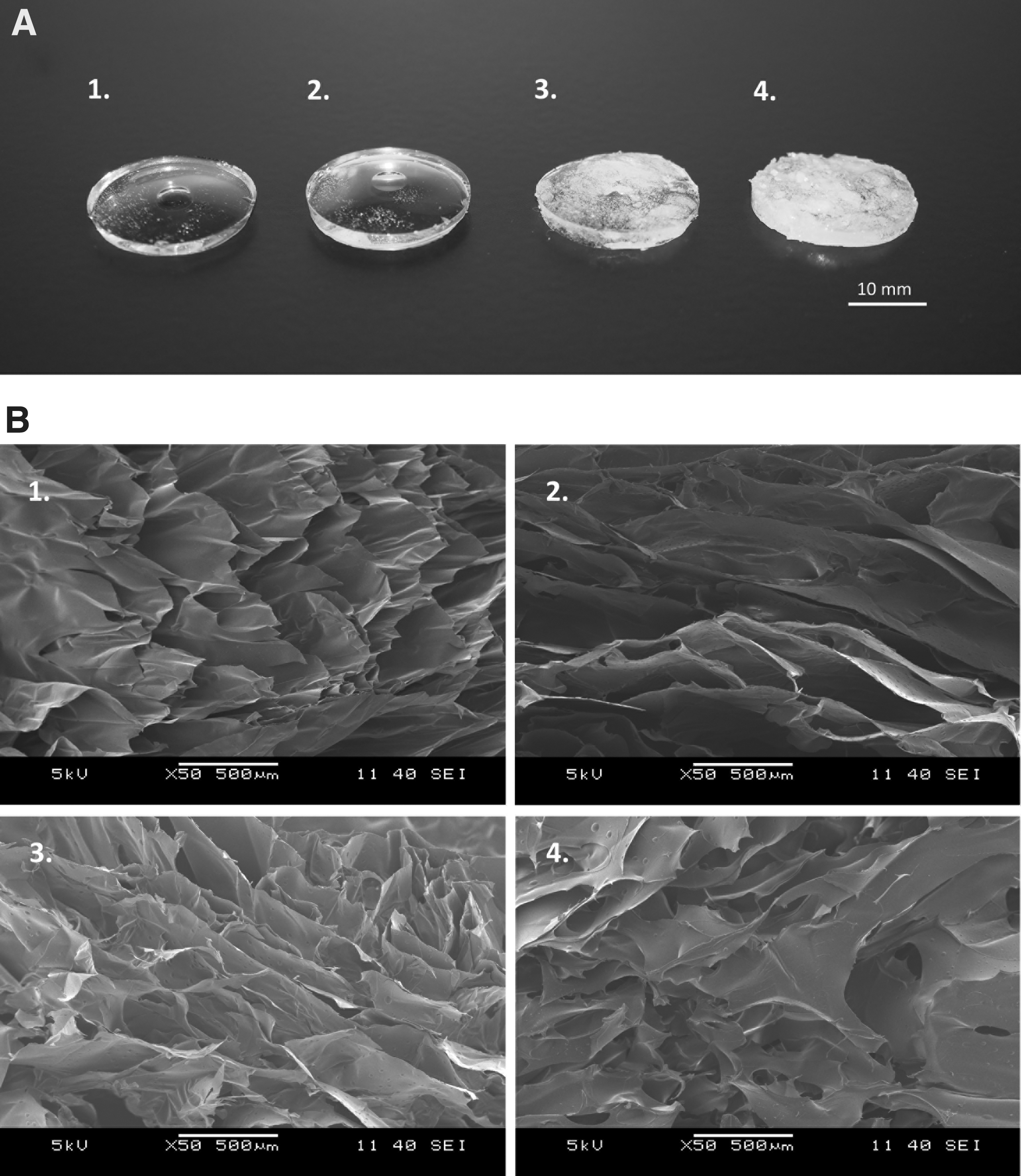
Appearance and structure of the crosslinked HA gels.
Swelling ratio
The swelling ratio is the quotient of the swollen and the freeze-dried gels' weight. It is inversely proportional to the degree of crosslinking; a strongly crosslinked hydrogel has a lower water uptake capacity and swells less than a weaker crosslinked gel. One-way ANOVA with Tukey's post hoc test was performed, and it was observed that the gels containing 2% crosslinker had significantly higher swelling ratio than 5% DVS- or BDDE-containing gels. In addition, the gels crosslinked with DVS were significantly less swollen than BDDE gels containing the same amount of crosslinker (Fig. 3A).

(
Enzymatic degradation
In vitro enzymatic degradation was examined with the help of Ehrlich's reagent, which determines the concentration of NAG, the product of HA degradation (Fig. 3B). HA gels were digested with hyaluronidase enzyme from bovine testes. NAG concentrations were calculated from the measured absorbances with the help of the calibration curve. The NAG content, which is directly proportional to HA degradation increased with time, but after 3 days, the degradation slowed down in each case, probably because the enzyme activity decreased. The 2% DVS-containing gel seemed to be degrading the fastest, although no significant difference could be found between 2% BDDE and 2% DVS gels. Significant difference was observed between 2% DVS and 5% DVS and between 2% DVS- and 5% BDDE-containing gels using one-way ANOVA with Tukey's post hoc test. Five percent BDDE and 5% DVS gels were the most resistant to enzymatic degradation, as their crosslinking density was higher, which also affects enzymatic degradation. None of the gel quarters was fully digested after 100 h; water-insoluble gel pieces were still visible.
FTIR analysis
The FTIR measurement results are shown in Figure 4, where the structure-related chemical changes are demonstrated. During the scaffold production steps, characteristic absorbance changes were visible in the IR spectra. The absorbances were normalized to the 1038 cm−1 peak in Figure 4A and B, and to the peak at 1533 cm−1 in Figure 4C, and the spectra were vertically shifted to prevent overlapping.

FTIR spectra of native sodium hyaluronate compared to crosslinked derivatives. (
In Figure 4A, the starting compound, sodium hyaluronate, was compared to the crosslinked derivatives. The main differences were that in all the crosslinked cases, the absorbance peaks related to the CH stretching vibrations of the CH2OH groups at 2931 and 2875 cm−1 increased. 46 Simultaneously, a new peak appeared at 1280 cm−1 if DVS was used as crosslinker, but not in the case of using BDDE. The C–OH vibration of alcohol sidebands of HA is seen at 1038 cm−1 in HA, but it shifts to the lower frequencies if crosslinker was used. At 1606 cm−1, the amide-related carbonyl peak is visible in all five derivatives.
In Figure 4B, the crosslinking with the blood separation derivative SPRF can be seen. The HA gels were prepared using BDDE or DVS crosslinkers and these gels were used to bind the serum product using only DVS as the second crosslinker. In the case when DVS was used as the first and second crosslinker, the spectra were similar to those in Figure 4A. When BDDE was used as the first crosslinker, then in the second step, new peaks occurred at 1238 and 1307 cm−1. The amide A band of the proteins (reflecting the N–H stretching) is also clearly visible around 3280 cm−1 on the top of the broad OH stretching band in the case when DVS was used as the second crosslinker.
In Figure 4C, the fibrin-coated crosslinked HA derivatives are shown. Two intense peaks appeared around 1643 and 1533 cm−1. These are the amide I and II bands of proteins. 47 The peak corresponding to the C–OH vibration of the alcohol sidebands is reduced considerably. probably due to the increased fibrin content. The two new peaks that appeared in the SPRF crosslinking have also appeared with the fibrin coating at 1238 and 1307 cm−1. Besides this, an intense peak appeared at 1390 cm−1.
SEM imaging of the cross-section of protein-containing hydrogels
The structure of the crosslinked gels was examined by SEM. In SPRF-containing gels (Fig. 5E–H), a protein film can be seen on the inner surfaces. In the case of fibrin-containing gels (Fig. 5M–P), the polymerized fibrin fibers can be observed. It is also visible that HA gels remained porous in all cases (Fig. 5A–D, I–L).

The cross-section of the crosslinked protein-containing HA gels.
Cytotoxicity measurement
Cytotoxicity measurement was performed to ascertain that the crosslinked hydrogels do not contain materials that are harmful to the living cells or hinder proliferation. Crosslinker reagents, such as BDDE and DVS, are poisonous and if the gels contain unreacted amounts of them, they can cause cell death. Thus, the hydrogels were washed several times after crosslinking. Cytotoxicity test showed that the viability of cells cultured on the bottom of the well in the presence of differently crosslinked hydrogels was as high as the viability of control cells, which were cultured in the wells without HA gels. Viability was calculated as the quotient of absorbance of cells cultured in the gel-containing wells and the absorbance of the control wells. No significant difference was detected performing one-way ANOVA with Tukey's post hoc test; therefore, it was observed that none of the crosslinked gels was cytotoxic (Fig. 6). Cytotoxicity of fibrin-containing gels was not measured, only the initial crosslinked HA gels, because by the linking of fibrinogen, only biocompatible materials were used, namely cryoprecipitate from human plasma, CaCl2, and thrombin, and the procedure occurred under aseptic conditions.
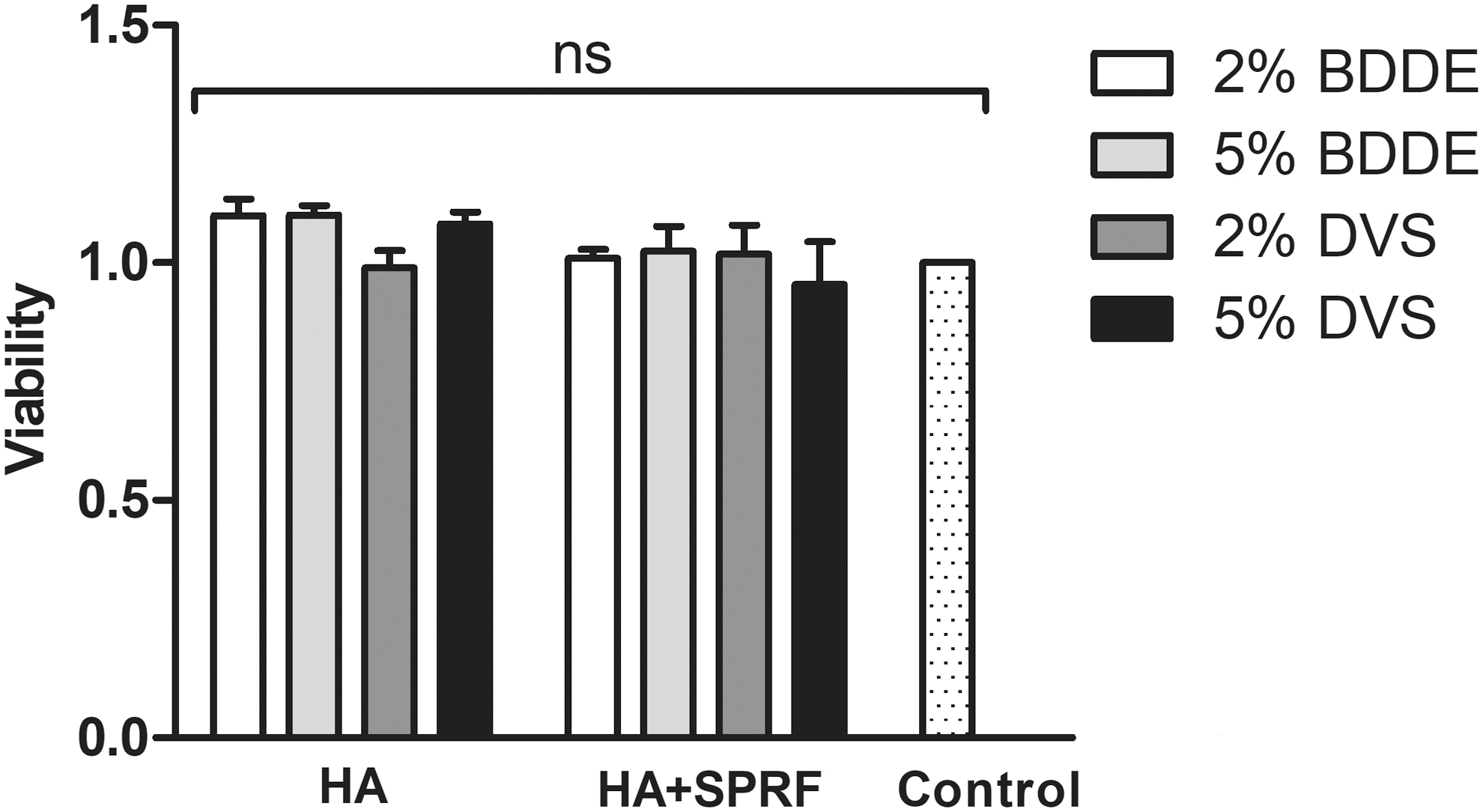
Cytotoxicity measurement of the crosslinked hydrogels. Cytotoxicity was determined by viability measurement of human MSCs. There is no significant (ns) difference between the sample groups and the control group; therefore, none of the gels was cytotoxic (n = 4). MSC, mesenchymal stem cell.
Cell attachment
We investigated if MSCs are capable of attaching onto the crosslinked gels. MSCs cultured on the hydrogels were visualized by live–dead staining. On the HA gels, which did not contain crosslinked proteins, no cells could be observed, which is in good accordance with previous studies.26–28 On the SPRF-containing hydrogels, a small number of attached living cells were visible, although the amount of the adhered MSCs was highly variable and cells attached only onto gels, which were crosslinked with DVS in the first step. If the SPRF-containing gels were crosslinked using BDDE, no cells adhered to the gel; thus, these images are not shown. The most cells could be observed on the fibrin-containing gels, especially if DVS was used in the crosslinking step (Fig. 7I, II). Besides, cell attachment was not homogenous; there were preferred regions on the gels where the surface structure could have been more beneficial for the MSCs. The nuclei were visualized with Hoechst (blue), living cells were stained with Calcein-AM (green), and dead cells with Ethidium-homodimer; however, only very few dead cells were present on each gel, thus this channel was not performed (Fig. 7I).

The percentage of the areas covered by the living cells and nuclei calculated by ImageJ software is shown in Figure 7II, where it can be observed that also crosslinker concentration seemed to affect cell attachment; more cells are visible on 5% DVS- and fibrin-containing gels than on the 2% version of this gel, and the same effect can be observed if BDDE was the crosslinker.
In vivo biocompatibiliy testing
The 5% DVS-crosslinked fibrin-containing hydrogel was found to be the most promising material based on previous experiments described above: in vitro degradation was the slowest, indicating that this matrix is the most resistant against enzymatic degradation, and also swelling ratio was found to be the lowest among the examined matrices, showing that HA concentration is the highest. Besides, the most attached cells were visible on this sample by live–dead staining; thus, this material composition was tested in vivo.
The homogenized gel was injected into the mice to investigate biocompatibility and degradation over 12 weeks and to examine the vascularization and remodeling of the gels. After 12 weeks, the mice were anesthetized, HA was harvested, and vascularization was observed. No inflammation was found, and the gels were intact and hard, but elastic. No significant weight loss was measurable; thus, it was suggested, that parallel with degradation, tissue growth occurred. It was observed that the surrounding tissues attached stronger to the fibrin-containing gels than to control gels. More blood vessels could be seen on fibrin-containing gels (Fig. 8). The harvested fibrin-containing HA matrices were also more reddish colored, which also indicates that those were more interlaced with blood vessels (Fig. 8C, D).
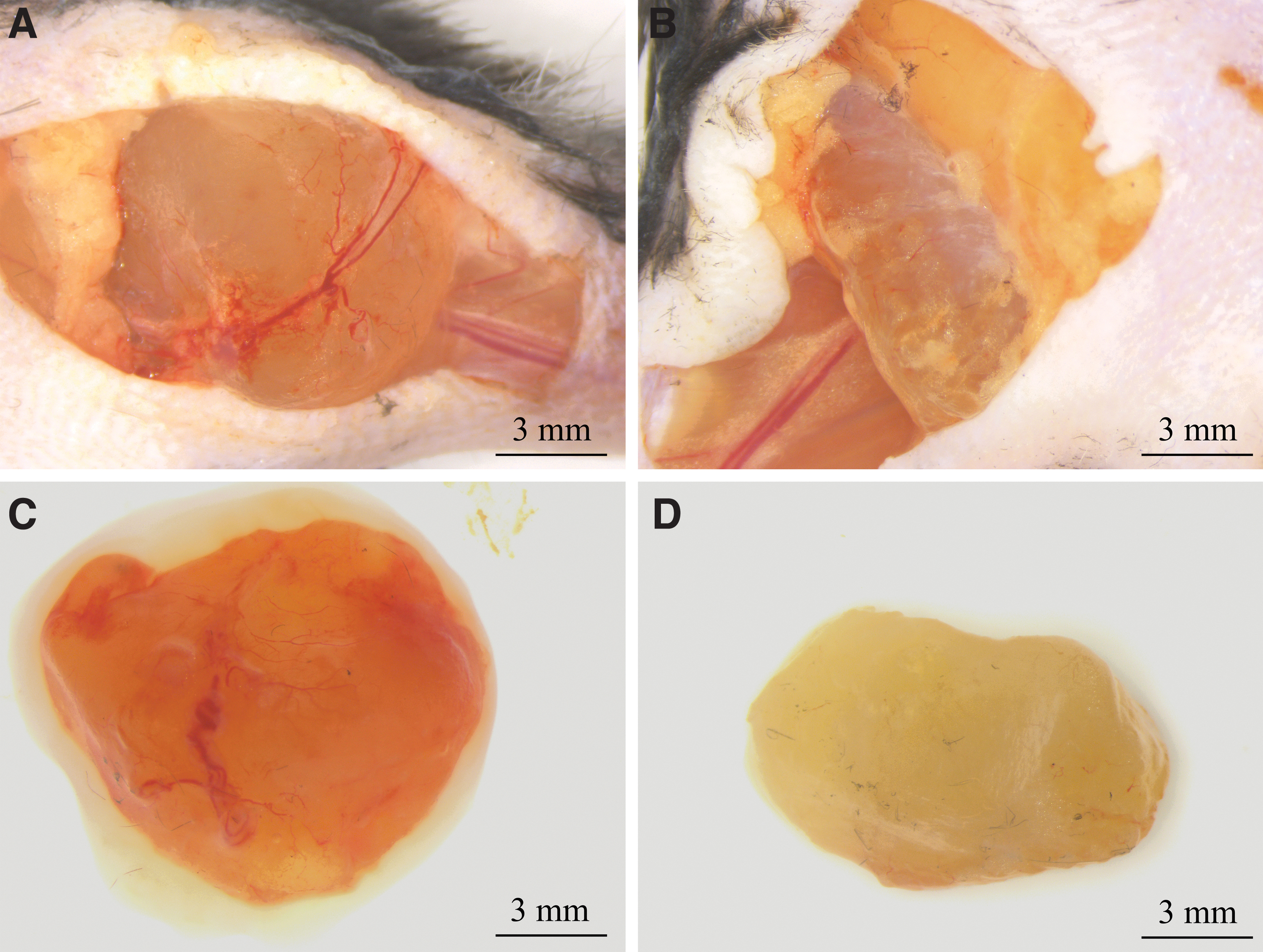
A typical image of the HA scaffolds in the mouse and the harvested matrices.
Histological analysis and hematoxylin–eosin staining showed that the scaffolds were partially remodeled and the matrices were embedded in connective tissue (Fig. 9A, C). HA gel pieces were also surrounded by connective tissue. Small blood vessels were visible, containing red blood cells (Fig. 9B, D) especially in the fibrin-containing gels. The number of nuclei was found to be roughly the same in fibrin-containing and control gels compared by ImageJ software (data not shown).

Histological analysis of the fibrin-containing and control 5% DVS scaffolds collected from mice after 12 weeks. The samples were stained with hematoxylin–eosin, the cells and nuclei are visible.
Discussion
HA is an outstanding base material for preparing scaffolds, as it naturally occurs in many parts of the human body, it is water soluble, biocompatible, biodegradable, and resorbable, and has regulative roles in angiogenic and inflammatory passages, proliferation, and cell motility, among others. 48 To extend its presence in the body when implanted, HA can be chemically modified with different crosslinker reagents.
In this study, water-insoluble gels were prepared with two different crosslinking reagents, DVS and BDDE, used in 2% and 5% concentration. As it was claimed by Ghosh et al., 49 we also observed that the swelling ratio decreases with increasing crosslinker concentration.
The speed of enzymatic degradation is an important property of the gels if the aim is to produce a biodegradable scaffold, which is remodeled by the surrounding cells, and is not resorbed until the new tissue is formed. When measuring the degradation with the help of Ehrlich's reagent, it was found that strongly crosslinked gels, which contain 5% crosslinker, degraded slower than 2% both in DVS- or BDDE-containing gels.
Crosslinked HA gels alone do not support cell adhesion on the gels and thereby tissue remodeling,26–28 so they are not applicable as scaffolds. In this study, the HA gels were supplemented with two kinds of human blood derivatives; SPRF was linked with DVS and fibrin chains were polymerized into the structure of the gels to improve cell attachment onto the gels.
The FTIR analysis revealed the newly formed chemical bonds in the derivatives of HA, allowing us to identify relevant changes in the structure. When we added fibrin, two new intense peaks appeared around 1645 and 1530 cm−1. The new peaks were probably due to the new amine groups of the serum fraction that are also present in fibrin 50 as both absorbances are characteristic for fibrin; it is probable that these are not to be confused with the 1606 cm−1 peak indicating the amide group in native HA. 51 It is also important to note that when we used fibrin, the peak at 1030 cm−1 decreased, which probably means that the fibrin provided an effective coating.
The cross-section of the crosslinked and protein-containing gels was analyzed using SEM. Each gel type has a porous structure, even if they contained SPRF or fibrin. On the inner surfaces of SPRF-containing gels, a protein film can be seen, which seems to be connecting only physically, or through weak secondary bonds, forming a coating inside the HA matrices. On fibrin-containing gels, the fibrin polymers are visible, which seem to be strongly attached to the HA matrix; the filaments appear to be an integral part of the scaffold.
Cytotoxicity of the matrices is also an important property of scaffolds; therefore the crosslinked and protein-containing gels were cultured together with MSCs, and viability was measured with XTT. It was observed that the gels were not cytotoxic; therefore, we could examine cell adherence.
Performing live–dead staining, it was observed that MSCs were capable of attaching onto the rough surface of DVS gels if they contained SPRF, but with altering efficiency. If the gels contained fibrin polymers, a low number of MSCs adhered even on the smoother BDDE-containing gels, although most cells could be observed if the crosslinking of HA occurred with DVS and fibrin was polymerized into them. Therefore, to improve MSC adhesion on the hydrogels, it is preferred to crosslink the HA with DVS and then supplement it with fibrin polymers.
Based on enzymatic degradation, swelling ratio, and cell attachment tests, 5% DVS-crosslinked HA gels with fibrin were chosen to be the best candidate to be tested in vivo to assess vascularization and remodeling. The gels were removed after 12 weeks from the mice and based on the hematoxylin–eosin staining, HA fragments were visible, which were surrounded with connective tissue. The number of red blood cells was higher in the fibrin-containing implants; however, cell quantification did not lead to significant differences. It is also probable that the fibrin content was the first to be utilized by the cells involved in remodeling; thus, the role of fibrin could not be assessed after 12 weeks of implantation.
This article aimed to develop a potential product that is novel, but contains only well-described materials, and thus has reduced regulatory risks and can be stored and transported, while preserving stability. The addition of HA as soft tissue implantation has been successfully used over decades; the results were promising in similar studies using noncrosslinked, 18 crosslinked, 52 chemically modified, 53 and interconnected composites, 54 and hyaluronic gels were also combined with living cells, for cell seeding. 9 To place our findings among these derivatives, our results have supported that plasma- or serum-derived contents are advantageous for cell viability 18 ; however, we generally aimed for space-filling and slow degradation; thus, crosslinking seemed beneficial for our preparation from the abovementioned methods.
Although chemical modification was observed to be effective in stabilization and functionalization of HA, this was only effective for a few days or weeks 53 ; thus, we chose crosslinking instead. The idea of creating a composite HA–fibrin gel was also described earlier; however, in these cases, HA was not crosslinked, only fibrin formation lead to gel formation, and in these cases, the degradation also took only 4 weeks.53,54 Since our preference was to create a potential product, that is a novel combination of known materials, not the creation of novel materials, we chose crosslinking the HA monomers and then add fibrin as cell attachment enhancing, without mixing the gel with cells.
The study that examined a similar degradation profile preparation was described to degrade in the 10–36 week interval, which was comparable to our results. 52 The authors indicated that there was no tissue growth within the scaffold; however, we found cells and connective tissue in both the control and the fibrin-containing scaffolds. The effect of blood-derived protein is also in accordance with our results; probably, the cytokine content of plasma or serum took their effect in the first weeks, similar to what is stated by Okabe et al. 18 After 12 weeks, the higher number of present blood vessels in the scaffold is the only thing that can support the vascularization effect of the cytokines that were present; however, this theory is yet to be proven.
Conclusion
To conclude this work, we investigated a well-known polymer as a candidate for wound-dressing and soft tissue implantation. The main drawback of HA is swift degradation and the lack of supporting cell proliferation. Thus, to overcome these limitations, we crosslinked HA and enhanced cell attachment and proliferation using human blood-derived protein sources. The resulting materials were examined using FTIR, SEM, in vitro cell adherence, and in vivo biocompatibility experiments. According to our results, the surface of 2% and 5% DVS-crosslinked HA gels was the most advantageous and the fibrin network supported the attachment of cells most effectively. The most promising composition was tested in vivo and it has shown good vascularization and connective tissue formation, while maintaining shape and integrity. The materials that were used are medicinal products; however, the formulation is novel and seems to have beneficial properties to be applicable with the intended use to be a soft tissue implant or a wound dressing. As a next step, extensive assessment of regulatory requirements is essential for further advancement of the development of an applicable medical device or pharmaceutical.
Disclaimer
Authors Z.L. and I.H. are employees, stockholders, or advisory board members of OrthoSera GmbH, a startup company developing hyperacute serum technology toward clinical applications. The research was financed by the Higher Education Institutional Excellence Programme of the Ministry of Human Capacities in Hungary, within the framework of the Molecular Biology thematic programme of the Semmelweis University.
Footnotes
Acknowledgments
The authors are thankful to OrthoSera GmbH for the research support to Semmelweis and Danube Universities.
Disclosure Statement
No competing financial interests exist.
Funding Information
This research received no external funding.