
Abstract
Biomaterial scaffolds have been used successfully to promote the regenerative repair of small endometrial lesions in small rodents, providing partial restoration of gestational function. The use of rabbits in this study allowed us to investigate a larger endometrial tissue defect and myometrial injury model. A gelatin/polycaprolactone (GT/PCL) gradient-layer biofilm was sutured at the defect to guide the reconstruction of the original tissue structure. Twenty-eight days postimplantation, the uterine cavity had been restored to its original morphology, endometrial growth was accompanied by the formation of glands and blood vessels, and the fragmented myofibers of the uterine smooth muscle had begun to resemble the normal structure of the lagomorph uterine cavity, arranging in a circular luminal pattern and a longitudinal serosal pattern. In addition, the repair site supported both embryonic implantation into the placenta and normal embryonic development. Four-dimensional label-free proteomic analysis identified the cell adhesion molecules, phagosome, ferroptosis, rap1 signaling pathways, hematopoietic cell lineage, complement and coagulation cascades, tricarboxylic acid cycle, carbon metabolism, and hypoxia inducible factor (HIF)-1 signaling pathways as important in the endogenous repair process of uterine tissue injury, and acetylation of protein modification sites upregulated these signaling pathways.
Impact Statement
Current tissue engineering and regenerative medicine strategies utilize advanced biomaterials to assist in the treatment of tissue injury to achieve good repair outcomes. At present, there are few studies that investigate the mechanism that determines how biomaterial scaffolds guide endogenous regeneration and repair of uterine tissue. This study is the first to investigate how 3D electrospun gelatin/polycaprolactone (GT/PCL) biofilms can be used to repair large endometrial and myometrial injuries and to use proteomics to explore endogenous repair mechanisms that may be activated during biofilm-guided tissue repair. This study provides a theoretical basis for further research on the endogenous regeneration and repair mechanism of the uterus.
Introduction
The uterus is the reproductive organ in which fetal gestation takes place, and its structural and functional integrity are the basis for maintaining pregnancy. 1 The end treatment for common gynecologic conditions and associated morbidities such as uterine fibroids, adenomyosis, and endometriosis is often surgical removal of the lesion, or in the case of pregnancy, surgical delivery by cesarean section.2,3 Surgical interventions can destroy uterine structural integrity and disrupt uterine function in pregnancy. 4 Therefore, exploring compensatory management strategies after uterine surgery, hysterectomy, or cesarean section could potentially benefit the reproductive capacity of women with uterine disorders.
Recent developments in tissue engineering technology have led to the production of biomaterial scaffolds capable of assisting the regeneration of injured uterine tissue. 5 The general approach involves the implantation of biomaterials that are critical to promoting the repair of the endometrium and myometrium into the surgically resected area of the uterus. 6 Biomaterials are required to be nonimmunogenic and to gradually degrade after their implantation while meeting conditions such as affordability and accessibility. 7 Gelatin is a naturally derived material that meets the above criteria and has been widely employed in the field of tissue engineering in recent years. 8
Owing to its biocompatibility and biodegradability, as well as its special physicochemical and mechanical properties such as viscoelasticity and ease of synthesis, polycaprolactone (PCL) has also been used safely in biomedical capacities for over 70 years.9–12 Furthermore, investigators have successfully applied gelatin and poly (d-glyceride)-synthesized scaffolds for the regenerative repair of tissue injuries. 13
Three-dimensional (3D) electrospinning combines traditional electrospinning with conventional 3D printing technology, merging the advantages of precise control over the diameter of the generated nano- and submicron-sized fibers with in situ cross-linking technology, which ultimately provides a more stable and adaptable scaffold structure to promote normal cellular behavior. The 3D electrospinning technique can also achieve stereotactic tailoring of product components, complex profiles, and mass production according to the requirements of organ regeneration and repair of different tissues. 14
Researchers have previously used 3D electrospinning technology to fabricate gelatin and poly (inner lipid) materials into gradient-layer biopolymers; the close association between the two native (unaltered) constitutions of the gelatin and polymeric materials and tissue structures would achieve physical isolation and thus meet the differential tissue environment needs in tissue/organ regeneration and repair. Then, by precisely regulating the voids and various densities between biomaterial molecules, the degradation time of the material in vivo can be determined. 15 Varying the layers and their stacking sequences offers unique opportunities for designing gradients and tailoring mechanical properties, biodegradation rates, and even biological functions.
In this study, we explore whether 3D electrospun gelatin/polycaprolactone(GT/PCL)gradient-layer biofilms can promote the repair of large endometrial defects and damaged uterine smooth muscles fibers. New Zealand white rabbits were used to establish a wide range of models of endometrial defects and full-thickness rupture of the myometrium. 16 Using different bilateral treatments of the same uterus, we determined that a GT/PCL gradient-layer biofilm scaffold manufactured by 3D electrospinning technology guided the regeneration and repair of damaged endometrium and broken myometrial fibers. We further investigated the endogenous uterine repair mechanisms using 4D proteomics.
Materials and Methods
Fabrication of 3D electrospun GT/PCL gradient-layer biofilms
GT/PCL was prepared by Nuoymeier (Suzhou) (Medical Technology Co., Ltd., Suzhou City, Jiangsu Province, P. R. China). The preparation plan for GT/PCL biofilm has been previously published by other colleagues in our team. 17 Before in vitro preclinical validation experiments, biohybrid membranes were sterilized with 70% ethanol (Millipore) for 20 min and equilibrated in sterile physiologic sodium chloride solution for 3–5 min.
Animals
All experimental procedures were performed in accordance with the Guide for the Care and Use of Laboratory Animals published by the National Institutes of Health, USA. A total of 21 female New Zealand white rabbits (body weight, 2500–3000 g; age, 6–8 months) were used to prepare animal models, and 8 male New Zealand white rabbits (body weight, 2500–3000 g; age, 6–8 months) were used for the breeding test.
All experimental animals were provided by the Xiling Horn Culture and Breeding Center (Ji’nan City, Shandong Province, P. R. China) and were housed under standard conditions. The temperature was between 20°C and 25°C, and the humidity was between 40% and 70%. The animals were maintained under 12-hour light/12-hour dark cycles. The feeding method was single-cage feeding, with one animal in each cage. They were fed 5% of their body weight twice a day, with free access to drinking water. At the end of each experimental phase, the experimental animals were euthanized by intravenous injection of sodium pentobarbital (100 mg/kg). All protocols were approved by the Experimental Animal Administration, and ethics committee approval was granted by the experimental animal ethics committee of the Provincial Hospital Affiliated to Shandong First Medical University (No. 2020-002).
Cell culture
Endometrial cells were extracted from the endometrial tissue scraped during the creation of a uterine injury model. Cells were cultured in Dulbecco’s modified Eagle’s medium (DMEM, Gibco) supplemented with 10% fetal bovine serum (FBS, Gibco), as well as streptomycin and penicillin (Gibco), under a humidified air atmosphere with 5% CO2 and 95% humidity at 37°C. The endometrial cells from passages 8–12 were used for in vitro experiments.
Cell cultivation on the GT/PCL gradient-layer biofilms
First, the biofilms were sectioned and placed in a 6-well plate. Then, the cells were seeded on top of GT/PCL at a density of 1 × 10⁴ cells/mL and cultured at 37°C, 97% humidity, and 5% CO2. Before cell seeding, the GT/PCL biofilms were first soaked in DMEM supplemented with 10% phosphate buffer saline (PBS; Biosharp) for 3 min.
Scanning electron microscopy and analysis
The morphology and microstructure of the endometrial cells grown on GT/PCL were analyzed using scanning electron microscopy (SEM) at 3 and 7 days.
Uterine damage/regenerative model preparation
All experimental procedures were performed in accordance with the Guide for the Care and Use of Laboratory Animals published by the National Institutes of Health, USA. A total of 21 female New Zealand white rabbits with 42 uteri were used to create the uterine injury model, adopting a paired-sample design, with the right horn as the GT/PCL biofilm implantation group and the left as the spontaneous regeneration group. The right uterus of each rabbit is the experimental group and the left uterus is the control group. Rabbits showing 3 consecutive days of heat behavior were selected for the study.
General anesthesia was induced with intravenous sodium pentobarbital (TINA, 57-33-0, Germany; diluted 1 part in 1000 parts of normal saline, 1 mL/kg), administered through the ear vein. The abdominal hair was scraped and disinfected twice with iodophor (LIRCON, China), and lidocaine (lidocaine hydrochloride, 5 mL/0.1 g, Shanghai Pujin Linzhou Pharmaceutical Co., Ltd., China) was infused by local abdominal infiltration following induction. The uterus was exposed through a midline abdominal incision, and the organ surface was kept moist by covering it with gauze soaked in physiological saline (0.5% sodium chloride injection, 250 mL, Sichuan Kelun Pharmaceutical Co., Ltd., China) during surgery.
A 4-cm full-thickness incision was made at each miduterine segment bilaterally; the incision was located contralaterally to avoid damaging the mesentery. The endometrial tissue 0.5 cm from the incised edge on each side of the incision was removed to preserve the muscular layer and serosa (Fig. 1E and G). The GT/PCL biofilm was implanted in one horn at the incision injury site, and the serosal layer was sutured with 4-0 absorbable thread (JOSON) (Fig. 1F and G) through simple interrupted suture. In addition, while suturing the serous layer, a needle was inserted slightly deeper to fix the biofilm on the uterine cavity surface of the serous layer.

Images of the GT/PCL biofilm scaffold and the establishment of an animal model of uterine injury.
The uterus on the spontaneously regenerated side was not implanted with GT/PCL biofilm, and the serous layer was sutured using 4-0 absorbable thread. To mark the position of the cut, both ends of the serosal incision were sutured 0.5 cm away from the incision with 2-0 silk thread (JOSON) (Fig. 1G and J). Postoperatively, the abdominal cavity was irrigated with normal saline, and the rectus fascia and skin were sutured with 3-0 silk sutures using a simple interrupted pattern. Once the rabbits could stand after the operation, they were immediately given some fresh grass to supplement nutrition. Rabbits that did not feed autonomously 2–4 h after the operation were syringe-fed dilutions of powdered food. All animals received oral administration of 0.1 mg/kg of meloxicam (meloxicam tablets, 7.5 mg * 10s, Jiangsu Feima Pharmaceutical Co., Ltd., China) once a day for 5 consecutive days.
Pregnancy evaluation
Six female rabbits were mated with male rabbits 90 days after the operation, and pregnancy was confirmed by abdominal color ultrasonography (MINDRAY, Vetus 7) 10 days after mating. All pregnant rabbits underwent cesarean section the day before expected delivery to examine uterine appearance, count the number of fetuses, and evaluate the positions of the placental implantation sites in the uterine incision segment.
Gross examination and uterine patency testing
Four observation points were established at 7, 14, 28, and 90 days postsurgery, and 3 rabbits were observed at each time point. The abdominal cavity was explored on postoperative days 7, 14, and 28 to evaluate for healing, adhesions, and inflammation of the uterine incision; when the surgical incision was made without redness or swelling, without purulent exudate, without hydrops, and without adhesion to the surrounding tissues, the modeling procedure was considered to be successful. During the exploratory surgery, incision segment damaged by the previous modeling surgery was found by examining the uterine segment between the 2-0 silk thread knots.
The uterine tissue of this segment was marked with the GT/PCL side and non-GT/PCL side, and the tissue was marked and stored right way. On postoperative day 90, the experimental rabbits underwent contrast-enhanced bilateral radiography by injecting 3 mL of contrast agent (ioversol injection, 50 mL, Jiangsu Hengrui Pharmaceuticals Co., Ltd., China) into the uterus through the cervical canal, and exploratory laparotomy was performed on the same day. Uterine repair-site tissue samples were acquired at each observation point, and then, all animals were euthanized by intravenous injection of sodium pentobarbital (100 mg/kg).
Histologic and molecular analyses
Selected uterine cross-sections of tissue specimens obtained at 7, 14, 28, and 90 days after surgical injury from successfully pregnant rabbits were sectioned in paraffin for hematoxylin and eosin (H&E) and Masson trichrome staining. Subjects of evaluation included uterine cavity morphology, endometrial thickness, number of glands and blood vessels, collagen content, and spatial arrangement of uterine smooth muscle at the repair site. On postoperative days 28 and 90, histologic sections of three randomly selected repair sites were observed microscopically at 400×, and the number of endometrial microvessels and glands was counted. To evaluate myometrial morphology and integrity (i.e., the presence or absence of smooth muscle fiber fragmentation) in rabbits with recovered pregnancy function after surgery, we performed the following assessments: Immunohistochemical (IHC) staining was performed using the streptavidin-perosidase method and color was added using the 3,3-N-diaminobenzidine tertrahydrochloride (DAB) method.
The secondary antibodies used include antibodies against Ki67 (1:1200, Millipore), vimentin (1:1000, Genetex), CD31 (1:200, Novusbio), Transforming Growth Factor-Beta (TGF-β) (1:1000, Genetex), Insulin-like growth factor (IGF)-1 (1:1000, Servicebio), vascular endothelial growth factor (VEGF) (1:200, Servicebio), Estrogen Receptor-α (ER-α) (1:1000, Genetex), and progesterone receptor (PR) (1:1000, Genetex). Three visual fields were randomly selected for each slice for analysis to calculate the mean optical density value of the aforementioned indicator proteins on the GT/PCL biofilm-implanted and spontaneously repaired uterine sides; the levels of positive expression were compared. 18
Protein samples from postoperative days 14 and 28 were also extracted for Western immunoblotting analysis and incubated with antibodies against ER-α (1:1000), PR (1:1000), VEGF (1:1000), IGF-1 (1:1000), and actin (house-keeping gene, 1:1000, Servicebio) to detect the levels of these molecules at the site of repair. The expression levels in uterine tissue from the implanted side of the GT/PCL biofilm were compared with that of the spontaneously repaired side. 18 Uterine tissue proteins from 7, 14, and 28 days postoperation were also extracted for analysis by enzyme-linked immunosorbent assay (ELISA); samples were incubated with antiKi67 (Jianglaibio), α-SMA (BIOSWAP), platelet-derived growth factor (PDGF) (Jianglaibio), or b-FGF-2 (Jianglaibio) to examine the expression levels of these molecules at the repair site, and the results were compared between the GT/PCL biofilm-implanted side and the uterine tissue on the spontaneously repaired side.
4D Label-free whole-protein quantitative analysis and verification
Uterine tissues were retrieved on postoperative days 14 and 28 and subjected to 4D label-free whole-protein quantitative analysis. The principal experimental procedures included protein extraction, trypsin digestion, liquid chromatography–tandem mass spectrometry (LC/MS/MS) analysis, and data analysis. A search of the tandem mass spectrometry proteomics database for the genus Oryctolagus (41,459 sequences) and the reverse-decoy database resulted in the identification of overall differences in peptide and protein numbers in the repaired tissues after data filtering.
The identified proteins were annotated with Gene Ontology (GO), Kyoto Encyclopedia of Genes and Genomes (KEGG), Protein Domain, Clusters of Orthologous Groups of proteins/Clusters of orthologous groups for eukaryotic complete genomes (COG/KOG) common functions database annotation; Quantitative analysis of protein, including quantitative distribution and repeatability analysis; According to the quantitative results, the differences were screened, and the statistical maps related to the difference analysis were drawn; The functional classification and statistical analysis of the differential proteins, including GO secondary classification, subcellular localization classification, COG/KOG classification and statistics; Based on the statistical results obtained by different classification methods, Fisher’s exact test method was used for enrichment analysis; the functional relationships of different proteins under different experimental conditions are compared by enrichment clustering analysis; Through protein–protein interaction (PPI) analysis, the key regulatory proteins under specific experimental conditions were screened out.
Differential proteins were subjected to functional classification and functional enrichment analysis, and according to results from the primary assessment, a total of nine peptides were selected and added to the inclusion list for determination in the parallel reaction monitoring (PRM) assay,19,20 with the proteomics analysis in our study supported by the Biology Laboratory of Jingjie PTM (Hangzhou, China).
Statistical analysis
We used GraphPad Prism 9 software (GraphPad Software, USA) to analyze and plot data. A p value of <0.05 was considered statistically significant.
Results
Endometrial cells could be successfully grown on GT/PCL membranes
GT/PCL membranes were generated in laboratory and this was confirmed by SEM before preimplantation hydration (Fig. 1A–C). SEM of cell growth on the biofilms demonstrated that after 3 days of culture, the endometrial cells adhered to the GT/PCL biofilm fibers with normal morphology (Fig. 1D), and on day 7 after implantation, the endometrial cells had grown in good condition and were evenly distributed inside and on the surface of the biofilm (Fig. 1E).
GT/PCL biofilm implantation resulted in enhanced recovery with stereotypic features of normal uterine tissue
Macroscopic observations performed on days 7, 14, 28, and 90 after surgery showed that the uterine morphology on the side receiving GT/PCL biofilm implantation and the side with spontaneous regeneration largely recovered to their original anatomy, with no adhesion to adjacent organs (bladder and intestine); marked line knots at both ends of the uterine surgical site were clearly visible (Fig. 1J–M). Rabbits underwent contrast-enhanced abdominal radiography 90 days after the operation. Contrast medium was injected into the cervical canal for a bilateral uterine patency test, which revealed that the uterine cavity repair in the GT/PCL biofilm implantation group was complete and unobstructed; the entire uterine cavity was completely filled by contrast medium and the bicornuate uterine horns remained patent. In contrast, the uterine cavity on the spontaneous regeneration side was not patent after repair; the proximal functional cavity was blocked and the movement of contrast medium was interrupted (Fig. 1Q).
In the earliest stages of recovery, both the sites of GT/PCL implantation and spontaneous recovery were generally similar in histological distribution of glandular structures, vascular infiltration, and collagen production (Fig. 2A–C, F–H, K–M, and P–R). Ninety days after surgery, the regenerated tissue at the injury site on the GT/PCL biofilm implantation side showed a more mature endometrium with more numerous glandular structures and blood vessels compared with the regenerated tissue from the uterus on the spontaneous regeneration side (Fig. 2D and 1; compared with Fig. 2N and S).

H&E and Masson staining were performed to observe the morphologic changes in the injured uterus on the surgical site at different times after GT/PCL implantation.
Endometrial thickness was also greater on the GT/PCL biofilm implantation side compared with that on the spontaneous regeneration side 90 days, but not 28 days, after surgical injury (p = 0.0333, Fig. 3C–E), as was endometrial gland density (p = 0.0496, Fig. 3H–J). When we evaluated neovascularization by IHC staining for CD31, we noted no difference in vessel density between the biofilm implantation side and the spontaneous regeneration side at the site of uterine repair at either time point (p > 0.05, Fig. 3M–O). The collagen content in the GT/PCL biofilm implantation group also did not differ from that in the same spontaneous regeneration group by Masson staining (p > 0.05, Fig. 3R–T).
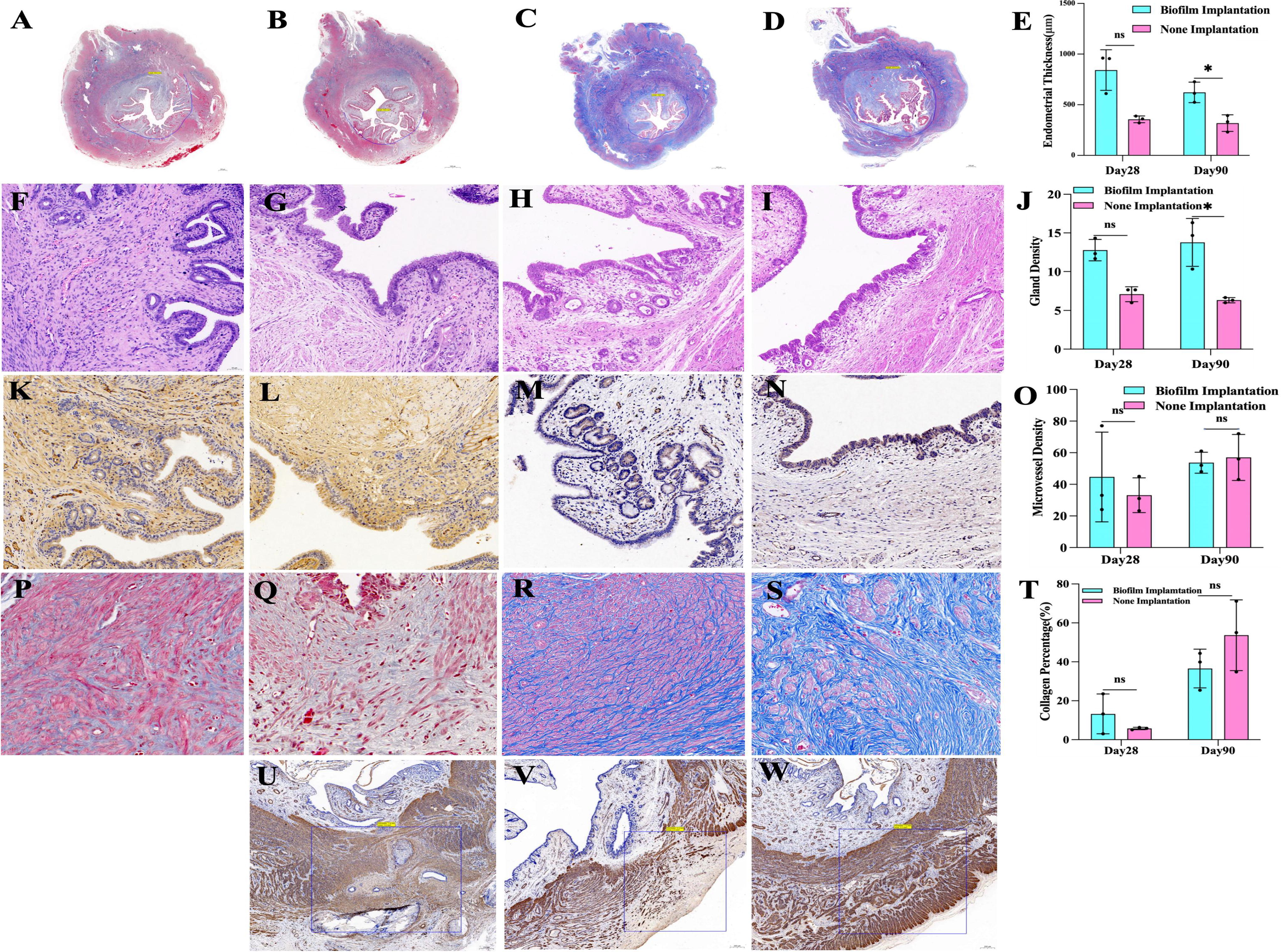
Morphological evaluation of regenerated endometrium and myometrial end-repair evaluation.
When we analyzed ER-α, PR, and α-SMA protein expression by IHC 90 days after surgery, we noted no difference between their levels in the repair sites of the GT/PCL biofilm implantation and the spontaneous regeneration groups (p > 0.05, Fig. 5H). The uteri from rabbits showing viable pregnancy were processed for analysis of ER-α and PR levels, and IHC revealed that the repair sites in the GT/PCL biofilm implantation group did not differ from those in the spontaneous regeneration group (p > 0.05, Fig. 5I).
No difference in immunohistochemical expression of key protein markers at 7, 14, and 28 days postoperation
Ki67 (Fig. 4A–B′), VEGF (Fig. 4C–D′), TGF-β (Fig. 4E–G′), and IGF-1 (Fig. 4H–J′) levels were measured by IHC in the tissues at the uterine repair site on days 7, 14, and 28 after surgery. Statistical analysis revealed that the mean optical density values for the four molecules at the repair site in the GT/PCL biofilm implantation group were not different from those in the spontaneous regeneration group (p > 0.05, Fig. 4K–M).
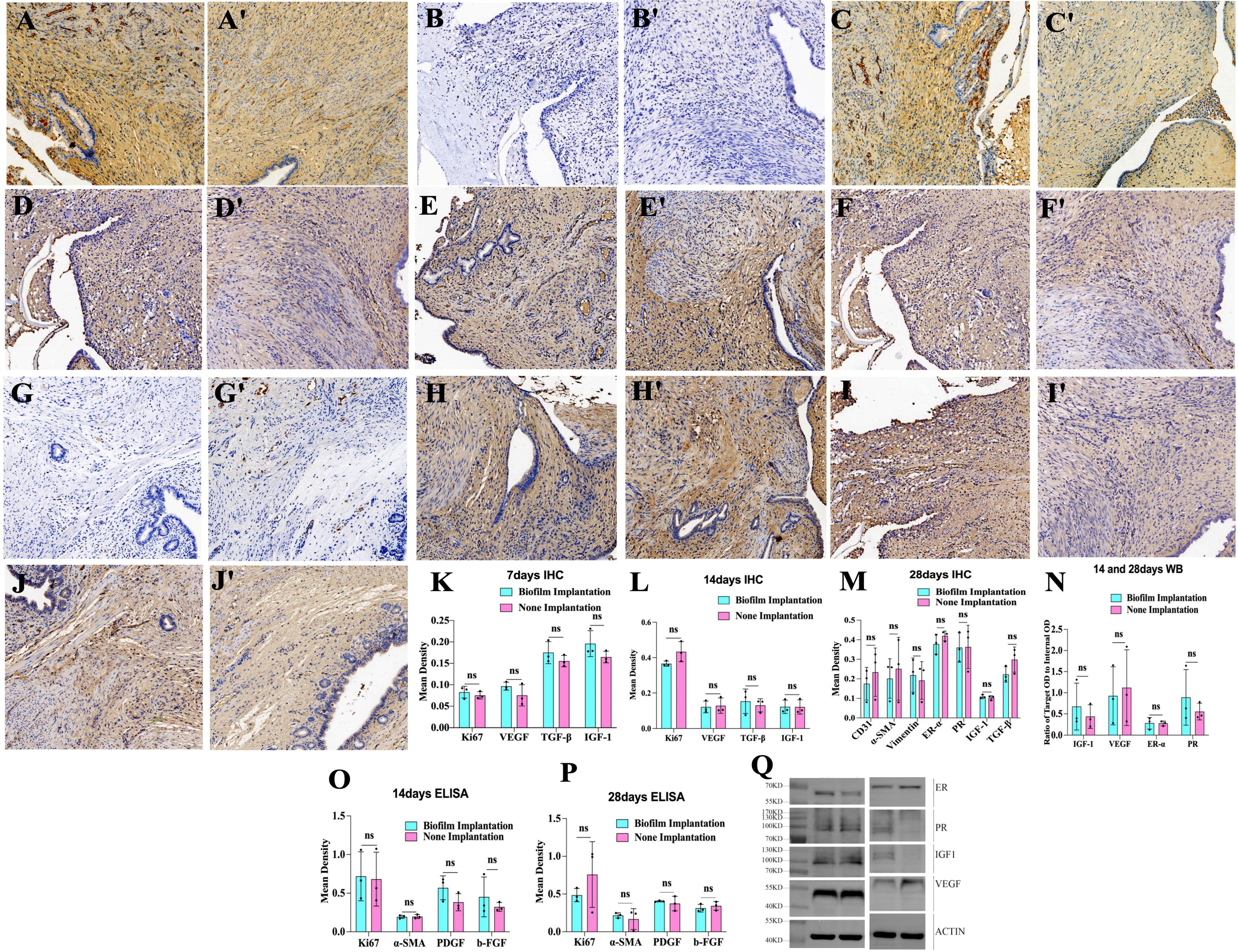
The expression levels of related protein molecules in the tissues after repair of the nodal uterus were observed at different times in the injury site after surgical modeling using IHC staining, Western blotting, and enzyme-linked immunoassay.

Endometrial maturity was quantified by IHC staining.
Western blot analysis confirmed that ER-α, PR, IGF-1, and VEGF expression in uterine repair site tissues were not different between the two groups at 14 and 28 days postoperatively (p > 0.05, Fig. 4N). ELISA analysis of b-FGF2, PDGF, α-SMA, and Ki67 in repaired uterine tissues 14 and 28 days postoperation depicted expression levels in the GT/PCL biofilm implantation side as not significantly different from those in the spontaneous regeneration side (p > 0.05; Fig. 4O and P).
GT/PCL implantation supported health pregnancies and normal fetal development
Fertile male rabbits (bucks) were mated naturally with 6 experimental female rabbits (does) 90 days after uterine injury surgery. To confirm fetal heartbeat, color ultrasonographic examination of the lower abdomen 10 days after mating revealed that 5 rabbits conceived successfully with uneventful pregnancies. Well-developed altricial rabbit fetuses were delivered by cesarean section at the expected time on the 28th day after mating (Fig. 1P); the placental attachment position was determined intraoperatively by marking with silk thread (Fig. 1L and M).
The functional recovery of pregnancy was determined to be grossly superior in the repaired uterine tissues with GT/PCL biofilm implantation compared with the spontaneous regeneration side. In those that underwent successful pregnancy, the number of embryos on the GT/PCL biofilm implantation side was significantly greater than that on the spontaneous regeneration side (p = 0.0008, Fig. 5J). On the biofilm side, there were also placental attachment points located within the implanted segment of the material itself, integrated within the intact myometrium that had developed within the scaffold. There was no visible abnormal protrusion or bulge at the implantation site, indicating that the elastic strain capacity of the myometrial smooth muscle fibers in the implanted biofilm segment was restored well enough to withstand the tension exerted on the myometrium by the growing fetus over the course of gestation (Fig. 2E and J).
4D Label-free whole-protein quantification revealed upregulation of immunoregulatory and metabolic pathways in biofilm-assisted uterine repair sites
The repaired uterine tissues were harvested on days 14 and 28 after injury for use in a 4D label-free whole-protein quantitative analysis assay. There were significant differences in protein expression levels between the experimental groups on postoperative day 28 (Fig. 6B) compared with day 14 (Fig. 6A). There were 159 differentially upregulated proteins and 45 downregulated proteins in the GT/PCL biofilm implantation side compared with the spontaneous regeneration group (Fig. 6B–G), and 76 upregulated proteins were expressed after acetylation of modification sites in differential proteins (Fig. 7A).
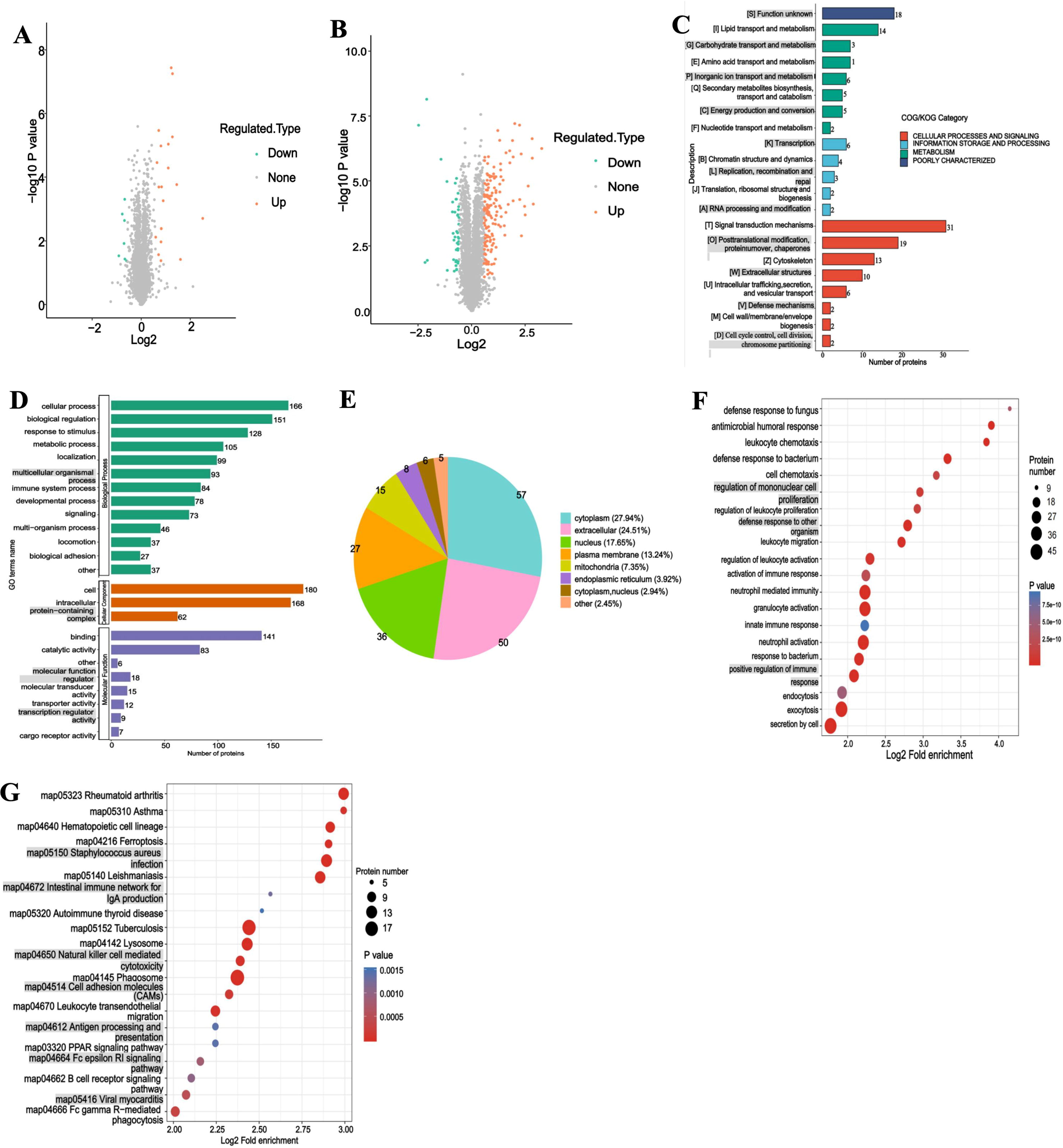
4D label-free, whole-protein quantification results of protein expression changes in the uterine tissue of the repaired site on the side implanted with GT/PCL biofilm versus the spontaneously repaired side.

4D label-free, whole-protein quantification results of protein expression changes after acetylation of modification sites in the uterine tissue of the repaired site on the side implanted with GT/PCL biofilm versus the spontaneously repaired side 28 days after hysterotomy injury.
Subcellular structure-localization analysis of the differential proteins showed that 56.58% of the proteins were distributed to cytoplasm, 22.37% to mitochondria, 10.53% to nucleus, 5.26% to extracellular matrix, 2.63% nucleus and plasma membrane, and 2.63% to other localization (Fig. 7D). The differentially expressed proteins preacetylation (Fig. 7A–F) were subjected to COG/KOG functional classification and GO secondary annotation functions, which revealed that the differentially upregulated proteins were involved in cellular processes (such as tricarboxylic acid [TCA] metabolic process, regulation of transmembrane transporter activity, actin cytoskeleton organization, and endocytosis), signaling, and metabolism (Fig. 6D and F). Further biological process analysis revealed that the differentially expressed proteins after acetylation of modification sites were principally involved in immune system activation and positive regulation of immune defense responses (Fig. 7E).
In addition, we found from the KEGG pathway enrichment analysis of the differential proteins that the upregulated proteins after acetylation of modification sites were significantly enriched in cellular adhesion molecules (CAMs), phagosome, ferroptosis, rap1 signaling pathways, hematopoietic cell lineage, complement and coagulation cascades, TCA cycle, carbon metabolism, and HIF-1 signaling pathways (Fig. 6G and 7F). PRM quantification experiments revealed that the nine proteins (HK2, CTSS, ITGB2, MPEG1, TFRC, CYBB, CORO1A, CD14, and ITGAM) were significantly upregulated in the uterine tissues of the GT/PCL biofilm implantation group compared with tissues of the spontaneously regenerated side (Table 1).
Upregulated Proteins in GT/PCL Biofilm Implantation Group vs Spontaneous Regeneration Group
GT/PCL, gelatin/polycaprolactone. Values of p < 0.05 for comparisons between GT/PCL biofilm implantation group and spontaneous regeneration group.
Discussion
A variety of materials have been applied in biomedical research for the regenerative repair of endometrial injury, and studies have achieved favorable pregnancy outcomes when assisting in the repair of small-area uterine injury in small-animal models.21,22 However, when the endometrium incurs a larger area of damage, such as when the area affected is over half of the organ, the regenerated endometrium cannot recover its original structure and normal pregnancy function without intervention.23,24 When a large incision is made in the myometrium, the proliferation and regeneration of smooth muscle cells (SMCs) and their arrangement pattern comprise the most problematic issues facing the recovery of the injured uterus to its original structure and function in pregnancy.25,26
Importantly, SMCs can undergo self-regeneration when placed in a proper environment or scaffold27,28; this provides a rationale for the optimization of biomaterials to promote SMC proliferation and alignment into a native conformation, thus guiding recovery of the injury site into a more normal and functional spatial structure.27,28 In this study, the alignment of myofibers in the repair site was more similar to that of the outer longitudinal shape of the inner ring of myofibers in normal myometrium with the GT/PCL membrane and theorizes that this ultimately led to the recovery of pregnancy function, and the regenerated endometrium was thicker and had more glands than the ipsilateral nonimplanted sites. Endometrial thickness and gland sufficiency are critical for the functional recovery of pregnancy after endometrial damage.29,30
There was no significant difference in the expression of ER-a, PR, a-SMA, vimentin, and CD31 protein between the two groups, confirming that the repair process of uterine tissue damage is spontaneous and that the implantation of GT/PCL biofilm will not have a negative effect on the endogenous regeneration and repair mechanism. In addition, there was no difference in the number of endometrial microvessels between the implanted GT/PCL biofilm and the nonimplanted sides. Therefore, it is possible that when uterine tissue injury occurs, the regenerated endometrial tissue and myofibers on the spontaneous repair side undergo self-proliferation, and that the various functional proteins and microvessel numbers attain levels that support pregnancy.
However, the original intercellular framework is destroyed and no longer supports endometrial proliferation, and the arrangement of regenerated myofibers loses its original order, resulting in reduced fertility. In contrast, implantable nano/micron-sized GT/PCL biofilms are able to provide spatial structures more suitable for cellular proliferation and thus allow the endometrial cells to self-proliferate.
The repair site with the implant then forms a structure that closely emulates the normal endometrium, enabling the repaired myofibers at the damaged ends of the myometrium to become arranged in a manner comparable to normal myometrium. These findings illustrate that, although collagen is more abundant in damaged myometrial repair sites than in normal myometrium, the repaired site is sufficient to support fetal growth to full term as long as the myofiber arrangement can tolerate the tension produced by an enlarged uterus during pregnancy. In this experimental paradigm, there was no change in the expression levels of VEGF, PDGF, TGF-β b-FGF2, and IGF-1 after biofilm implantation at the injury site, indicating that implanted GT/PCL played no major role in the signaling pathways in which these factors participated, nor did GT/PCL restore the original tissue structure through the signaling pathways involved by these factors.
Thus, the spatial structure and arrangement pattern of sodium micrometers possessed by the biofilm may have activated an endogenous signaling pathway that modulated the regenerative response to promote the proliferation of endometrial cells and uterine SMCs in a conformation typical of native, uninjured tissue. 31 A variety of natural and artificial materials have been applied to tissue injury sites with acceptable outcomes, suggesting that the provision of a spatial structure, such as engineered scaffolds, may be more important than the raw materials themselves in promoting injury repair.32–34 Proper nano- or micron-scale spatial structures provided to cells are more conducive to cellular growth and replication.35,36
To identify potential endogenous signaling pathways, we executed 4D label-free proteomic analysis of the treated uterine tissue; there were 76 differentially upregulated proteins after acetylation of modification sites in the tissue implanted with the GT/PCL biofilm compared with the spontaneously regenerated side. These postacetylation upregulated proteins were significantly enriched in the CAMs, phagosome, ferroptosis, and rap1 signaling pathways, the complement and coagulation cascades, and hematopoietic cell lineage, suggesting that the GT/PCL biofilms provided a suitable microenvironment for organizing the cells to display diverse functions.
Acetylation is a conservative and reversible protein modification in vivo, and nonhistone acetylation is involved in the regulation of metabolic pathways and metabolic enzyme activities. Signal transduction and energy metabolism exert both activating and potentiating regulatory effects on the immune system. 37 Investigators have demonstrated that cells conduct space sensing in nano- and submicron-sized materials, activating integrin-mediated signaling pathways to promote cellular proliferation and growth.31,35,38 In this study, the functional proteins in the phagosome signaling pathway were also upregulated after acetylation of modification sites, indicating that immunophagocytes are important in the regenerative repair of the injured uterus and the remodeling process.
Postacetylation upregulation of ferroptosis-related signaling pathway proteins provides insight into how ferroptosis functions during regenerative repair of the uterus.39,40 During the process of tissue damage repair, oxidative stress reactions occur, and iron homeostasis is closely related to oxidative stress reactions.41,42 The rap1 signaling pathway, which was also upregulated, is involved in a variety of cellular activities, such as the signal transduction of plasma membrane receptors, cytoskeleton rearrangement required for cell division, intracellular and matrix adhesion, and cell extravasation or fusion.43–45 In this study, the postacetylation upregulation of proteins involved in hemopoietic cell lineage signaling suggests that the structure and microenvironment provided by GT/PCL biofilm can provide a niche for hematopoietic stem cell maintenance and function.46,47
In addition, in this study, the upregulated acetylation of the complex and coagulation cascades signal pathway indicates that biofilm implantation enhanced the activation of a pathway known to be involved in tissue repair, compared with the nonimplanted side to the repair of uterine tissue after injury. 48 In summary, these data suggest that the immune system was activated within the nano- and submicrometer space provided by the GT/PCL biofilm, and that various immune cells exerted immunomodulatory and signaling effects and activated energy-metabolic pathways—including the TCA cycle, carbon metabolism, and HIF-1 signaling pathways. This provides a more favorable biologic environment for cellular proliferation and the growth of uterine tissue. The identification of specific mechanisms of action underlying the immunomodulation observed in our model requires further investigation.
In previous studies, while biological tissue engineering scaffolds loaded with bioactive substances and stem cells achieved favorable repair outcomes, the arrangement of the scaffolds concealed the biological roles occupied by biomaterials, growth factors, and the cells themselves as independent elements of injury repair, and the roles of scaffolds could not be investigated independently.49–51 This raises questions regarding precise interventions in regenerative repair processes. In addition, although the extant studies suggest a common underlying mechanism of action for the applied factors and cells in promoting tissue repair of uterine injury, there has yet to be any major breakthrough in this area, and further research is therefore needed.
Conclusions
In this study, it was demonstrated that a gradient layer biofilm fabricated by a 3D electrospinning technique using gelatin and polycaprolactone as raw materials could guide the regeneration and repair of a large-area endometrial injury and full-thickness fracture injury of the myometrium in rabbits, and that it restored the damaged endometrial and myometrial structures and pregnancy function. These results demonstrated that GT/PCL biofilms provided a favorable supporting structure and a microenvironment conducive to activating endogenous repair mechanisms. And the activated endogenous repair mechanisms involved the key proteins with functionality in immunomodulation and metabolism, and most of these upregulated protein modification sites have undergone acetylation modification in these signaling pathways, and can be used to elucidate the mechanism of endogenous tissue repair activation. The results of this study provide a basis for studying the roles of immune regulation, energy metabolism, and nonhistone acetylation in endogenous regeneration and repair mechanisms.
Footnotes
Acknowledgments
The authors acknowledge the laboratory animals that were sacrificed in this experiment. The authors also thank the experimental sites provided by the Laboratory Animal Center of Jinan Central Hospital, Shandong, China, and are grateful to friends for their assistance and support. Special thanks are due to Prof. Fuchang Li for his guidance, to Prof. Chunhong Song and to Prof. Hua Li, Lingzhi Ning (technician) at the Seed Rabbit Experimental Base of Shandong Agricultural University. The authors thank LetPub (![]() ) for linguistic assistance and presubmission expert review.
) for linguistic assistance and presubmission expert review.
Data Availability Statement
The raw/processed data required to reproduce these findings are available from the corresponding author on reasonable request.
Ethical Statement
All protocols were approved by the experimental animal ethics committee of the Provincial Hospital Affiliated to Shandong First Medical University (No. 2020-002).
Conflict of Interest
The authors declare that they have no known competing financial interests or personal relationships that could have appeared to influence the work reported in this article.
Disclosures Statement
No competing financial interests exist.
Funding Information
This work was supported by Shenzhen High-level Hospital Construction Fund (YBH2019-260), Shenzhen Key Medical Discipline Construction Fund (No. SZXK027), Sanming Project of Medicine in Shenzhen (No. SZSM202011016), and General Project of Shenzhen Science and Technology Innovation Commission (No. JCYJ20220531094012027).