
Abstract
Scaffolds made from cartilage extracellular matrix are promising materials for articular cartilage repair, attributed to their intrinsic bioactivity that may promote chondrogenesis. While several cartilage matrix-based scaffolds have supported chondrogenesis in vitro and/or in vivo, it remains a challenge to balance the biological response (e.g., chondroinductivity) with structural (e.g., robust mechanical performance, >1 MPa in compressive stiffness) and translational (e.g., ease of surgical implantation) considerations. Few studies have evaluated encapsulated cell viability within high-stiffness (>1 MPa) hydrogels. We previously fabricated one formulation of a high-stiffness (>3 MPa) pentenoate-functionalized, solubilized, devitalized cartilage (PSDVC) hydrogel that possessed an injectable, paste-like precursor for easy surgical application. In the current study, the characterization of the PSDVC material was expanded by varying the degree of functionalization (i.e., 0.45–1.09 mmol/g) and amount of crosslinker, dithiothreitol (DTT), to improve the reproducibility of the high compressive moduli and evaluate the viability of encapsulated human bone marrow-derived mesenchymal stem cells (hBMSCs) in high-stiffness cartilage matrix hydrogels. Prior to crosslinking, specific formulations functionalized with 0.80 mmol/g or less of pentenoate groups retained a paste-like precursor rheology. After crosslinking, these formulations produced hydrogels with greater than 1 MPa compressive stiffness. However, hBMSCs encapsulated in PSDVC hydrogels with lower functionalization (i.e., 0.57 mmol/g, no crosslinker) had a higher stiffness (i.e., 1.4 MPa) but the lowest viability of encapsulated hBMSCs (i.e., 5%). The middle PSDVC functionalization (i.e., 0.70 mmol/g) with DTT (i.e., 0.50 mmol thiols/g) demonstrated high cell viability (77%), high mechanical performance (1.65 MPa, 31% failure strain), and translational features (i.e., paste-like precursor, 1.5 min crosslinking time). For future evaluations of PSDVC hydrogels in cartilage repair, a middle functionalization (i.e., 0.70–0.80 mmol/g) with the addition of a crosslinker (i.e., 0.50 mmol thiols/g) had a desirable balance of high mechanical performance (i.e., >1 MPa compressive stiffness), high viability, and paste-like precursor for surgical translation.
Impact Statement
Cartilage matrix scaffolds are promising materials for cartilage repair. While cartilage matrix scaffolds have been chondroinductive, many have poor mechanical performance, or may lack the paste-like precursor rheology for surgical placement in irregular cartilage defects, factors that may limit impact in clinical translation. Improving upon high-stiffness pentenoate-functionalized, solubilized, devitalized cartilage (PSDVC) hydrogels from prior studies, several new formulations were investigated. The refined hydrogel balanced desirable cell viability (77%), high mechanical performance (1.65 MPa, 31% failure strain), and translational features (paste-like precursor, 1.5 min crosslinking). The new PSDVC hydrogel formulation may be a translational platform for supporting chondrogenesis/cartilage repair.
Introduction
Cartilage matrix is a promising raw material for promoting cartilage regeneration after acute injuries or osteoarthritic damage to cartilage. Cartilage matrix that has been devitalized (DVC) via freeze-thaw cycles, but not intentionally decellularized, may improve bioactivity and chondroinductivity compared with decellularized cartilage matrix.1–3 The major challenges of developing biomaterials for cartilage regeneration are developing materials that are translational in a surgical context (e.g., easy to deliver, good retention in an injury), while balancing structural (e.g., high mechanical performance) and biological considerations (e.g., biocompatible, chondroinductive).
While many cartilage matrix scaffolds are chondroinductive in vitro and in vivo, they have typically had low stiffness (i.e., <300 kPa compressive moduli2,4–7) compared with native cartilage (i.e., 1.8 MPa 8 ) and/or were more challenging to deliver surgically (i.e., low viscosity precursors that may leak after injection, pre-formed rigid scaffolds). For example, recent work with lyophilized, solubilized, crosslinked (e.g., carbodiimide, dihydrothermal, and/or glyoxal) cartilage matrix scaffolds, which has progressed into large animal models, demonstrated low stiffness and mechanical performance (∼1 kPa).2,5 Natural hydrogels in general have not typically been able to achieve >1 MPa in compressive stiffnesses without synthetic polymer reinforcements, 9 which adds complexity without adding value to chondroinductivity. Robust mechanical performance is vital for survival of an implant in an articulating joint space, 10 and may enable early weight-bearing activities that accelerate cartilage repair. 11
We previously focused on improving the mechanical performance and clinical translatability of cartilage matrix-based hydrogels.12,13 A high-stiffness (>3 MPa) pentenoate-functionalized, solubilized, devitalized cartilage (PSDVC) hydrogel was fabricated, with a paste-like, fast crosslinking precursor. 12 The injectable, paste-like precursor may enhance translation by simplifying surgical placement into articular cartilage defects (no leakage), 14 and the fast in situ ultraviolet (UV) crosslinking (1.7 min) may enhance material retention within the defect. The focus has now turned to improving the reproducibility of the high compressive modulus and biological activity. In previous work, the addition of raw DVC particles into cartilage3,15 and synthetic 16 hydrogels improved chondrogenesis. However, the DVC particles in bioprinted PSDVC hydrogels reduced the compressive modulus and reduced the cell viability from high shear rates. 12 Therefore, we looked at alternate variables to improve the reproducibility of the stiffness and improve cell viability. Given that the degree of functionalization can considerably alter the precursor rheology and hydrogel performance, 13 there is a need to characterize PSDVC formulations of varying functionalization to improve reproducibility of obtaining stiff hydrogels with paste-like precursors. Second, there is a need to understand the effects of high-stiffness hydrogels on encapsulated bone marrow-derived mesenchymal stem cell (BMSC) viability. Few studies have evaluated encapsulated cell viability in stiff hydrogels (i.e., >1 MPa in compressive stiffness), and such studies were performed using synthetic hydrogels.17,18 Therefore, assessing viability in cartilage matrix hydrogels that are greater than 1 MPa is a critical next step toward tuning the translational PSDVC hydrogel platform to better support chondrogenesis.
The goals of the current study were to (1) fabricate several PSDVC formulations [i.e., varying functionalization and crosslinker, dithiothreitol (DTT)], (2) identify formulations that maintain translational features in a surgical context (i.e., paste-like precursor, fast-crosslinking, high-stiffness hydrogel, high failure strain), and (3) evaluate the viability of human BMSCs (hBMSCs) encapsulated within the new formulations of high-stiffness PSDVC hydrogels.
Materials and Methods
Materials
A condensed list of materials is listed here, with the full list in the Supplementary Data S1. The following were all from Millipore Sigma (Burlington, MA): hydrochloric acid (HCl, HX0603-75), N,N-dimethylformamide (DMF, 319937), 4-(dimethylamino)pyridine (DMAP, 107700), 4-pentenoic anhydride (PA, 471801), 3-(trimethylsilyl)propionic-2,2,3,3-d4 acid sodium salt (TMSP; 269913), phosphate-buffered saline (PBS; P3813), DL-DTT (D0632), sodium chloride (NaCl, 746398), dimethylmethylene blue (DMMB, 341088), and 4′,6-Diamidino-2-phenylindole (DAPI, D9542). The following were from Thermo Fisher Scientific (Waltham, MA): sodium hydroxide (NaOH, S318), ethylenediaminetetraacetic acid, disodium salt (EDTA, 17892), the Quant-iT PicoGreen dsDNA Assay Kit (P7589), PrestoBlue HS Cell Viability Reagent (P50201), and LIVE/DEAD® Viability/Cytotoxicity Kit for mammalian cells (L3224). Deuterium oxide (D2O, DLM-4–100) was from Cambridge Isotope Laboratories, Inc. (Andover, MA). The Chondrex Type II Collagen Detection Kit Multi-Species (NC203401) was from Chondrex (Woodinville, WA). Lithium phenyl-2,4,6-trimethylbenzoylphosphinate (LAP, TCL0290-1G) was from TCI America (Portland, OR). hBMSCs (Donor: 310310, 25 years, male, MSC-003) were from RoosterBio® (Frederick, MD). Transforming growth factor (TGF)-β3 (8420-B3) was from R&D Biosystems (Minneapolis, MN). The Phalloidin-iFluor 488 Reagent (ab176753) was from Abcam (Boston, MA).
Synthesis of PSDVC
Porcine knees (mixed Hampshire or Duroc cross, 14–18 kg, females, 6–8 weeks, 38 knees) were used for harvesting articular cartilage and synthesizing PSDVC (Fig. 1a), as previously described.12,13 Briefly, to form solubilized and devitalized cartilage (SDVC), cartilage was coarse-ground, cryoground (SPEX 6770 Freezer/Mill; SPEX SamplePrep, Metuchen, NJ), and solubilized with pepsin (10 mg/mL DVC, 1 mg/mL pepsin in 0.1 M HCl, 2 or 27 days). After neutralizing with NaOH and centrifuging, the solubilized DVC in the supernatant was retained and dialyzed, while any remaining insoluble solid DVC particles were discarded. After lyophilizing the SDVC, PSDVC was synthesized by reacting SDVC (1 g in 150 mL deionized water [DI], 100 mL DMF, 250 mg DMAP) with varying amounts of PA (0.96, 2.4, 4.8, or 9.6 mL) while maintaining the pH between 8 and 9 with NaOH (4 M) for 1 h. The reaction was precipitated in acetone, and then centrifuged (6000 × g, 3 min) pellets were dissolved in DI (150 mL) before dialysis, lyophilization, and storage at −20°C.

Graphic illustration of the synthesis of pentenoate-functionalized, solubilized, devitalized cartilage (PSDVC) and hydrogel formation from a paste-like precursor to a photocrosslinked hydrogel.
Nuclear magnetic resonance and biochemical characterization of PSDVC
Nuclear magnetic resonance (NMR) spectra of SDVC or PSDVC samples (∼2 mg in 0.7 mL D2O, 1 mg/mL TMSP) were collected on a VNMRS-500 MHz Spectrometer (Varian, Palo Alto, CA) or on a Jeol 500 MHz spectrometer (Jeol USA, Peabody, MA), and analyzed using MestReNova software v.12.0.1 (Mestrelab Research, Santiago de Compostela, Spain), as previously described.12,13 Briefly, 1 H NMR spectra were collected at 80°C (16 scans, 35 s recycle delay, 90-degree pulse width, 60 s preacquisition delay).
For collagen II quantification of DVC, SDVC, or PSDVC materials (n = 3), the collagen was solubilized by five pepsin digestions and an elastase digestion (see Supplementary Data S1 for solubilization details), and then quantified via enzyme-linked immunosorbent assay using the Chondrex Type II Collagen Detection Kit.
For determining the DNA and glycosaminoglycan (GAG) content in DVC, SDVC, or PSDVC samples (n = 3), the samples (1–3 mg) were digested in papain (5.79 U/mL, 5 mM N-acetyl-L-cysteine, 5 mM EDTA, in PBS) overnight at 60°C. DNA was quantified using the PicoGreen assay and the DMMB assay, as previously described. 19
Hydrogel precursor rheology
The viscosity, yield stress, and storage modulus recovery (n = 3) of the hydrogel precursors were evaluated using a 20-mm crosshatched plate (500 µm gap, room temperature) on a Discovery Hybrid Rheometer-2 (DHR-2, TA Instruments, New Castle, DE), as previously described. 13 Details are in the Supplementary Data S1.
Hydrogel formulations and fabrication
PSDVC materials (10 w/v%) were formulated from batches of varying pentenoate functionalization (i.e., 0.57, 0.70, 0.80, 1.09 mmol/g). PSDVCs were dissolved in PBS containing a photoinitiator, LAP (2.2 mM), and varying amounts of crosslinker, DTT (Table 1). To make bulk hydrogels for mechanical testing and in vitro studies, precursors were prepared in a rubber mold (1 mm depth) between two glass slides, and then crosslinked with a handheld UV light (EB-160C, 365 nm bulb, Spectro-UV, Farmingdale, NY) (Fig. 1b) according to the determined crosslinking times (Table 1). Cylindrical hydrogels (6 mm diameter) were punched out with a biopsy punch.
DTT Concentrations and Hydrogel Crosslinking Times
DTT, dithiothreitol; PSDVC, pentenoate-functionalized, solubilized, devitalized cartilage.
Hydrogel crosslinking time, stiffness, failure, and swelling
The aforementioned DHR-2 rheometer with an 8-mm geometry was used to determine the crosslinking times (n = 3–5) of PSDVC hydrogel precursors and the mechanical performance (n = 4–6) of the fully crosslinked hydrogels. For crosslinking time, the base plate was a handheld UV light, as previously described. 12 Briefly, hydrogel precursor was loaded and trimmed (1 mm gap) and the storage moduli were measured during low oscillatory shear (10 Pa) for 30 s and then for 10 min while the UV light was on. The crosslinking time was calculated as the time at which samples reached 95% of their final storage moduli.
The compressive stiffnesses, absorption ratios, swelling ratios, and failure measurement of fully crosslinked hydrogels (n = 4–6) were determined as previously described.16,20 After hydrogels were swollen in PBS at 37°C overnight, the diameters were measured on a micrometer (Mitutoyo, Kawasaki, Japan). A tare load was applied (0.01 N, height: 1.05 ± 0.06 mm) and hydrogels were compressed (5 µm/s, 0.48% strain/s) at room temperature under dry conditions until failure. A custom MATLAB® app (MathWorks, Natick, MA) (available as “Compressive Moduli App” in the MATLAB Add-On Explorer, and online: https://github.com/ekiyotake/Compressive-Moduli-App) was employed to calculate compressive moduli, shear moduli, Ogden parameters, and ultimate stresses and strains (see Supplementary Data S1 for details).16,20
The absorption ratios (swollen mass divided by fabricated mass) and swelling ratios (swollen mass divided by dry mass) were calculated from the hydrogel masses weighed immediately after fabrication, after swelling in PBS overnight at 37°C, and after lyophilization.
Cell viability studies
PSDVC materials were sterilized and prepared with final concentrations: 10 w/v% PSDVC, 2.2 mM LAP, 10 × 106 cells/mL, and varying DTT (Table 1). See Supplementary Data S1 for precursor preparation and hBMSC expansion methods. In the first study, three PSDVC materials of different functionalization with three different stiffnesses (i.e., 239, 594, 1456 kPa) were selected to fabricate bulk hydrogels (i.e., crosslinked between glass slides in a sterilized rubber mold and cylindrically punched) using no DTT (n = 3–4). Bulk hydrogels with cells were cultured in control or TGF-β3 medium for 1 or 14 days in hypoxia (5% O2) (see Supplementary Data S1 for media compositions). In the second study, the hydrogel precursors of the two highest stiffness formulations (i.e., 594, 1456 kPa, no DTT) were bioprinted (n = 3) into cylindrical grids (10 mm diameter, 1 mm tall, 4 layers) on a BioAssemblyBot® 400 (Advanced Solutions, Louisville, KY) using printing parameters listed in Table 2. Bioprinted scaffolds with cells were UV crosslinked and cultured for 1, 7, or 14 days in control medium under hypoxia. In the third study, a medium-stiff PSDVC (604 kPa, no DTT) was fabricated into bulk hydrogels with varying amounts of DTT (0, 0.25, or 0.5 mmol/g DTT) (n = 3) and cultured for 1 or 10 days in control medium under hypoxia.
Bioprinting Parameters
Metabolic activity and DNA content were measured using the PrestoBlue reagent and the PicoGreen assay (see Supplementary Data S1 for details). In the first study, metabolic activity for each sample was divided by its corresponding DNA content, and then normalized to the average metabolic activity/DNA content of the stiffest hydrogel group in control medium (see Supplementary Fig. S1 for metabolic activity and DNA content alone). In the second study, metabolic activities were normalized to the blank PrestoBlue solution. Live/dead and F-actin/DAPI staining were performed with the LIVE/DEAD® kit, the Phalloidin-iFluor 488 reagent, and DAPI as we have done previously. 13 Hydrogels were imaged using a Leica TCS SP8 confocal laser scanning microscope (Leica Microsystems, Wetzlar, Germany). Fiji 21 and the Analyze Particles tool were used to count the number of live or dead cells for determining viability, and quantify cell morphology parameters (i.e., circularity, where 1 is a perfect circle) using custom macros. See Supplementary Data S1 for imaging parameters and Fiji macros used.
Statistics
All statistical analyses were performed in GraphPad Prism version 10.3.1 for macOS (GraphPad Software, San Diego, CA, www.graphpad.com). A one-way or two-way Analysis of Variance (ANOVA) with Tukey’s post hoc test were used to analyze all data. A level of p < 0.05 was considered significant. All results were reported as mean ± standard deviation.
Results
Characterization of PSDVC
Pentenoate-functionalization of PSDVC batches increased from 0.45 to 1.09 mmol/g of dry material (Fig. 2a) with increasing amounts of PA. The collagen II contents (Fig. 2b) of the SDVC and PSDVC batches were 43% and 2–39% of the original DVC collagen II content (p < 0.0001), respectively. The DNA contents (Fig. 2c) of all the SDVC and PSDVC batches were 5–15% of the DNA content of the original DVC (p < 0.0001). The GAG contents (Fig. 2d) of all the PSDVC batches were 18–40% greater than that of the DVC (p < 0.05), and the 0.45, 0.65, and 1.09 mmol/g PSDVC batches had 11–31% greater GAG content than that of the SDVC (p < 0.05).
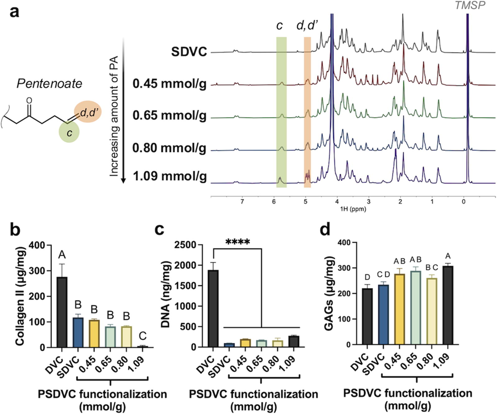
PSDVC batches were synthesized with varying functionalization. Compared with DVC, PSDVC batches had decreased collagen II content, but increased glycosaminoglycan (GAG) content and decreased DNA content (per mg of material).
Precursor rheology and crosslinking time
The 0.45, 0.65, and 0.80 mmol/g PSDVC precursors had a paste-like consistency (Fig. 3a) and had 612–1228 times higher viscosities (Fig. 3b) than that of the 1.09 mmol/g precursor (p < 0.0001). The yield stress (Fig. 3c) of the 0.80 mmol/g precursor was 28% greater than that of the 0.45 mmol/g precursor (p < 0.05), which in turn was 34% greater than that of the 0.65 mmol/g precursor (p < 0.05). The 1.09 mmol/g precursor did not possess a yield stress or a storage modulus recovery. For the other functionalizations, storage modulus recoveries (Fig. 3d) ranged from 64% to 70%, with no significant differences among those groups. The crosslinking time of the PSDVC precursors (Fig. 3e) decreased from 7.4 ± 0.9 to 1.5 ± 0.4 min as the amount of DTT crosslinker increased, regardless of functionalization (p < 0.0001).

The rheology of PSDVC precursors (i.e., before crosslinking) were paste-like in consistency with yield stresses (>450 Pa) and storage modulus recoveries (>60%), except for the highest functionalized PSDVC (1.09 mmol/g), which had low viscosity and no yield stress. The crosslinking time of all PSDVC batches decreased from ∼8 min down to ∼1.5 min with increasing amounts of DTT.
Hydrogel compressive moduli and swelling
Increasing the functionalization decreased the compressive moduli (Fig. 4a), where the 0.57 mmol/g PSDVC hydrogels had compressive moduli 1.6 times greater than those of the 0.70 and 0.80 mmol/g hydrogels (p < 0.0001), which in turn were 47 times greater than those of the 1.09 mmol/g hydrogels (p < 0.0001). With increased amounts of DTT (i.e., 0.25 and 0.50 DTT), the compressive moduli were 1.5–2.1 times greater than those crosslinked with lower amounts of DTT (i.e., 0 or 0.10 DTT) (p < 0.001). For water absorption ratios (Fig. 4b), the highest functionalization of 1.09 mmol/g hydrogels had 32–43% greater absorption than those of all other hydrogels (p < 0.0001). DTT minimally affected the absorption ratios, except with the 1.09 mmol/g hydrogels, where the hydrogels with 0.25 DTT had 12–44% greater absorption than those crosslinked with other amounts of DTT (p < 0.0001). Similar to absorption ratios, the swelling ratios (Fig. 4c) of the 1.09 mmol/g hydrogels were 27–34% greater than those of all other PSDVC functionalizations. DTT had a greater effect on swelling ratio than the absorption ratios, where hydrogels with no DTT had 4–18% greater swelling ratios that those of hydrogels crosslinked with other DTT amounts (p < 0.01).

As functionalization of PSDVC increased, the compressive moduli of fully crosslinked hydrogels decreased, while the absorption and swelling ratios increased. As DTT concentration increased, generally the compressive moduli increased and swelling ratio decreased.
Hydrogel failure performance
Both the Ogden fits (fitted to the majority of the strain region) and the linear fits (fitted to a more conventional linear strain region, i.e., 15–20% strain) accurately fit the respective data range (>0.99 R2 values) (Fig. 5a). Generally, the shear moduli (Fig. 5b) followed the trends of the compressive moduli, where the shear moduli of the 0.57 mmol/g PSDVC hydrogels were 1.4 times greater than those of the 0.70 mmol/g hydrogels (p < 0.0001), which in turn were 5 times greater than those of the 1.09 mmol/g hydrogels (p < 0.0001). In terms of DTT, the shear moduli of hydrogels crosslinked with most DTT (i.e., 0.50 DTT) were 1.3–2.3 times greater than those of hydrogels crosslinked with less DTT (p < 0.001). Shear moduli of hydrogels crosslinked with 0.25 DTT were 29–78% greater than those crosslinked with no or 0.10 DTT (p < 0.05). All hydrogels had high nonlinearity parameters (i.e., α > 10, Fig. 5c). Hydrogels with lower functionalization (i.e., <0.70 mmol/g), had 33–47% higher nonlinearity parameters than those of the highest functionalization (i.e., 1.09 mmol/g) (p < 0.0001). For ultimate strains at failure (Fig. 5d), the highest functionalized 1.09 mmol/g hydrogels had 45–52% greater failure strains than those of the lower functionalized hydrogels (i.e., 0.57 and 0.70 mmol/g) (p < 0.0001). The ultimate stresses (Fig. 5e) of the lowest functionalized 0.57 mmol/g hydrogels were 1.2 times greater than those of the 0.70 mmol/g hydrogels (p < 0.0001), which in turn had 2.7 times greater ultimate stresses than those of the highest functionalized 1.09 mmol/g hydrogels (p < 0.0001).

While PSDVC hydrogels had high nonlinearity (>10), the shear moduli from the Ogden model of the majority of the strain region (until failure) still resulted in the same trends as the compressive moduli determined from the more conventional linear fit in a limited strain region (15–20%). All PSDVC hydrogels had ultimate strains above 33%.
Viability of hBMSCs in bulk hydrogels with no DTT
The metabolic activity (normalized to DNA content and to the stiffest group in control medium) (Fig. 6a) and viability (Fig. 6b) of encapsulated hBMSCs in bulk PSDVC hydrogels (no DTT) both decreased with increasing stiffness. There were minimal effects of control versus TGF-β3 supplemented media on metabolic activity (per ng DNA) and viability. After 1 day and regardless of the medium, hBMSCs in the softest hydrogel (239 kPa) had 12–177 times greater metabolic activities (per ng DNA) than those in all other hydrogels (p < 0.0001). Comparably, the day 1 viabilities of hBMSCs in the softest hydrogel were ∼4 times greater than those in the medium stiffness hydrogel (p < 0.0001), which in turn were 4–9 times greater than those in the stiffest hydrogel (p < 0.01). On day 14, the cells in the softest hydrogels had ∼4 times greater metabolic activities (per ng DNA) than those in the medium stiffness hydrogel (p < 0.0001), which in turn were ∼7 times greater than those in the stiffest hydrogel (p < 0.05). Comparably, the day 14 viabilities of cells in the softest and medium stiffness hydrogels were three and seven times greater than those in the stiffest hydrogel (p < 0.001). The live/dead staining (Fig. 6c) supported the metabolic activity (per ng DNA) and viability analyses with few live cells and mostly dead cells in the stiffest hydrogel. On day 1, all the cells had rounded morphologies, and on day 14, the live cells in all groups had spread morphologies.

The viability and metabolic activity (per ng of DNA) of encapsulated human bone marrow-derived mesenchymal stem cells (hBMSCs) decreased within PSDVC hydrogels of increasing stiffness.
Viability of hBMSCs in bioprinted hydrogels with no DTT
The metabolic activities of encapsulated hBMSCs (Fig. 7a) immediately after printing (day 0) and after 1 day, were ∼2.2 times greater in the 594 kPa hydrogel than those in the 1456 kPa hydrogel (p < 0.001). By day 7, there was no metabolic activity, and no visible live cells, in the 1456 kPa hydrogels (confirmed with live/dead staining, Fig. 7b). However, in the 594 kPa hydrogel, a few live cells with spread morphology were present in the bioprinted strut. After 14 days, the 1456 kPa hydrogel struts appeared to have decreased in size and turned opaque, whereas the 594 kPa hydrogel struts appeared similar to their original widths (Fig. 7c). Macroscopically, some of the pores of the 594 kPa hydrogel were partially filled in with what appeared to be new matrix. F-actin/DAPI staining and imaging through the entire hydrogel (1 mm depth) revealed that the pores were filling in from the bottom with hBMSCs and potentially new matrix. In contrast, the 1456 kPa hydrogels showed no filled in pores or F-actin stained hBMSCs.
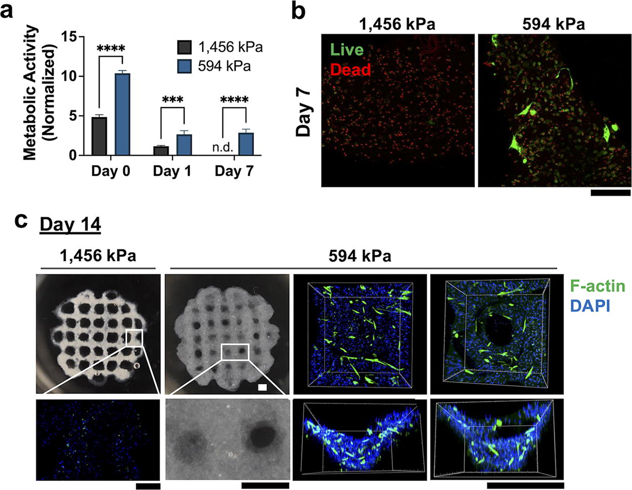
The encapsulated hBMSCs in the bioprinted 594 kPa PSDVC hydrogel had ∼twofold greater metabolic activity than the 1456 kPa PSDVC hydrogel immediately after printing (day 0) and after 1 and 7 days of culture.
Viability of hBMSCs in bulk hydrogels with varying DTT
The viabilities of hBMSCs encapsulated in bulk PSDVC hydrogels were 30% higher on day 1 (>77%) than those on day 10 (>60%) (p < 0.05) but showed no differences across crosslinker concentrations (i.e., 0, 0.25, or 0.5 DTT) on either day 1 or 10 (Fig. 8a). From the live/dead imaging of a 200 µm z-stack inside the hydrogel (Fig. 8b, left two columns), the encapsulated cells in all hydrogels exhibited a rounded morphology on day 1 with similar circularities (Fig. 8c) across groups (0.63 ± 0.03). On day 10, the cell morphology became more spread compared with day 1, where cells on day 1 had 91% greater circularity than those of cells on day 10 (p < 0.0001). On day 10, the highest concentration of DTT (i.e., 0.50) had 63–71% greater circularity (0.45 ± 0.07) compared with those in hydrogels with 0.25 DTT or no DTT (p < 0.001), respectively. In contrast, F-actin/DAPI staining of cells from a single image on the hydrogel surface (Fig. 8b, right column) showed increased cytoskeletal spreading as the DTT/stiffness increased.
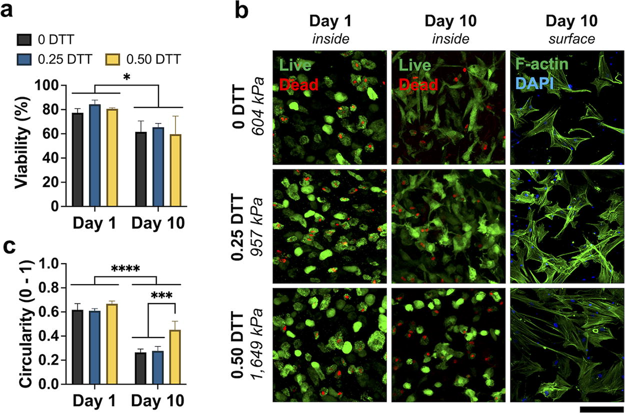
Encapsulated hBMSCs had high viability in bulk PSDVC hydrogels crosslinked with DTT over 10 days, with cell morphologies that were less spread inside stiffer (i.e., crosslinked with more DTT) hydrogels, but more spread on the exterior of the stiff hydrogels compared with the softer hydrogels.
Discussion
To broaden the characterization of our high-stiffness, fast-crosslinking PSDVC hydrogel, several PSDVC formulations were fabricated and characterized by varying functionalization and crosslinker concentrations. With clinical translation in mind, an approximate maximum functionalization (i.e., ≤ ∼0.80 mmol/g) was identified that maintained paste-like precursor rheology beneficial for both extrusion bioprinting and future surgical translation. The trend of high functionalization (i.e., 1.09 mmol/g) reducing the paste-like rheology is consistent with our past observation of a methacryloylated SDVC (MeSDVC). 13 While low viscosity formulations may still be beneficial for other bioprinting formats (e.g., ink-jet, digital light processing printing), 22 our criteria to surgically fill cartilage defects without leakage required an injectable, paste-like precursor.
We previously synthesized the stiffest cartilage matrix hydrogels to date with our 3 MPa PSDVC hydrogels (compressive stiffness, 0.32 mmol/g of functionalization, 0.50 DTT). 12 The current study found that increasing the PSDVC functionalization to more than 0.32 mmol/g decreased the stiffness, which followed the same trend as our previous MeSDVC hydrogels. 13 It was evident that the synthesis resulted in batch-to-batch variability, which decreased the reproducibility of the final hydrogel functionalization, stiffness, and other characteristics. In the current study, the rigor of the PSDVC synthesis was improved by identifying an approximate upper bound of functionalization (≤0.80 mmol/g) that resulted in hydrogel formulations that exceed 1 MPa in compressive stiffness, which was remarkable considering most cartilage matrix scaffolds are typically under 0.3 MPa.
Stiffness is a common measure of mechanical performance, but failure testing may be just as important as a clinical outcome and is not routinely performed for cartilage matrix scaffolds. 23 While compression testing is subject to higher variability than other recommended tests for characterizing cartilage failure (e.g., tensile testing with single edge notch test 23 ), the compressive test is straightforward, where both failure and stiffness can be characterized from a single compressive test. The Ogden model is a valuable analysis tool for failure data because it enabled an evaluation of mechanical performance over the full range of strain instead of an arbitrarily selected range for a linear fit. The Ogden model was selected over other nonlinear models because of its ability to accurately characterize the nonlinear behavior of the PSDVC hydrogels while being relatively easy to model with two parameters of the shear modulus, G, and nonlinear parameter, α. Other nonlinear models such as the neo-Hookean model (which is a simpler Ogden model when α = 216,24) had high accuracy for synthetic hydrogels (e.g., polyethylene glycol (PEG)/agarose interpenetrating network hydrogels). 24 However, for natural hydrogels (e.g., hyaluronic acid/PEG/DVC hydrogels), the accuracy of the Ogden model was greater for the full range of data compared with the more limited neo-Hookean model. 16 As we discussed in Nedrelow et al., 16 other models with many parameters may improve accuracy of stress predictions, but at the cost of increasing complexity, which may hinder widespread adoption. The one-term Ogden model has been used for over 50 years for modeling a range of materials such as rubber, several types of tissues, and most notably, hydrogels. While the Ogden is a versatile and accurate model for a variety of hydrogels, the implementation is more complicated than the conventional linear modeling, therefore, to aid widespread use, we have published the MATLAB app used in the current study in the MATLAB Add-On Explorer.
In the current study, all PSDVC hydrogels had improved failure strains (i.e., 31–56% strain) compared with previous MeSDVC hydrogels (i.e., ∼7.5% ultimate strain, 8 or at least 20% strain 13 ), possibly from the increased flexibility of the five-carbon chain of the pentenoate–DTT–pentenoate bonds compared with the shorter methacryloyl–methacryloyl bonds. The higher failure strains of PSDVC hydrogels may support limited weight-bearing activities, such as standing and walking (i.e., 5–10% strains 25 ) after implantation to potentially facilitate cartilage repair. 11 It is recommended to incorporate failure testing for all biomaterials developed for cartilage repair in the future. Additionally, other properties may need to be characterized, such as wear resistance to mimic the shear forces that occur during walking.
Utilizing a small di-functional crosslinker, DTT, for crosslinking PSDVC hydrogels was beneficial for decreasing the crosslinking time and improving the compressive modulus. DTT enabled faster crosslinking (i.e., from 8 to 1.5 min) via thiol-ene click chemistry compared with the pentenoate–pentenoate crosslinking that occurs without DTT. To compare with other chemistries, in previous work, the pentenoate functionalization for SDVC materials resulted in faster crosslinking than methacryloylation. 12 However, the pentenoate’s thiol-ene click chemistry will likely result in slower crosslinking times compared with other thiol-ene click chemistries (norbornene 26 ), which could be used in the future to further decrease crosslinking time. In terms of compressive moduli, the degree of functionalization had a greater influence compared with DTT concentration. While the lowest functionalized materials did not need DTT to exceed 1 MPa in stiffness, DTT was beneficial in pushing the middle functionalized PSDVC materials (i.e., 0.70 and 0.80 mmol/g) above the 1 MPa threshold.
The evaluation of cell viability was the next vital step in further developing these translational, high-stiffness cartilage-based hydrogels for cartilage repair. To avoid confounding the viability results with the potential cytotoxicity of DTT, cells were encapsulated in PSDVC hydrogels without DTT and found that the lowest functionalized/stiffest PSDVC hydrogel (0.57 mmol/g, 1456 kPa) had low cell viability (∼5%). However, the high stiffness alone may not have directly resulted in lower viability because the viability on day 1 was greater than 77% when DTT was used to obtain stiffer hydrogels (1649 kPa, 0.70 mmol/g, 0.50 DTT). Additionally, others have found high BMSC viability (i.e., >85%) in synthetic hydrogels up to 2 MPa. 17 High shear from the paste-like precursor extrusion through a needle is known to reduce cell viabilities,12,27 which may be responsible for lower viabilities observed in the current study in the bioprinted hydrogels compared with the bulk hydrogels. Correspondingly, in the bulk hydrogel study it may be possible that the higher viscosity of the lower functionalized PSDVC precursor caused greater shearing during the precursor mixing process that reduced viability.
Looking forward, the medium functionalized PSDVC hydrogels (0.70 mmol/g) with 0.25 or 0.50 DTT were able to balance high cell viability and translational characteristics (i.e., paste-like precursor, 0.96–1.65 MPa stiffness, 1.5–3 min crosslinking time, 21–31% failure strain). The extensive characterization of numerous formulations improved quality control for reliably producing mechanically robust, translational, and biocompatible PSDVC hydrogels. Intriguingly, the stiffest medium functionalized PSDVC formulation (0.70 mmol/g, 0.50 DTT) promoted rounded morphologies of live hBMSCs within the hydrogels after 10 days, which is a characteristic of a chondrogenic phenotype. Several in vitro studies have explored the effect of stiffness on chondrogenesis, for a given material28–31 ; however, there are inconclusive results on which stiffness range may best promote chondrogenesis. Moreover, stiffness is not an independent variable, given that other variables are typically used to alter stiffness (e.g., degree of crosslinking, molecular weight, concentration). 32 Stiffness may not directly correlate with chondrogenesis, but faster relaxing hydrogels with greater viscous than elastic characteristics have promoted a more chondrogenic phenotype of MSCs in vitro on isoelastic hydrogels. 33 PSDVC hydrogels previously demonstrated stress-relaxing behavior, 12 which may be one of many plausible mechanisms for high-stiffness PSDVC hydrogels to enhance chondrogenesis. Further characterization of additional mechanical properties of the leading PSDVC hydrogel formulation (e.g., stress relaxation/equilibrium modulus) with correlation to mechanotransduction and chondrogenesis of encapsulated hBMSCs will help further tune PSDVC hydrogels for cartilage repair.
In addition to chondrogenesis, the analysis of new matrix synthesis will be essential in future studies. There are challenges in distinguishing new matrix production from the existing matrix already in cartilage-based hydrogels, 34 such as the current study’s PSDVC hydrogels and previously developed MeSDVC hydrogels.3,8,15 While conventional assays for collagen II and GAG content generally do not differentiate between new and existing matrix, recent methods using metabolic labeling elucidate spatial and temporal production of new proteins (e.g., azide-modified amino acids) and proteoglycans (e.g., azide-modified sugar analogs). Such analyses will enable determination of new matrix synthesis within PSDVC hydrogels in future studies.
Conclusions
The evaluation of PSDVC functionalization and crosslinker concentration revealed that the lower functionalized hydrogels reduced cell viability, and the highly functionalized precursors lost their paste-like precursor. However, the medium functionalized hydrogels (i.e., 0.70 mmol/g) crosslinked with DTT balanced the translational features (i.e., paste-like precursor, high-stiffness, fast-crosslinking, improved failure strain) with high cell viabilities. Specifically, one formulation of PSDVC hydrogels (i.e., 0.70 mmol/g, 0.50 DTT) promoted a rounded morphology of encapsulated cells after 10 days, which is characteristic of a chondrogenic phenotype. The new PSDVC hydrogel formulation developed in this study may become a valuable translational biomaterial platform for future cartilage repair.
Footnotes
Acknowledgments
The authors thank Dr. Lyndi Gillium at Oklahoma State University for supplying the porcine knees. The authors also thank the OU NMR Facility, and Drs. Anthony Burgett and Susan Nimmo at the University of Oklahoma Health Sciences (OUHSC) for providing the instrumentation to collect the NMR data. The microscopy data were collected at the Samuel Roberts Noble Microscopy Laboratory, an OU core facility supported by the Vice President for Research and Partnerships. We thank OUHSC for use of the BioAssemblyBot bioprinter.
Authors’ Contributions
E.A.K.: Conceptualization (lead), formal analysis (lead), investigation (lead), visualization (lead), writing—original draft, and review and editing (lead). C.I.: Formal analysis (equal) and investigation (equal). K.P.: Formal analysis (equal) and investigation (equal). T.G.: Formal analysis (equal), investigation (equal), and writing—original draft (equal). J.M.T.: Conceptualization (equal), visualization (supporting), and writing—review and editing (equal). M.S.D.: Conceptualization (equal), funding acquisition (lead), project administration (lead), resources (lead), supervision (lead), visualization (supporting), and writing—review and editing (equal).
Disclosure Statement
The authors declare that they have no known competing financial interests or personal relationships that could appear to influence the integrity of the submission.
Funding Information
This study was supported by the National Institute of Arthritis and Musculoskeletal and Skin Diseases of the National Institutes of Health (R21 AR077800, R21 AR079705).
References
Supplementary Material
Please find the following supplemental material available below.
For Open Access articles published under a Creative Commons License, all supplemental material carries the same license as the article it is associated with.
For non-Open Access articles published, all supplemental material carries a non-exclusive license, and permission requests for re-use of supplemental material or any part of supplemental material shall be sent directly to the copyright owner as specified in the copyright notice associated with the article.