




Abstract
Polyhydroxyalkanoates are promising biomaterials, but their application in cartilage repair is still limited. In this study, an injectable thermosensitive hydrogel poly(3-hydroxybutyrate-co-3-hydroxyvalerate-co-3-hydroxyhexanoate)-Polyethylene Glycol (PEG)/hyaluronic acid/kartogenin was prepared from 3-hydroxybutyrate, 3-hydroxyvalerate, 3-hydroxyhexanoate, hyaluronic acid, and kartogenin. The hydrogels are porous, temperature-sensitive, and hydrophilic and have good compressive modulus. Mesenchymal stem cells derived from peripheral blood can proliferate on the hydrogels under two- and three-dimensional cultures. In addition, the hydrogel has the ability to induce chondrogenic differentiation of stem cells and induce M2 differentiation of macrophages. The hydrogel loaded with peripheral blood mesenchymal stem cells can repair cartilage defects in the knee joints of New Zealand rabbits and the newly formed cartilage was identified as type II collagen. Overall, this newly developed system could provide a new treatment option for repairing cartilage defects.
Impact Statement
In this study, poly(3-hydroxybutyrate-co-3-hydroxyvalerate-co-3-hydroxyhexanoate) was modified with hyaluronic acid and kartogenin to synthesize a thermosensitive injectable hydrogel scaffold. The scaffold has anti-inflammatory and cartilage-promoting effects. This study used the scaffold to carry peripheral blood mesenchymal stem cells to repair cartilage defects in rabbit knee joints, providing a new idea for the treatment of cartilage defects.
Keywords
Introduction
Cartilage injuries are common in sports medicine and often result in pain, swelling, and joint dysfunction. 1 Due to the avascular nature of cartilage, its intrinsic repair capacity is limited, frequently leading to progressive degeneration and osteoarthritis, which imposes a substantial disease burden.2,3 In recent years, regenerative medicine and tissue engineering have emerged as promising strategies to restore cartilage structure and function. 4 Among various scaffold materials, hydrogels have attracted widespread interest owing to their hygroscopicity, porosity, biocompatibility, and biodegradability. 5
Polyhydroxyalkanoates (PHA), a family of bacterial biopolyesters, are known for their nontoxicity and good biocompatibility, making them attractive candidates for tissue engineering applications.6,7 Within this family, poly(3-hydroxybutyrate-co-3-hydroxyvalerate-co-3-hydroxyhexanoate) (PHBVHHx) shows enhanced compatibility with human bone marrow mesenchymal stem cells (BMSCs), thus offering the potential for cartilage repair. 8 Although PHBVHHx has been applied as a carrier in treating diseases like systemic lupus erythematosus, its application in orthopedic tissue engineering remains limited. 9
In terms of seed cells, mesenchymal stem cells (MSCs) are widely favored for cartilage regeneration due to their multilineage differentiation potential. While bone marrow, umbilical cord, and adipose-derived MSCs have shown efficacy,5,10 their clinical application is constrained by collection difficulties. Peripheral blood mesenchymal stem cells (PBMSCs) have recently gained attention for their ease of acquisition and comparable regenerative capacity. 11
To enhance the regenerative microenvironment, bioactive molecules such as hyaluronic acid (HA) and kartogenin (KGN) have been explored. HA, a key component of cartilage extracellular matrix (ECM), not only exhibits anti-inflammatory and lubricating properties but also serves as a supportive scaffold material.12–16 KGN, a small molecule compound first reported by Johnson, 17 promotes chondrogenic differentiation of MSCs by modulating the expression of hedgehog and TGF-β signaling pathways.18–21
Moreover, macrophages play a crucial role in the repair process. M1 macrophages exacerbate inflammation, whereas M2 macrophages promote tissue regeneration.22,23 Therefore, evaluating a scaffold’s immunomodulatory effect, especially its ability to promote M2 polarization, is essential in cartilage tissue engineering.
In this study, a novel injectable thermosensitive PHBVHHx-based hydrogel loaded with HA and KGN was designed to encapsulate PBMSCs for cartilage repair. The present study aimed not only to evaluate its mechanical and biological performance but also to explore its potential mechanisms of action, including chondrogenic induction and immunomodulation. This work seeks to provide a comprehensive preclinical investigation of a cell-laden bioactive hydrogel for cartilage tissue engineering.
Methods
Materials source
Preparation of hydrogels
Preparation of PHBVHHx-PEG/HA
PHBVHHx-PEG (0.500 g), 4-formylbenzoic acid (0.751 g), and 4-Dimethylaminopyridine (DMAP) (0.123 g) were dissolved in 30 mL tetrahydrofuran (THF), followed by the addition of Dicyclohexylcarbodiimide (DCC) (0.258 g/5 mL THF) under nitrogen. The mixture was stirred at room temperature for 72 h, precipitated in cold ether, and purified as PHBVHHx-PEG-diBA. HA (0.5 g) was dissolved in 50 mL deionized water with adipic dihydrazide (ADH) (25 g) and a catalytic amount of 1-ethyl-3-(3-dimethylaminopropyl) carbodiimide (EDC), adjusted to pH 5.0 and stirred at 25°C for 24 h. The solution was dialyzed (MWCO = 6–8 kDa) and lyophilized to obtain HA-ADH. The two components were dissolved in phosphate-buffered saline (PBS) and mixed at 37°C to form PHBVHHx-PEG/HA hydrogel.
Preparation of PHBVHHx-PEG/HA/KGN
KGN (0.317 mg) was dissolved in 0.1 mL dimethyl sulfoxide (DMSO), and then diluted with PBS to 1 mM. PHBVHHx-PEG/HA (0.4 g) was weighed into a 15 mL centrifuge tube, mixed with 9.9 mL PBS and 0.1 mL KGN solution, sealed with foil, and sterilized at 120°C for 40 min. The final PHBVHHx-PEG/HA/KGN hydrogel contained 10 μM KGN for in vitro experiments, while in vivo formulations used 100 μM KGN.
Characterization
Fourier transform infrared spectroscopy analysis
Hydrogels were frozen at −80°C, freeze-dried for 24 h, mixed with dry KBr powder, and analyzed by Fourier transform infrared spectroscopy (FTIR).
Internal microstructure
Samples were fixed in 2.5% glutaraldehyde overnight, washed, freeze-dried, quenched in liquid nitrogen, and sectioned for Scanning Electron Microscopy (SEM) imaging. After getting the picture, the porosity was evaluated by ImageJ software.
Hydrophilia of hydrogels
A water droplet was placed on hydrogel films, and the contact angle was measured 0.1 s after contact.
Biomechanical characteristics of hydrogels
Hydrogels were tested by dynamic mechanical analysis at loading frequencies of 1, 2, 5, and 10 Hz with 1 mN preload and 1% strain.
Rheological property
Hydrogel (0.5 mL) was heated to 80°C, subjected to oscillatory shear at 1 Hz, and cooled from 60°C to 30°C >155 s while recording storage (G′) and loss moduli (G″).
Isolation, culture, and identification of PBMSCs
PBMSCs were extracted from peripheral blood of 3-month-old New Zealand White rabbits and identified through trilineage-induced differentiation, as previously reported.24,25 The chondrogenic ability was verified via RT-PCR for chondrogenic gene expression. The procedure and primer sequences are in Supplementary Data S2.
In vitro experiments on hydrogels and PBMSCs
Cytotoxicity of scaffolds
Three hydrogels were soaked in Dulbecco’s Modified Eagle Medium (DMEM) medium for 24 h, to collect the extracts of them. Well-grown PBMSCs were selected and seeded into 96-well plates at a density of 5 × 10⁵/mL. The test was carried out by incubating PBMSCs with cell counting kit 8 (CCK-8) working solution in the dark for 1.5 h and detecting the absorbance (Optical Density value) of the working solution at 450 nm on a microplate reader, according to the instructions of CCK-8 kit.
Influence of hydrogels on cell migration
About 5 × 10⁵ PBMSCs were plated in a 6-well dish and cultured overnight in DMEM with 16% FBS. A 200 μL micropipette tip was used to create scratches along a ruler. Cells were then cultured in serum-free hydrogel leachate, and scratch area reduction was assessed every 12 h. Serum-free DMEM served as the control.
Three-dimensional culture and SEM scanning
PBMSCs (5 × 10⁷/mL) were resuspended in PBS at 44°C. A 900 μL preheated hydrogel precursor was mixed with 100 μL cell suspension, placed in a mold, and incubated at 37°C to reach 5 × 10⁶/mL. After 5 min, hydrogels were removed and cultured in DMEM with 16% FBS, with medium changes every 2 days. After 14 days, hydrogels were washed, fixed in 4% paraformaldehyde, and analyzed via SEM.
Cell viability of PBMSCs in hydrogels
On days 7 and 14, hydrogel-encapsulated PBMSCs were collected for viability assessment via live-dead staining. After PBS washing, Calcein acetoxymethyl ester/Propidium Iodide (AM/PI) staining reagent was applied per kit instructions. Samples were analyzed under confocal microscopy at 488 nm (green) and 561 nm (red). Random images were taken, and live/dead cells were quantified using ImageJ software.
Chondrogenic differentiation capacity of PBMSCs in hydrogels
PBMSCs in hydrogels (see the “Three-dimensional culture and SEM scanning” section) were cultured in chondrogenesis induction medium. Immunofluorescence staining for Col II and Sox9 was performed on days 14 and 28. Scaffold/cell complexes were fixed, permeabilized, and blocked before incubation with primary and fluorescent secondary antibodies. 4′,6-Diamidino-2-Phenylindole (DAPI) staining was conducted, and confocal microscopy captured images was used for ImageJ analysis.
Chondrogenesis-related gene expression was analyzed via PCR after homogenization in lysis buffer, following procedures from the “Isolation, culture, and identification of PBMSCs” section.
Macrophage polarization induced by hydrogels
RAW264.7 macrophages (10,000 cells/well) were cultured in a transwell system. M0 control received DMEM, while M1 and M2 controls were treated with lipopolysaccharide (LPS) (100 ng/mL) and Interleukin-4 (IL-4) (20 ng/mL). Experimental groups included PHBVHHx-PEG, PHBVHHx-PEG/HA, and PHBVHHx-PEG/HA/KGN hydrogels (100 μL) in the upper chamber. After 48 h, cells were stained with CD86 and CD206 antibodies, followed by fluorescent secondary antibodies and DAPI. CD86+ and CD206+ cells were quantified using ImageJ.
In vivo experiments
Macrophage polarization
Rats (3 per group, 250 g) received 200 μL of PHBVHHx-PEG, PHBVHHx-PEG/HA, or PHBVHHx-PEG/HA/KGN hydrogels subcutaneously, with saline as control. Tissues were collected at 7 and 14 days, fixed, sectioned (4 μm), and stained for iNOS, Arginase-1 (ARG-1), CD68/CD86, and CD68/CD206. Fluorescence analysis was performed using ImageJ.
Ectopic chondrogenesis in vivo
Ectopic cartilage formation was evaluated in nude mice. PBMSC-loaded hydrogels (300 μL, 106/mL) were implanted subcutaneously in 8-week-old male mice (n = 3/group). After 4 weeks, implants were harvested and stained with safranin O, toluidine blue, type II collagen, and aggrecan (AGG) for analysis.
In situ repair of cartilage defects
PHBVHHx-PEG hydrogel showed poor chondrogenic performance, so in situ cartilage defect repair was tested in five groups: control, PHBVHHx-PEG/HA, PHBVHHx-PEG/HA+PBMSC, PHBVHHx-PEG/HA/KGN, and PHBVHHx-PEG/HA/KGN+PBMSC, using 40 male New Zealand White rabbits (2.5 kg each).
Rabbits were anesthetized and a femoral condyle defect was created. Hydrogels with or without 5 × 10⁶/mL PBMSCs were injected. Incisions were closed, and postoperative monitoring was performed.
At 6 and 12 weeks, femurs were collected for assessment. Gross repair was evaluated via International Cartilage Repair Society (ICRS) scoring, and condyles were decalcified, sectioned, and stained with safranin O and type II collagen.
Statistical analysis
Statistical analysis was performed using SPSS 23.0 (IBM, Armonk, NY). Independent t-tests compared two groups, while one-way Analysis of Variance (ANOVA) analyzed multiple groups with LSD post hoc tests for pairwise comparisons. Statistical significance was set at p < 0.05.
Results
Characterization of hydrogels
Macroscopic appearance of hydrogels
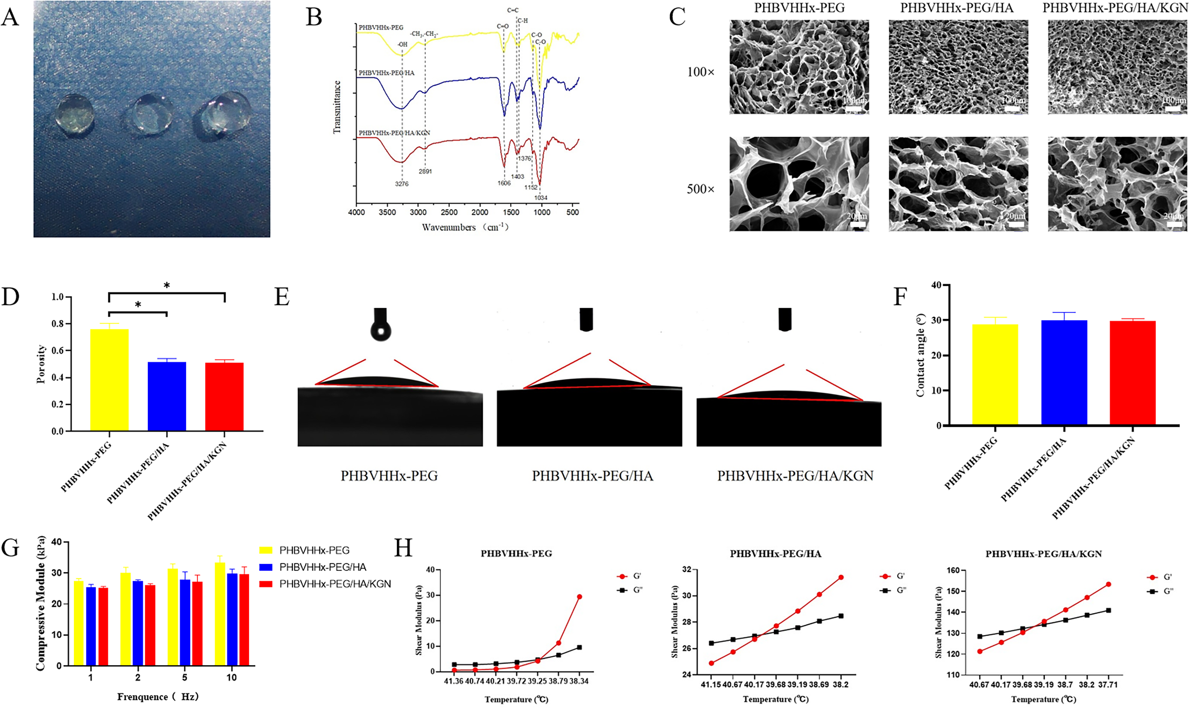
Upon storage at room temperature, the hydrogel solutions underwent gelation. The resulting hydrogels, including PHBVHHx-PEG/HA/KGN, PHBVHHx-PEG/HA, and PHBVHHx-PEG formulations, exhibited transparency after molding. Notably, the PHBVHHx-PEG hydrogel had a faint yellowish tint (Fig. 1A).
FTIR
The FTIR results showed that the intensity of peak around 3276 cm−1 in PHBVHHx-PEG/HA increased compared with PHBVHHx-PEG, indicating the content of hydroxyl groups in PHBVHHx-PEG/HA increased. The enhanced intensity near peak 1606 cm−1 in PHBVHHx-PEG/HA was due to the increase of carbonyl content after adding HA, while the C–O bond content was basically unchanged. In the infrared image of PHBVHHx-PEG/HA/KGN, the hydroxyl peak around 3276 cm−1 was broadened due to the addition of KGN. However, the intensity of the carbonyl peak at 1606 cm−1 decreased, which might be caused by the formation of hydrogen bonds between the carbonyl group in KGN and the carboxyl group in HA (Fig. 1B).
Internal microstructure and porosity
All hydrogels exhibited a loose, porous, and interconnected structure, ensuring even substance distribution. PHBVHHx-PEG had a slightly larger pore size than PHBVHHx-PEG/HA and PHBVHHx-PEG/HA/KGN (Fig. 1C). Statistical analysis confirmed that PHBVHHx-PEG had higher porosity (76.3% ± 4.2%) compared with PHBVHHx-PEG/HA (51.7% ± 2.5%) and PHBVHHx-PEG/HA/KGN (51.3% ± 2.1%) (all p < 0.05) (Fig. 1D).
Water contact angle
The water contact angles of PHBVHHx-PEG, PHBVHHx-PEG/HA, and PHBVHHx-PEG/HA/KGN hydrogels were 28.9 ± 2.0°, 30.0 ± 2.3°, and 29.8 ± 0.6°, respectively. No significant difference was found between the groups (p > 0.05), with all contact angles <90° (Fig. 1E and F).
Biomechanical properties of hydrogels
The compressive moduli of PHBVHHx-PEG hydrogels at 1, 2, 5, and 10 Hz were 27.4 ± 0.8, 30.0 ± 1.8, 31.3 ± 1.7, and 33.4 ± 2.2 kPa, respectively. For PHBVHHx-PEG/HA hydrogels, they were 25.5 ± 0.8, 27.4 ± 0.4, 27.9 ± 2.5, and 30.0 ± 1.3 kPa. The compressive moduli of PHBVHHx-PEG/HA/KGN hydrogels were 25.1 ± 0.5, 26.1 ± 0.4, 27.2 ± 2.2, and 30.0 ± 2.4 kPa. No significant differences in compressive modulus were observed among the hydrogels at 1, 2, 5, or 10 Hz (p > 0.05) (Fig. 1G).
Gelation temperature
The liquid–solid transition points of PHBVHHx-PEG, PHBVHHx-PEG/HA, and PHBVHHx-PEG/HA/KGN hydrogels were approximately 39.5°C, 40°C, and 39.5°C, respectively. As the temperature dropped below these points, the storage modulus exceeded the loss modulus, indicating a shift to a solid state (Fig. 1H).
Identification of PBMSCs
Morphology of PBMSCs
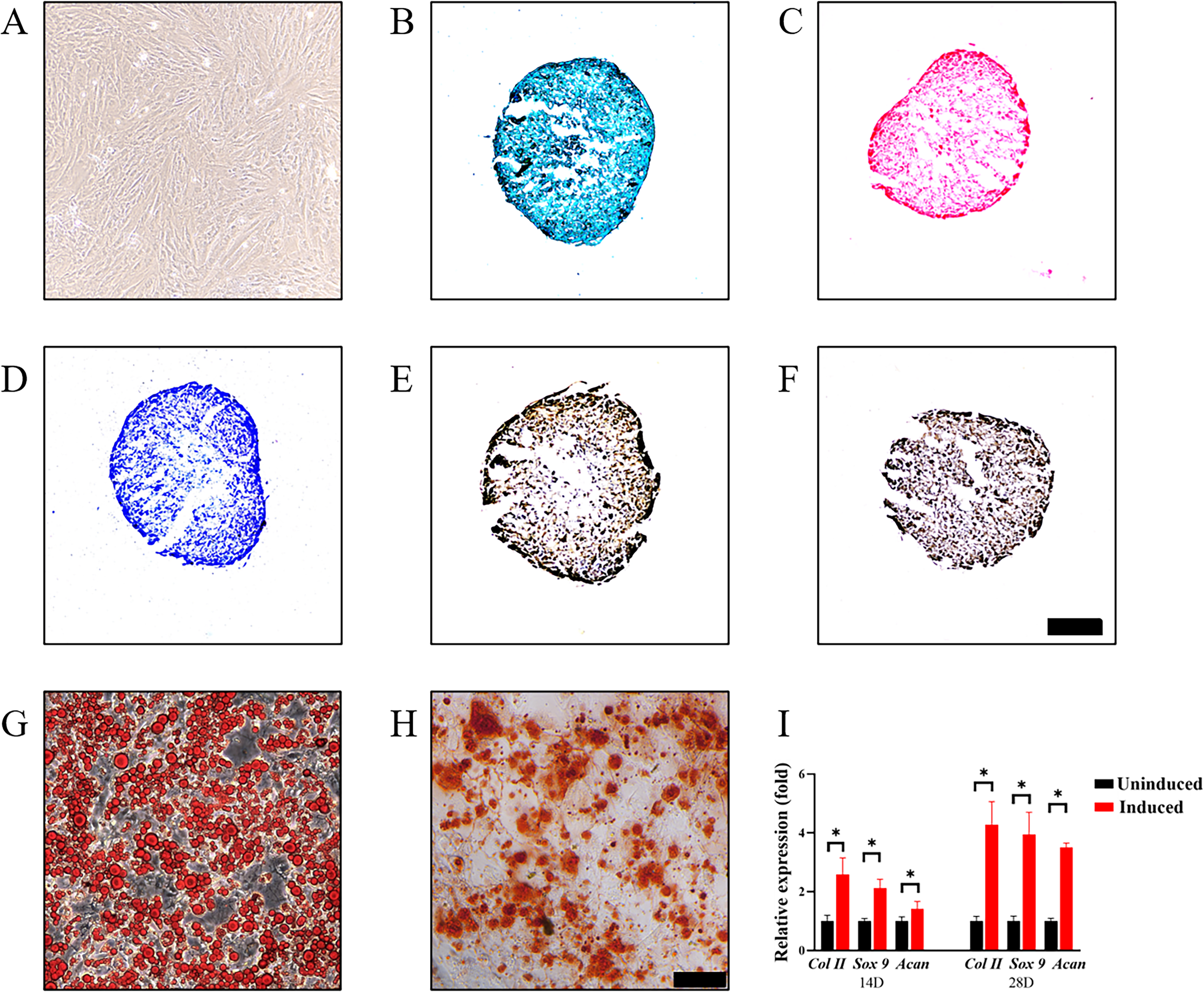
In the initial culture stages, primary rabbit peripheral blood cells showed a round morphology. By day 5, spindle cells appeared, and over time, these cells exhibited clonal growth. After the first passage, nearly all cells adopted a spindle shape and arranged in a spiral pattern (Fig. 2A).
Trilineage induction
Histological staining showed that PBMSCs have trilineage differentiation ability (Fig. 2B–H). After 14 and 28 days of chondrogenesis-induced culture, the expressions of chondrogenesis-related genes Col II, Acan, and Sox9 in PBMSCs were significantly upregulated compared with uninduced PBMSCs (all p < 0.05) (Fig. 2I).
In vitro experiments on hydrogels and cells
Cytotoxicity of scaffolds
Compared with PBMSCs cultured with DMEM, the viability of PBMSCs cultured with the extract medium did not change significantly (p > 0.05), and there was no significant difference among the experimental groups (Fig. 3A).

Cell migration
The initial scratch areas of the control group, PHBVHHx-PEG group, PHBVHHx-PEG/HA group and PHBVHHx-PEG/HA/KGN group were 831082.3 ± 8152.2, 839916.0 ± 18579.3, 843273.0 ± 17233.8, and 839566.3 ± 17314.9 μm2, respectively. After 48 h of cell migration, the remaining areas were 20799.7 ± 379.0, 20911.0 ± 928.5, 20747.7 ± 971.1, and 20160.3 ± 1457.2 μm2. At all detection time points, no significant differences were observed in the remaining scratch area among each group (p > 0.05) (Fig. 3B and C).
3D culture of PBMSCs encapsulated in hydrogels
Microscopic observation of PBMSCs encapsulated in hydrogels revealed that the cells were evenly distributed in the three hydrogels. After 14 days of culture, the cells maintained a round shape in the 3D culture (Fig. 3D).
SEM observation of 3D culture
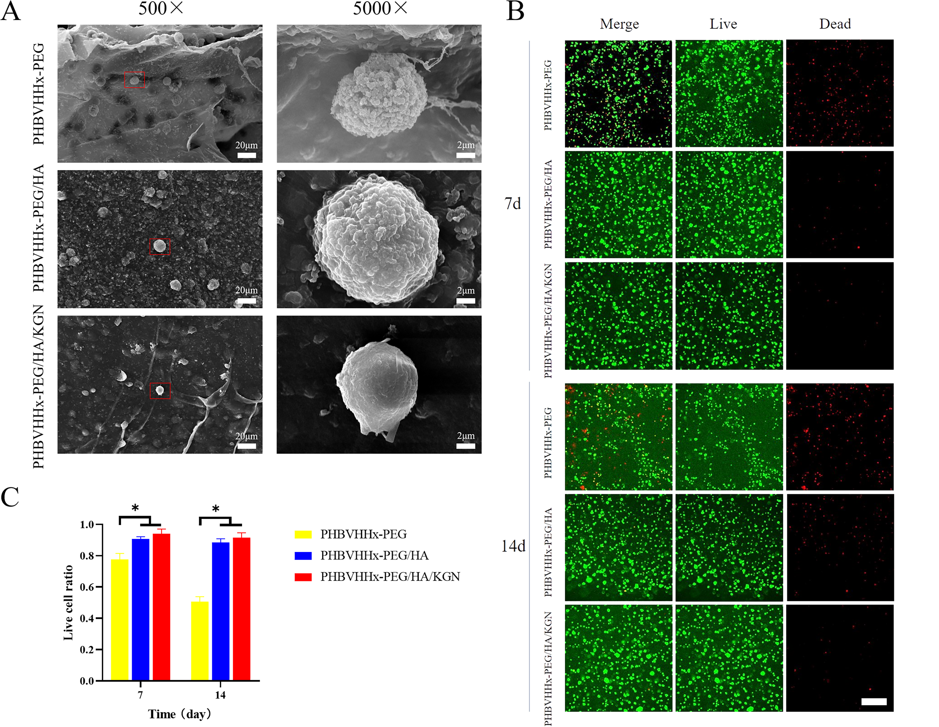
PBMSCs were uniformly distributed within three hydrogels. The cells were attached to the network structure inside the hydrogels, maintaining a round shape (Fig. 4A).
Viability of PBMSCs in hydrogels
Dead cells (red) appeared in the PHBVHHx-PEG group at 7 days, increasing at 14 days. Few dead cells were found in the PHBVHHx-PEG/HA and PHBVHHx-PEG/HA/KGN groups (Fig. 4B). Viable cell ratios at 7 days were 0.777 ± 0.038, 0.907 ± 0.015, and 0.940 ± 0.030 for the PHBVHHx-PEG, PHBVHHx-PEG/HA, and PHBVHHx-PEG/HA/KGN groups, respectively. At 14 days, ratios were 0.507 ± 0.032, 0.883 ± 0.025, and 0.917 ± 0.031. The PHBVHHx-PEG/HA and PHBVHHx-PEG/HA/KGN groups had higher viable cell ratios than PHBVHHx-PEG (p < 0.05), with no significant difference between PHBVHHx-PEG/HA and PHBVHHx-PEG/HA/KGN (p > 0.05) (Fig. 4C).
Chondrogenic differentiation capacity in 3D culture
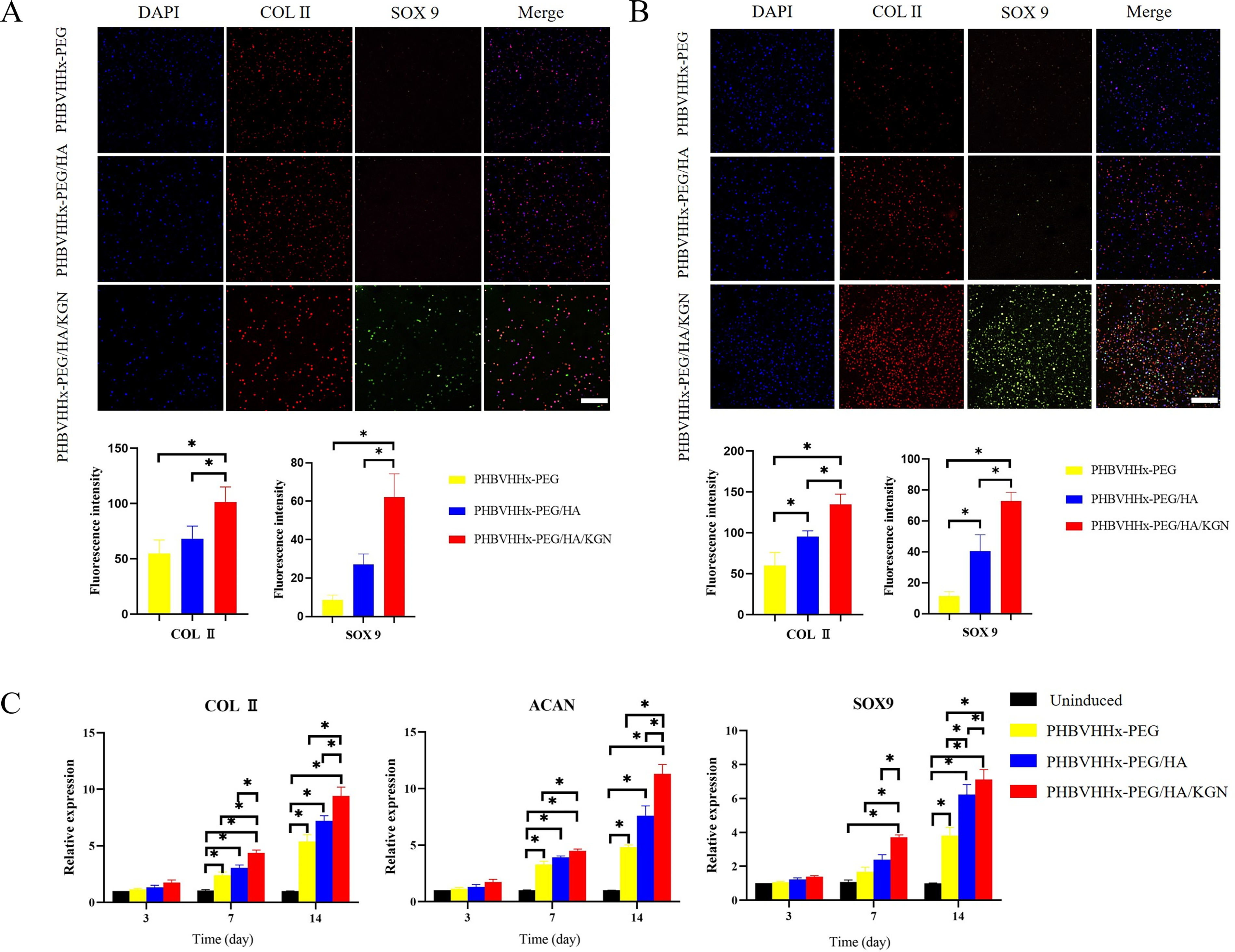
On day 14, the mean fluorescence intensity of COL II in the PHBVHHx-PEG, PHBVHHx-PEG/HA, and PHBVHHx-PEG/HA/KGN groups were 54.7 ± 12.2, 68.0 ± 11.5, and 101.3 ± 13.7, respectively. For SOX9, the intensities were 8.7 ± 2.5, 27.0 ± 5.6, and 62.0 ± 12.3. The PHBVHHx-PEG/HA/KGN group showed stronger fluorescence for both COL II and SOX9 compared with the other groups (all p < 0.05) (Fig. 5A).
Immunofluorescence staining of COL II and SOX9 and semiquantitative analysis on
On day 28, the mean fluorescence intensity of Col II was 60.0 ± 15.7, 95.3 ± 7.0, and 134.7 ± 12.5 for the three groups, and for Sox9, it was 11.3 ± 3.1, 40.3 ± 10.8, and 73 ± 5.6. The PHBVHHx-PEG/HA/KGN group still showed stronger fluorescence for both Col II and Sox9 compared with the others (all p < 0.05), with the PHBVHHx-PEG/HA group having higher fluorescence than the PHBVHHx-PEG group (p < 0.05) (Fig. 5B).
Changes of chondrogenic gene expressing
Chondrogenesis-related gene expression in PBMSCs encapsulated in hydrogels was assessed on days 3, 7, and 14, with PHBVHHx-PEG-coated PBMSCs in DMEM as the control. On day 7, Col II expression was upregulated in all hydrogel groups (p < 0.05), with the PHBVHHx-PEG/HA/KGN group showing the highest expression (p < 0.05). On day 14, this trend continued, with PHBVHHx-PEG/HA/KGN outperforming the others (p < 0.05). Acan expression was also higher on day 7 in all groups compared with the control (p < 0.05), with PHBVHHx-PEG/HA/KGN showing the strongest expression (p < 0.05). This continued on day 14, with PHBVHHx-PEG/HA/KGN having the highest expression (p < 0.05). Sox9 expression was upregulated in all groups on days 7 and 14 (p < 0.05), with PHBVHHx-PEG/HA/KGN showing the highest expression at both time points (p < 0.05) (Fig. 5C).
Macrophage polarization
The M2-positive control and PHBVHHx-PEG/HA/KGN groups had the highest number of CD206+ cells, followed by the PHBVHHx-PEG/HA group. The PHBVHHx-PEG group had fewer CD86+ cells than the M1-positive control group (Fig. 6A).

Cell count results showed that the proportion of CD86+ cells in the M1-positive control group was 0.660 ± 0.046, higher than in other groups (all p < 0.05). The PHBVHHx-PEG group had 0.340 ± 0.066, lower than M1 (p < 0.05) but higher than the other groups (all p < 0.05). The proportion of CD206+ cells in the M2-positive control group was 0.723 ± 0.071, in PHBVHHx-PEG/HA/KGN was 0.667 ± 0.038, and in PHBVHHx-PEG/HA was 0.570 ± 0.044, all higher than the other groups (all p < 0.05), with PHBVHHx-PEG/HA differing from the M2-positive control group (p < 0.05) (Fig. 6B).
In vivo experiment
Macrophage polarization
The materials in the PHBVHHx-PEG group were positive in large areas when stained by iNOS, there were a few scattered weak positives in the PHBVHHx-PEG/HA group, and the PHBVHHx-PEG/HA/KGN group was negative. The results of ARG-1 staining showed that the PHBVHHx-PEG group was negative, the materials in the PHBVHHx-PEG/HA group and the PHBVHHx-PEG/HA/KGN group were both positive, and the material in the PHBVHHx-PEG/HA/KGN group was strongly positive at the junction of the surrounding tissue. For the control group injected subcutaneously with normal saline, all stainings were negative (Fig. 6C and D).
CD68/CD86 immunofluorescence staining detected M1 macrophages. At 7 days, CD86+ cells were prominent in PHBVHHx-PEG, with a CD86+/CD68+ ratio of 0.407 ± 0.085, significantly higher than control, PHBVHHx-PEG/HA, and PHBVHHx-PEG/HA/KGN (p < 0.05). At 14 days, CD86+ cells remained elevated in PHBVHHx-PEG, with no significant differences among other groups (p > 0.05).
CD68/CD206 staining detected M2 macrophages. At 7 days, CD206+ cells appeared in PHBVHHx-PEG/HA and PHBVHHx-PEG/HA/KGN, with CD206+/CD68+ ratios of 0.483 ± 0.040 and 0.457 ± 0.025, significantly higher than PHBVHHx-PEG (p < 0.05). At 14 days, PHBVHHx-PEG/HA and PHBVHHx-PEG/HA/KGN maintained higher CD206+/CD68+ ratios, significantly surpassing PHBVHHx-PEG and control (p < 0.05) (Fig. 7A–D).
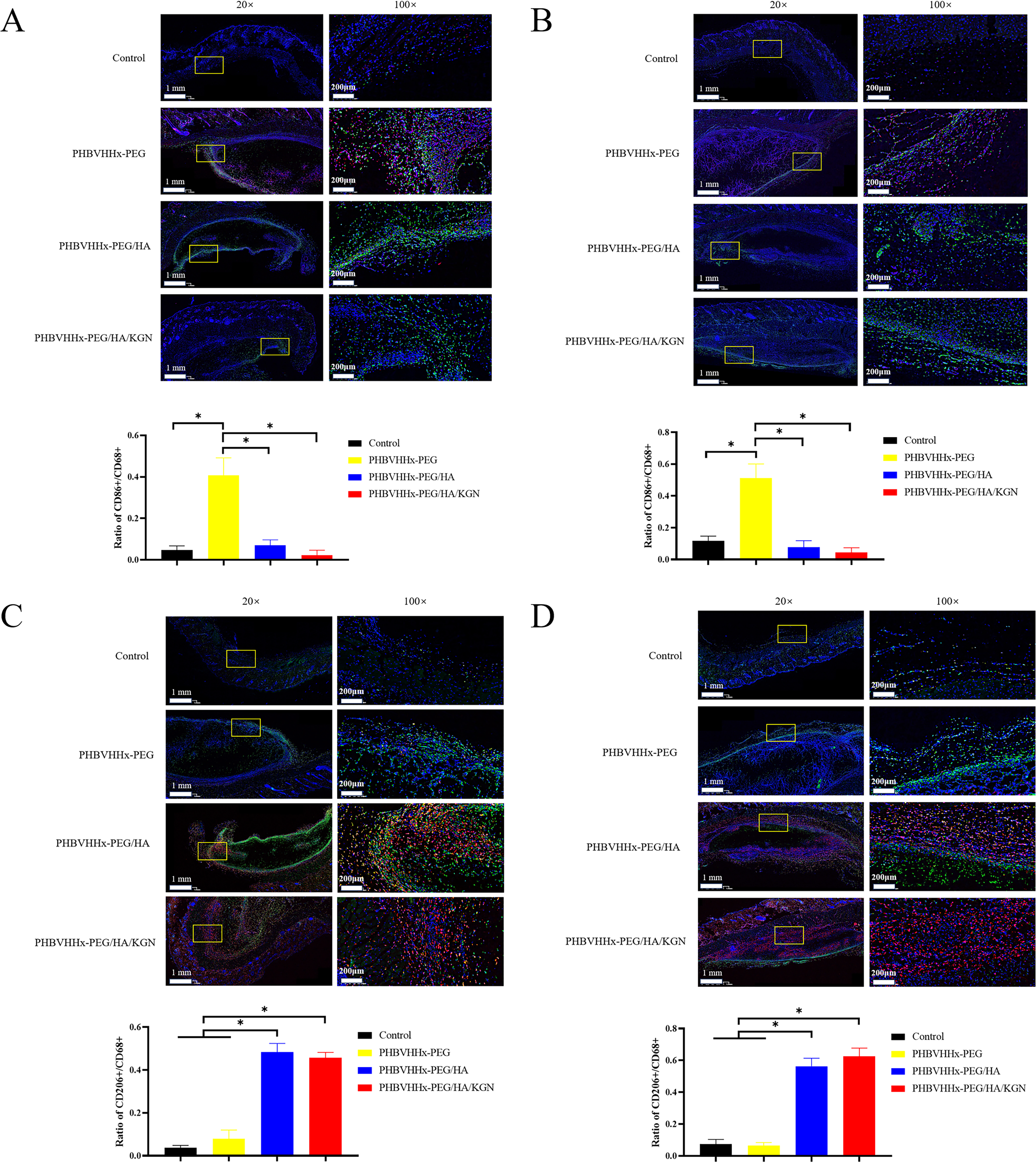
Immunofluorescence and semiquantitative analysis of M1 polarization in vivo on
Ectopic chondrogenesis
After implanting hydrogels subcutaneously in nude mice, the materials were still visible after 28 days. Grafts in PHBVHHx-PEG/HA and PHBVHHx-PEG/HA/KGN groups were slightly smaller than PHBVHHx-PEG. PHBVHHx-PEG grafts were soft and easily bendable, while PHBVHHx-PEG/HA/KGN and PHBVHHx-PEG/HA grafts were stiffer (Fig. 8A).
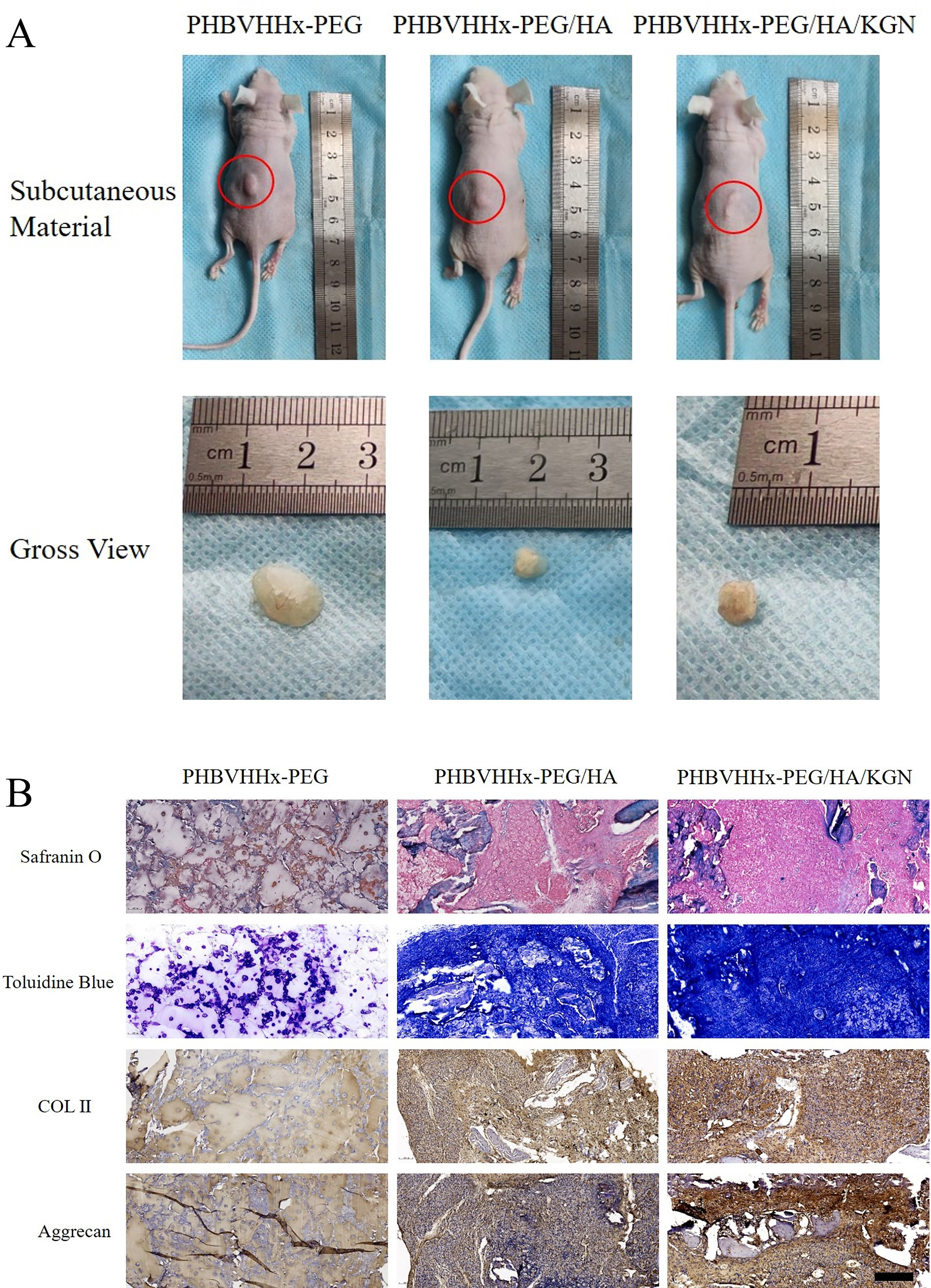
The cell-loaded material was implanted subcutaneously in nude mice and detected after 28 days.
Safranin fast green staining showed faint red coloration around PBMSCs, while PHBVHHx-PEG/HA and PHBVHHx-PEG/HA/KGN exhibited wider, deeper coloration. Toluidine blue staining gave similar results. Immunohistochemical staining for type II collagen and AGG showed positive results in PHBVHHx-PEG/HA and PHBVHHx-PEG/HA/KGN, with stronger positivity in PHBVHHx-PEG/HA/KGN. PHBVHHx-PEG showed no positive staining (Fig. 8B).
In situ repair of cartilage defects
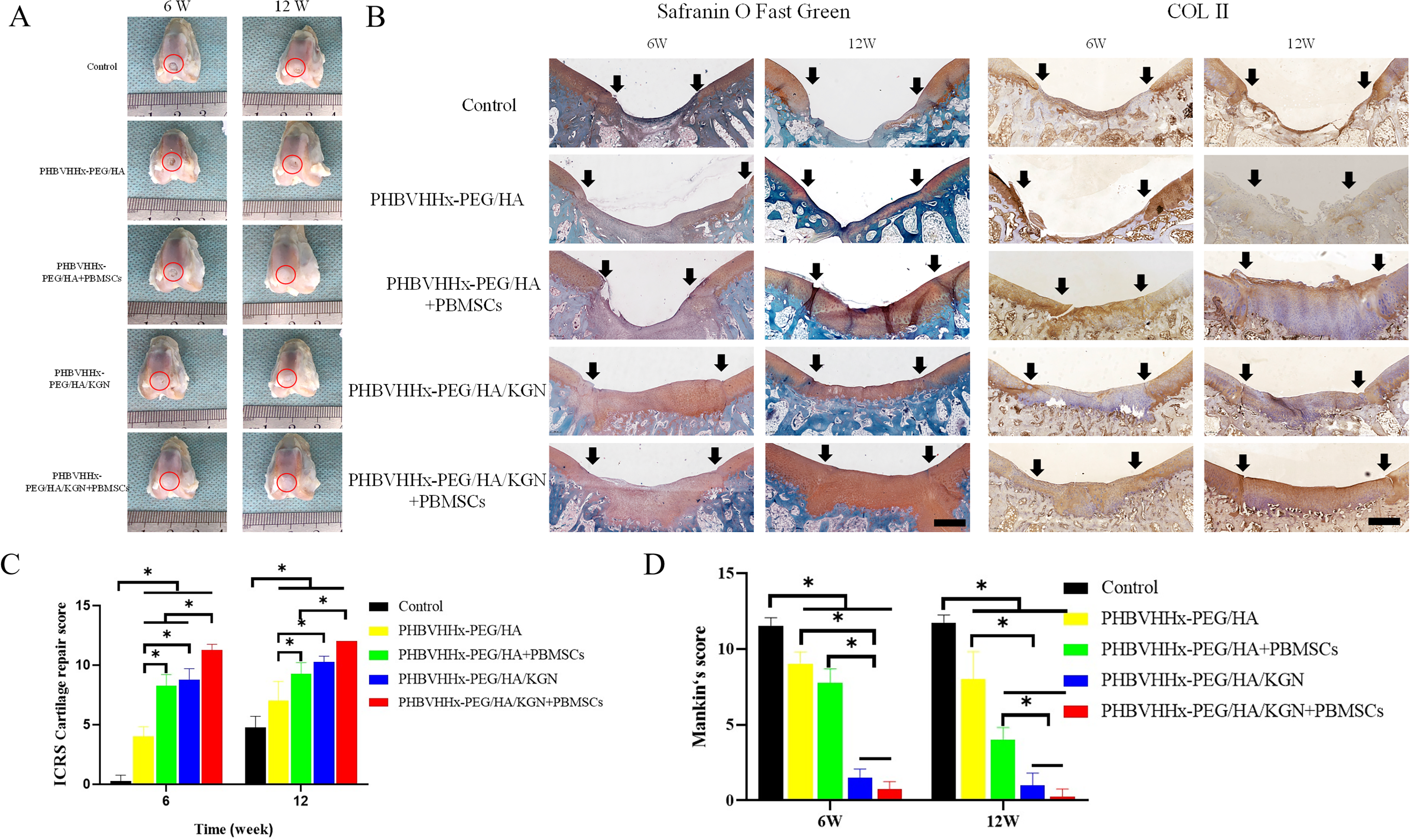
In the control group, cartilage defects persisted at 6 and 12 weeks. At 6 weeks, PHBVHHx-PEG/HA, PHBVHHx-PEG/HA+PBMSC, and PHBVHHx-PEG/HA/KGN groups showed partial filling, with PHBVHHx-PEG/HA defects remaining larger. PHBVHHx-PEG/HA/KGN+PBMSC group exhibited complete filling. At 12 weeks, incomplete repair remained in the PHBVHHx-PEG/HA group. Defects in other groups were filled but had issues like uneven surface. The PHBVHHx-PEG/HA+PBMSC+PBMSC group achieved complete repair.
Safranin fast green staining showed worsening defects in control, while PHBVHHx-PEG/HA and PHBVHHx-PEG/HA+PBMSC lacked safranin staining at 6 weeks. At 12 weeks, PHBVHHx-PEG/HA/KGN demonstrated clearer junctions, and PHBVHHx-PEG/HA/KGN+PBMSC had no significant differences in coloration, thickness, or smoothness compared with surrounding tissue.
Collagen II staining showed no production in control. PHBVHHx-PEG/HA+PBMSC and PHBVHHx-PEG/HA/KGN+PBMSC exhibited stronger positivity at both time points.
ICRS scores were based on gross view and Mankin’s score was used to quantitative safranin fast green (Fig. 9A–D).
Discussion
Characterization of scaffold
FTIR confirmed the successful synthesis of PHBVHHx-PEG, PHBVHHx-PEG/HA, and PHBVHHx-PEG/HA/KGN hydrogels. While PHBVHHx has been used in treating liver injury and systemic lupus erythematosus,9,26 it was modified here into an injectable thermosensitive hydrogel, which can allow sustained release of bioactive agents for cartilage repair. 27 SEM results showed that all hydrogels had a uniform distribution of components and a porous interconnected structure, which supports cell infiltration and growth. 28 Porosity analysis indicated that the addition of HA and KGN increased pore size, potentially improving cell–material interactions. 29 All hydrogels showed good hydrophilicity in water contact angle tests, which benefits cell adhesion and viability. 30 Rheological testing demonstrated similar gelling properties, with sol–gel transitions occurring at 39–40°C, close to the human physiological temperature of 36.8°C. 31 Thus, these hydrogels can be injected and form gels in situ, maintaining a stable state at body temperature.
Effect of scaffolds on seed cells
BMSCs and AMSCs are widely used in cartilage repair but face limitations such as invasive harvesting and variability. 5 In contrast, PBMSCs can be obtained via minimally invasive blood draws, making them more clinically feasible. 11
Under two-dimensional culture, the CCK-8 assays showed that the extracts from all three hydrogels did not impair cell metabolism, consistent with previous reports on the noncytotoxicity of their components.12,17,32 The present study further confirmed the biosafety of the synthesized hydrogels.
The 3D culture system was reported to be more suitable for the application of cartilage tissue engineering. 28 PBMSCs cultured in PHBVHHx-PEG/HA/KGN hydrogels showed superior performance. As PHBVHHx-PEG is a bacterial polyester not naturally found in mammals, 33 its cytocompatibility may be lower than that of HA, a native ECM component. 12 In addition, differences in porosity might also lead to this result, since PHBVHHx-PEG/HA and PHBVHHx-PEG/HA/KGN hydrogels provided more attachment sites for PBMSCs encapsulated in them.
In vitro chondrogenesis capacity of PBMSCs encapsulated in hydrogels was also evaluated. PBMSCs encapsulated in PHBVHHx-PEG/HA/KGN and PHBVHHx-PEG/HA hydrogels expressed higher Col II and Sox9 than those in PHBVHHx-PEG. HA contributes by supporting ECM secretion and maintaining chondrocyte phenotype. 34 The addition of KGN further enhanced this effect, as it promotes chondrogenesis via type II collagen induction in MSCs. 35 Upregulation of Col II and Acan in PHBVHHx-PEG/HA/KGN suggested enhanced cartilage matrix synthesis.36,37 Sox9 expression was also upregulated on day 14, known to facilitate chondrogenesis and suppress hypertrophy via Ihh and PTHrP pathways. 38 Overall, PHBVHHx-PEG/HA/KGN hydrogel showed the best support for PBMSC chondrogenic differentiation.
Effect of scaffolds on macrophage polarization
Macrophages recruited to injury sites can polarize into proinflammatory M1 or anti-inflammatory M2 types.39–42 M2 macrophages facilitate cartilage repair by secreting anti-inflammatory factors. 43 M1 macrophages typically express CD86 and iNOS, while M2 macrophages express CD206 and ARG-1.39–41,44 In this study, PHBVHHx-PEG/HA and PHBVHHx-PEG/HA/KGN hydrogels induced M2 polarization both in vitro and in vivo, whereas PHBVHHx-PEG did not. Previous studies suggest that high molecular weight HA promotes M2 polarization, contributing to anti-inflammatory effects. 45 This study confirmed that HA-containing hydrogels shared this capacity, aligning with HA’s established role in osteoarthritis treatment. 46
In vivo cartilage repair effect
Nude mice experiments confirmed that PHBVHHx-PEG/HA and PHBVHHx-PEG/HA/KGN hydrogels loaded with PBMSCs induced ectopic cartilage formation, while PHBVHHx-PEG hydrogels did not. A previous study demonstrated that HA hydrogels promoted MSC differentiation into chondroblasts, enhancing COL II, ACAN, and SOX9 expression. 15 Similarly, the chondrogenic effect of PHBVHHx-PEG/HA and PHBVHHx-PEG/HA/KGN may be attributed to HA. In addition, PHBVHHx-PEG/HA/KGN showed stronger collagen type II and AGG staining than PHBVHHx-PEG/HA, likely due to KGN promoting MSC chondrogenesis. 17
In situ repair results highlighted that PHBVHHx-PEG/HA/KGN+PBMSC achieved superior cartilage regeneration. At 6 weeks, neocartilage formation was significantly better than in other groups, and at 12 weeks, PHBVHHx-PEG/HA/KGN+PBMSC maintained the highest ICRS score, followed by PHBVHHx-PEG/HA/KGN. Type II collagen and AGG staining confirmed greater ECM deposition in PBMSC-containing groups. Mankin’s score further indicated that PHBVHHx-PEG/HA/KGN+PBMSC had the best repair outcomes at all time points.
Overall, PHBVHHx-PEG/HA/KGN+PBMSC demonstrated the best cartilage repair, with PHBVHHx-PEG/HA/KGN outperforming PHBVHHx-PEG/HA. The presence of PBMSCs further enhanced the repair process.
Comparison of similar studies
Different kinds of PHAs, including PHBV, PHB, and PHBHHx, have been used in the cartilage tissue engineering.28,29,47–49 However, few studies validated in vivo effects, and none developed injectable thermosensitive hydrogels. This study introduced the first PHBVHHx-based injectable hydrogel, enhanced with HA and KGN for bioactivity, demonstrating effective cartilage repair in vitro and in vivo.
While PBMSCs’ chondrogenic potential has been widely studied, their application in cartilage defect repair remains limited. 5 A clinical study showed PBMSCs in HA hydrogel improved cartilage repair more than PBMSCs alone. 50 Since they have already proved that PBMSCs encapsulated in hydrogels provided better therapeutic effect, we did not set up a separate PBMSCs group. Compared with them, this study used PHBVHHx-PEG as the matrix and demonstrated HA’s chondrogenic induction effect as an additive.
Limitations
This study confirmed that scaffolds induced M2 macrophage polarization, but PBMSC–macrophage interactions were not analyzed, although the exosomes of MSCs have been proven to be able to induce macrophages to polarize toward M2. 51 The primary purpose of adding PBMSCs is chondrogenesis and matrix secretion, thus their role in macrophage polarization was not evaluated. In addition, macrophage polarization in repaired cartilage was not assessed. Although macrophage polarization plays a role throughout cartilage repair, its main effect was in the early inflammatory response phase, so this was not assessed in the long-term animal experiments. 52
Conclusion
PHBVHHx-PEG/HA/KGN injectable hydrogel-loaded PBMSCs demonstrated effective knee cartilage defect repair.
Authors’ Contributions
M.B.: Data acquisition, data analysis, writing, and funding. T.M.: Interpretation of the results and writing. Y.Y.: Data acquisition. L.L.: Data acquisition. L.J.: Interpretation of the results and supervision. W.D.: Conception, material synthesis, and supervision. F.W.: Conception, supervision, and funding. All authors approved the submission of this study to this journal.
Footnotes
Author Disclosure Statement
The authors declare no conflicts of interest.
Funding Information
The work was funded by the National Natural Science Foundation of China (82172508 and 82372490), Sichuan Science and Technology Program (2024NSFJQ0041), 1.3.5 Project for Disciplines of Excellence of West China Hospital Sichuan University (ZYJC21030), and Guangdong Basic and Applied Basic Research Foundation (2023A1515110336).