
Abstract
Bone graft material is often required for the treatment of osseous defects. However, due to limitations and risks associated with autologous as well as allogenic bone grafting procedures, alternative strategies are needed. In this context, ex vivo tissue engineering (TE) strategies for de novo generation of bone tissue include the combined use of autologous bone-forming cells and three-dimensional (3D) porous scaffold materials serving as structural support for the cells. Three-dimensional cultivation of osteoprogenitor cells presents several challenges, for example, insufficient nutrient and oxygen transport to and removal of waste products from the cells at the interior of the scaffold. By providing physical stimulation of tissue-engineered constructs and resolving mass transport limitations bioreactor systems denote key components for bone TE strategies. A variety of dynamic 3D bioreactor concepts mimicking the native microenvironment in bone tissue, for example, spinner flasks, rotating wall vessel constructs, perfusion bioreactors, and systems based on mechanical or electromagnetic stimulation of cell/scaffold composites, have been developed. These techniques differ considerably with respect to ease of use, cost-effectiveness, and degree of additional osteogenic stimuli, as well as monitoring and manipulation options. This review provides an overview of the concepts, advantages, challenges, and potential future applications associated with current bioreactor systems for bone TE.
Introduction
Bone tissue engineering
The discipline of bone TE involves the combined use of osteoconductive matrices, bone-forming cells, and osteogenic growth factors. Notably, the tissue constructs need to be maintained in a suitable cultivation environment. Cells are the living component of such a construct, capable of producing matrix-forming neotissue. Osteoblasts have been widely used for generating mineralized cell/scaffold constructs in vitro.13–15 However, osteoblasts are in an almost mature stage therefore showing less proliferative potential compared to osteoblast precursor cells. In contrast, mesenchymal stromal cells (MSCs) represent a proliferating and undifferentiated cell source. MSCs are mostly isolated from bone marrow aspirates, but can also be obtained from other tissues, for example, adipose tissue or cord blood. 16 MSCs have the potential to differentiate toward diverse mesenchymal lineages, including osteoblasts, chondrocytes, adipocytes, and myocytes. 17 Drawbacks of the application of MSCs are their limited availability and their in vitro replicative senescence. 18 In this context, the lifespan of human MSCs can be extended by ectopic expression of human telomerase reverse transcriptase (hTERT). In various in vitro studies hTERT-MSCs were used for seeding scaffolds and cultivating the cell/scaffold constructs under dynamic conditions.19–21
The ideal scaffold should possess mechanical properties comparable to bone and should be easily fabricated in a desired shape. A suitable scaffold material for generating mineralized cell/scaffold constructs should support cell attachment and ingrowth by the presence of an interconnected pore network. Further, the biomaterial should be biodegradable to facilitate natural bone formation and remodeling. Several types of synthetic or naturally occurring scaffold materials, including corals, bioceramics, biopolymers, and metals, have been suggested for generating mineralized cell/scaffold constructs.22–24 According to in vivo studies an optimal scaffold pore size for osteoblasts ranges from 200 to 400 μm.25,26 In addition, an interconnected pore network of the scaffold is needed to support vascularization. Using a computer-based simulation model, Khayyeri et al. showed that higher scaffold stiffness keeping the pore structure constant enhanced bone healing. 27 Further, scaffold internal pore architecture may influence the distribution of shear stress, the range of mechanical stimuli, and the proliferation and differentiation of osteoprogenitor cells.28–30
Besides their biophysical stimulation, osteoprogenitor cells are biochemically stimulated in vivo by specific cytokines and growth factors. A number of these factors are available for osteogenic differentiation of bone-forming cells in vitro. 31 Bone morphogenetic proteins (BMPs) are the most relevant factors in bone morphogenesis. 32 They belong to the transforming growth factor beta (TGF-β) superfamily of polypeptides displaying extensive conservation among species. BMP-2, −4, and −6 are the most readily detectable BMPs in osteoblast cultures. Currently, only BMP-2 and BMP-7 are approved for clinical application. Recombinant human BMP-2 (INFUSE® Bone Graft; Medtronic Spinal and Biologics) is endorsed for the treatment of spinal fusion, fresh tibial fractures, and for oral and maxillofacial bone grafting procedures. 33 BMP-7 (OP-1/BMP-7; Stryker®) is approved for the therapy of long bone fractures, nonunions, and for spinal fusions. 34 Another strategy to stimulate osteoprogenitor cells by growth factors is the use of autologous platelet-rich plasma, which has been reported to be an effective bioactive supplement for both soft- and hard-TE applications.35–37 Platelet-rich plasma contains osteogenic and angiogenic growth factors such as TGF-β1, platelet-derived growth factor insulin-like growth factor-1, and vascular endothelial growth factor. 38
Challenges in bioreactor-based bone tissue engineering
When using bioreactor systems, technical prerequisites and requirements related to the equipment used need to be considered. Bioreactors are classically used to facilitate, monitor, and control biological or biochemical processes, for example, in the context of industrial fermentation, waste water treatment, food processing, and production of pharmaceuticals. 39 The parameters that modulate growth in complex bioreactors include temperature, oxygen concentration, pH, nutrient concentration, and biochemical and mechanical stimuli. Closed bioreactor systems offer major advantages with respect to good manufacturing practice (GMP)-conform manufacturing of tissue-engineered products. The devices are usually composed of biologically inert and noncorrosive material to prevent toxic reactions or corrosion under a humidified atmosphere. The dimensions of a bioreactor are generally adapted to the spatial proportions of conventional incubators to guarantee favorable culturing conditions, for example, 99% humidity, 37°C, and 5% CO2. Bioreactors are frequently assembled by components consisting of synthetic polymers, for example, polymethylmethacrylate (PMMA), polyoxymethylene, or polysulfone that can withstand sterilization techniques. Using current new-generation bioreactor systems, crucial parameters can be monitored and controlled online.
Another crucial aspect that needs to be addressed during the design of a bioreactor is the diffusion limit. Systems used should provide the cell/scaffold constructs with efficient nutrients, oxygen, and a biophysical stimulus to direct cellular differentiation. The supply of oxygen and soluble nutrients becomes critical when the diffusion distance exceeds a distance of 100–200 μm. 40 Studies demonstrated low expression of osteogenic marker proteins and decreased proliferation of bone marrow stromal cells under static cultivation conditions in large cell/scaffold constructs. 41 Static cultivation methods can lead to an inhomogeneous concentration of nutrients and oxygen and consequently to an under-supply of cells in the interior of the scaffold potentially inducing cell death. Thus, the size of the scaffold that can be sucessfully used for static cultivation is restricted.
Uniform cell distribution and enhanced cellularity within a scaffold are prerequisites for the engineering of functional bone substitutes. Static seeding in a dropwise manner results in low seeding efficiencies and inhomogeneous spacial cell distribution.42,43 The use of bioreactor systems based on fluid flow has been proven beneficial for cell seeding compared to static seeding methods.44–47 Current dynamic seeding techniques include convection of medium using spinner flasks, 48 centrifugation, 49 perfusion, 50 and oscillatory perfusion. 51 By comparing two dynamic seeding methods, Godbey et al. showed that the centrifugation method led to more homogeneous cellular distribution throughout the scaffold as opposed to the spinner flask method. 49 Du et al. compared unidirectional perfusion and oscillatory flow as dynamic cell seeding methods for porous beta-tricalcium phosphate (β-TCP) ceramic scaffolds. 51 Only the back-and-forth nature of oscillatory flow resulted in a uniform proliferation of engineered bone in vitro compared to both unidirectional perfusion and static seeding.
In addition to the seeding technique another important factor in bone TE is the number of cells used for seeding. Cell–cell recognition and adhesion are essential for successful osteogenic differentiation of human osteoblasts. 52 Van den Dolder et al. observed higher calcium contents in rat bone marrow cells when being seeded with higher cellularity on titanium fiber mesh scaffolds. 53 In contrast, Wiedmann-Al-Ahmad and co-workers showed that the lowest cell density of 1×10⁵ human primary osteoblasts/mL used in their study showed the best results with respect to proliferation, cell distribution, and vitality compared to higher seeding densities. 54 Kruyt et al. investigated in vivo bone formation after using different cell seeding densities on porous biphasic calcium phosphate implanted in dutch milk goats. 55 A minimum of 8×10⁴ and an optimum of 8×10⁶ bone marrow stromal cells/cm3 scaffold were determined for successful bone formation. Further, Impens et al. analyzed various parameters potentially influencing cell seeding efficiency. 56 The authors reported that besides the cell density also the volume of seeding medium-to-free scaffold volume ratio, the seeding incubation time, and the scaffold morphology affected the cell seeding efficiency.
Further, bioreactor systems may be used to enhance osteogenic cellular differentiation by simulation of biophysical forces mimicking those physiologically occurring in vivo. In this respect, the following aspects have to be considered during the design phase. In vivo, bone is constantly exposed to mechanical stimulation in vivo by muscular contraction and body movements. Forces applied to bone during body movement result in changes of hydrostatic pressure, direct cell strain, fluid flow-induced shear stress, and electric fields. 57 Bone cells are more sensitive to mechanical deformation than most other cell types. 58 Mechanical loading stimulates bone formation and leads to an overall increase in bone mass. Shear stress generated by turbulence flow or perfusion stimulates proliferation and differentiation of human osteoblasts by activation of extracellular signal-regulated kinases-dependent and other pathways. 59 Ando and co-workers were the first to demonstrate that fluid-induced shear stress stimulates intracellular Ca2+ release in vascular endothelial cells. 60 In fact, Ca2+ acts as a second messenger by activating other proteins of signaling pathways. The activation of intracellular Ca2+ correlates with applied cell strain in single osteocytes in response to fluid flow. 61 Different signaling pathways, for example, those including wingless-type MMTV integration site family (WNT), estrogen receptor (ER), insulin-like growth factor-I, and BMPs, seem to be involved in the process of mechanotransduction. 62 If shear stress exceeds a certain limit, alkaline phosphatase (ALP) activity can be downregulated. 63 Weinbaum et al. predicted shear stress levels between 8 and 30 dyn cm2 in an in vitro model for excitation of osteocytes by bone fluid shear stress induced by mechanical load. 64 The authors describe that the magnitude of hydrodynamic shear stress is comparable with that observed in osteoblasts and other intracellular Ca2+ shear stress responses.
Providing mechanical stimulation and resolving mass transport limitations bioreactor systems are key components for bone TE strategies. This article intends to give an overview of the concepts, advantages, challenges, and potential future applications associated with current bioreactor systems for bone TE.
Bioreactor Systems for Bone Tissue Engineering
In this section bioreactor-based concepts for bone TE are outlined. Systems using hydrodynamic shear stress, including spinner flasks, and rotating and perfusion bioreactors, are introduced. Moreover, bioreactors with direct mechanical strain, pulsed electromagnetic fields (PEMFs), and the concept of in vivo bioreactors are discussed. Finally, selected commercially available systems will be presented.
Systems using hydrodynamic shear stress
Local internal shear stress created by systems using hydrodynamic forces and experienced by the cells on three-dimensional (3D) matrices is influenced not only by the medium flow rate but also by other parameters that have to be considered, for example, the porosity, the dimensions, the material and the geometry of the scaffold, the size, the anisotropy, and the degree of interconnectivity of pores, as well as the viscosity of the medium.
The fluid flow and surface shear stress in 3D tissue-engineered constructs cultivated in bioreactor systems can be characterized by optical measuring techniques, for example, particle image velocimetry or calculated by computational fluid dynamic (CFD) modeling, for example, Lattice–Boltzmann method and finite element analysis. 65 A detailed overview of the rapidly growing field of CFD modeling for analyzing and observing the impact of fluidic forces is given by Hutmacher and Singh.66,67 Further, studies often combine practical approaches and computer simulation to characterize flow fields, shear stress, and cell responses. The dynamic environment within a spinner flask system, for example, was characterized by Sucosky et al. combining laboratory (particle-image velocimetry) and numerical experimentation with emphasis on, for example, rotating turbulent flow and porosity of scaffolds. 68 The calculated results of maximum shear stress generated by the numerical model were in agreement with the experimental results. Finite element simulations were used by Pollack et al. combining the simulation and real-time optical techniques to describe motions of microcarriers in a rotating bioreactor. 69 Likewise, a bi-modular fluid characterization using CFD simulations and microparticle image velocimetry measurements on realistic conditions was performed by De Boodt and co-workers. 70 Porter et al., for instance, used the Lattice–Boltzmann method as described by Martys and Chen 71 for the simulation of flow conditions in combination with microcomputed tomography imaging to define the scaffold microarchitecture in a perfusion bioreactor. 72 The authors observed that an average surface shear stress of 5×10⁻⁵ Pa corresponds to increased cell proliferation, whereas higher shear stress levels were associated with the upregulation of bone marker genes. A prediction of the micro-fluid dynamic environment imposed to three-dimensional engineered cell systems in bioreactors has been published by Boschetti et al. addressing the influence of pore size and scaffold porosity. 73 The authors found that pore size is a variable strongly influencing the predicted shear stress level, whereas the porosity is a parameter strongly affecting the statistical distribution of the shear stresses, but not their magnitude.
To determine shear-stress-related cell responses in vitro, Bancroft et al. observed that rat marrow stromal osteoblasts cultivated on fiber mesh titanium scaffolds at continuous low, medium, and high media flow rates of 0.3, 1, and 3 mL/min, respectively, in a perfusion system demonstrated that a 3-fold increase in flow rate was associated with an over sixfold increase in calcium content, indicating enhanced extracellular matrix (ECM) mineralization. 13 In a study using flow rates of 0.01, 0.1, 0.2, and 1.0 mL/min, the authors concluded that cultures of MC3T3-E1 osteoblast-like cells on human trabecular bone scaffolds at a flow rate of 1.0 mL/min resulted in substantial cell death, whereas lowering the flow rate led to an increasing proportion of viable cells, particularly at the center of the constructs. 74 However, as the degree of shear stress resulting from fluid flow sensed by 3D cultivated osteoprogenitor cells is influenced by scaffold material characteristics, for example, pore size and porosity, the mentioned studies are not comparable. Instead, CDF modeling for the prediction of shear stress levels adapted to the particular scaffolds and bioreactor system used may be a beneficial additional tool for developing optimized tissue-engineered constructs. In this context, a combined practical and theoretical approach is an appealing strategy. Likewise, to establish predictive correlations between perfusion rates and osteogenesis of human MSCs, Grayson et al. examined the effects of a wide range of medium flow rates (80–1800 mm/s) on the formation of engineered bone constructs. 75 Based on histological analyses and protein-based assays the authors found that increasing the flow velocity significantly affected cell morphology, cell–cell interactions, matrix production and composition, and the expression of osteogenic genes and that the linear velocity of medium perfusion in the range of 400–800 mm/s resulted in the highest matrix deposition.
Spinner flask bioreactor
The spinner flask is a simple and inexpensive bioreactor system. Convective forces are provided by a stirrer and medium flows around the cell/scaffold constructs that are positioned in the center of the flask (Fig. 1). Scaffolds are attached to a needle connected to the lid of the flask. Two angled side arms equipped with filters guarantee oxygenation of the medium. The whole system is placed in an incubator controlling temperature and oxygen content. The degree of shear stress depends on the stirring speed. A stirring rate of 50 rpm was used in a study with collagen (Col) and silk scaffolds seeded with human MSCs. 76 Sikavitsas et al. used a stirring speed of 30 rpm in a 120 mL flask. 77 Various studies showed positive osteogenic effects by an increased level of the early osteoblastic differentiation marker ALP in rat osteoblasts, 23 rat 77 or human MSCs,76,78 and hTERT-MSCs.20,21 Additionally, in the majority of the cases, proliferation, expression of osteogenic marker genes, and mineralization were increased compared to static controls (Table 1A). Spinner flasks are offered by various companies in different sizes (Table 2). However, minor modifications of the system have to be carried out by customers to ensure proper attachment of the scaffolds within the flask. This type of bioreactor is also used as a dynamic cell seeding device.48,79
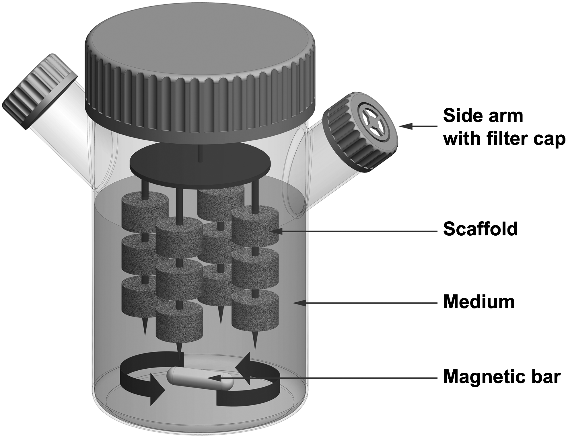
Schematic view of a spinner flask bioreactor. The cell/scaffold constructs are attached to a needle, and shear stress is applied by convection of medium.
Arrows indicate the effects by the systems compared to static cultivation:↑, positive effect;↔, no effect;↓, negative effect.
ALP, alkaline phosphatase; BSP, bone sialoprotein; Col1, collagen 1; MSC, mesenchymal stromal cell; OC, osteocalcin; OP, osteopontin; PLGA, poly(lactic-co-glycolic acid; TERT, telomerase reverse transcriptase.
Besides the beneficial effects with respect to differentiation and proliferation, another advantage of the system is its low cost of acquisition. A drawback of cultivating cell/scaffold constructs in a spinner flask systems is the possible formation of a dense superficial cell layer, which may hamper oxygen and nutrient supply of the cells in the center of the scaffolds. 21 In addition, shear stress is not applied homogenously as there appears to be a gradient of convective forces within a spinner flask with the highest level on the bottom of the vessel in proximity to the stirrer.
Rotating bioreactor systems
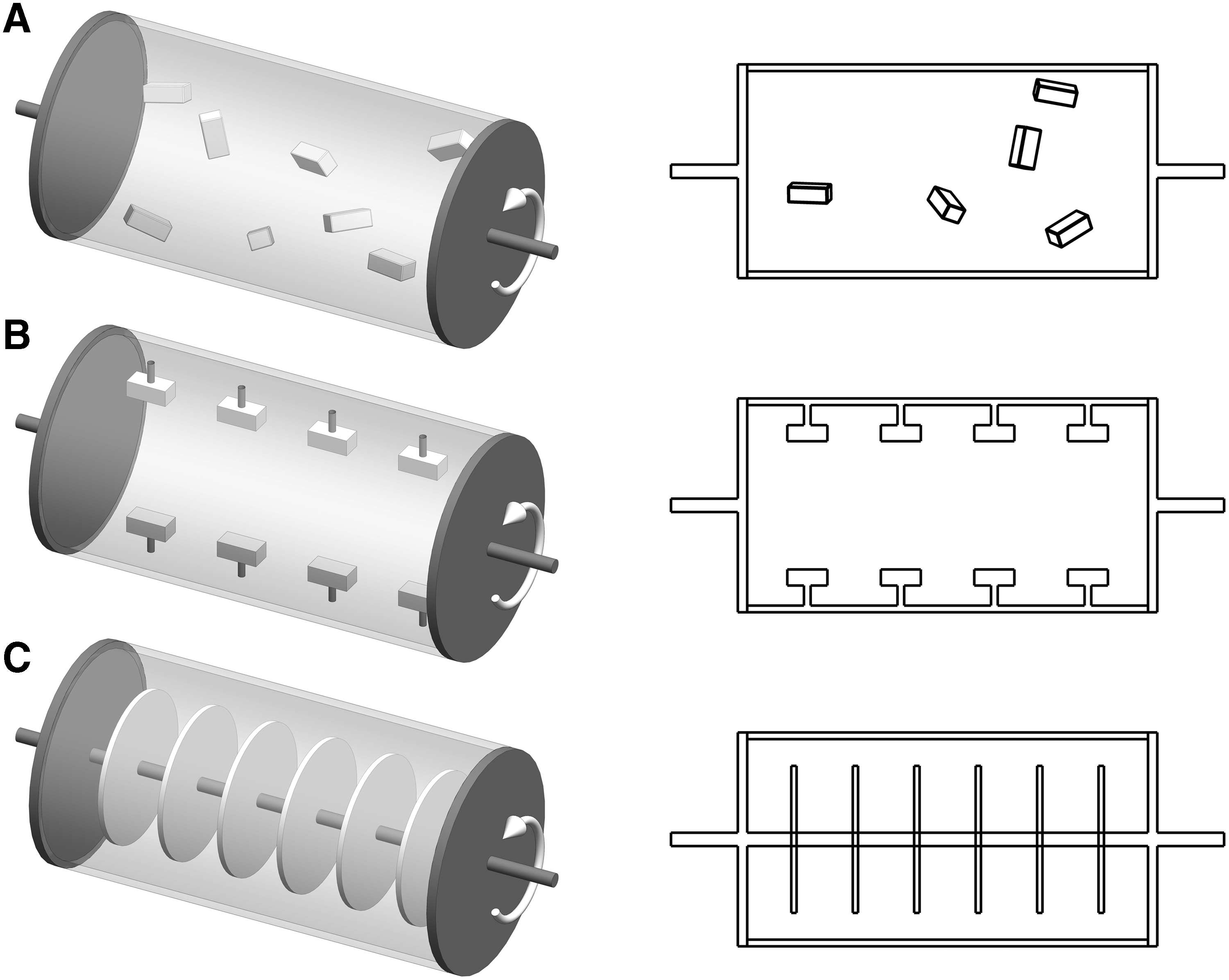
Rotating wall vessel (RWV) devices were originally designed by the National Aeronautics and Space Administration to simulate microgravity.80–82 The low levels of shear stress generated by the laminar flow of a rotating vessel along a horizontal axis are efficient to reduce diffusional limitations of nutrients and waste products. To date, different derivative designs of rotating bioreactor systems have been developed for dynamic 3D bone TE (Fig. 2A–C). Studies using dynamic rotation-based cultivation methods are specified in Table 1B. Qiu et al. applied an RWV system along with hydroxyapatite-coated hollow microcarrier scaffolds exhibiting a density similar to the medium, thereby avoiding collision with the wall of the bioreactor 83 (Fig. 2A). This cultivation method induced ECM formation in rat MSCs and cells of the osteosarcoma cell line ROS 17/2.8. Botchwey and co-workers employed a commercially available system (Synthecon Inc.) with poly(D,L-lactic-co-glycolide) “lighter-than-water” microcarriers. 84 Motions of microspheres were characterized by direct measurement using an in situ particle tracking system, originally developed by Pollack et al. 85 Seeding of scaffolds with human sarcoma osteogenic cells was performed in the bioreactor. An increase in ALP activity and enhanced mineralization compared to static culture was observed. In contrast, Goldstein and co-workers demonstrated decreased ALP activity and no change in OC level using the same bioreactor system. 86 These unfavorable results were confirmed by Sikavitsas et al., who reported decreased levels of ALP activity and Ca2+ as well as no change in proliferation and OC expression levels in rat MSCs using an RWV system. 77 Other studies proved enhanced differentiation of osteoprogenitor cells by RWV-based bioreactor systems.87–89 Song et al. developed an RWV bioreactor with the scaffolds attached to the outer wall by use of stainless steel clamps. 88 Outer and inner cylinders were driven by step motors (Fig. 2B). Compared to the cultivation in spinner flasks or static culture, the RWV resulted in better cell proliferation and differentiation. A different design of a rotating system was developed by Zellwerk GmbH in form of a rotating bed bioreactor (RBB). The schematic view of the bioreactor is shown in Figure 2C. In an RBB cell/scaffold constructs are attached directly on the axis and moved between gas and liquid phases in an alternating manner.90–92 One major benefit of the system besides the positive effects in terms of proliferation and differentiation is its compliance with GMP standards. Disadvantages of RWV systems, for example, collision of scaffolds with the bioreactor wall, which may damage scaffolds and disrupt attached cells, can be omitted by use of the RBB concept. Regardless of the advantages, however, one major potential disadvantage of the rotating system is that mineralization effects and culturing benefits are limited to the outside of the scaffolds. Internal nutrient transport limitations could not be eliminated by rotation-based bioreactor systems. 77
ECM, extracellular matrix; LDH, lactate dehydrogenase; PLAGA, poly(lactide-coglycolide); RBS, rotating bed system; SAOS, sarcoma osteogenic; RWV, rotating wall vessel.
Perfusion-based bioreactor systems
The knowledge of diffusional limitations involved in rotation-based bioreactor systems has implicated the development of perfusion bioreactors creating a laminar fluid flow and enabling mass transport of nutrients and oxygen through the entire scaffold. Investigations using either custom-made or commercially available perfusion bioreactor systems are specified in Table 1C. A recent review by McCoy and O'Brien provides a detailed overview to the influence of fluid shear stress in perfusion bioreactor cultures for bone tissue constructs. 93 Perfusion systems generally consist of containers, chambers, or cartridges harboring the cell/scaffold constructs. Cell culture medium is piped through tubes by a peristaltic roller pump to the scaffold and can either flow in a closed loop or the system provides a reservoir and a waste vessel (Fig. 3).

Flow chart of a perfusion bioreactor system. Arrows indicate flow direction in a closed loop.
BCP, biphasic calcium phosphate; BMP2, bone morphogenetic protein 2; HA, hydroxyapatite; PCL, polycaprolactone; PDMA, polydimethylsiloxane; PGE2, prostaglandin E2; PMMA, polymethylmethacrylate; RUNX2, runt-related transcription factor 2; SPCL, starch with polycaprolactone; SEVA-C, starch with ethylene vinyl alcohol; β-TCP, beta-tricalcium phosphate.
Oxygenation of the medium is ensured by either gas-permeable silicon tubes 13 or by an oxygenator device. 50 The mode of fluid flow can influence the effects of osteogenic stimulation. Jacobs et al. found that oscillating flow was a much less potent stimulator of bone cells than either steady or pulsing flow. Further, a decrease in responsiveness with increasing frequency was observed for the dynamic flow mode. 94 Enhanced levels of prostaglandin E2 (PGE2) in MC3T3 osteoblastic cells were measured upon oscillating flow type compared to perfusion-based and static culture systems. 95 Bakker et al. demonstrated that fluid flow-induced shear stress induces PGE2 production and release by bone cells in vitro. 96
Perfusion-based bioreactors can be divided into systems using indirect and direct medium perfusion (Fig. 4A, B). In indirect perfusion systems the scaffold attached to the cassette is not tightly sealed, thereby enabling medium to follow the path of least resistance around the scaffold. Thus, flow-derived shear stress may not reach the cells in the interior of the construct (Fig. 4A). Using Minucells perfusion containers (MINUCELLS and MINUTISSUE Vertriebs GmbH; Table 2), an indirect perfusion system, Wang et al. and Uemura and co-workers observed an increase in ALP activity and osteocalcin (OC) protein expression levels97,98 by dynamic cultivation of rat osteoblasts on β-TCP scaffolds. Further, bone formation was determined in a subcutaneous rat model by these authors: perfusion-stimulated cell/scaffold composites showed significantly enhanced bone formation compared to statically cultivated controls. Using a similar perfusion system human MSCs were cultivated on membranes made of mineralized Col1. 99 Lower levels of ALP activity and a decreased proliferation rate were observed as compared with statically cultivated cell/scaffold constructs. Volkmer et al. modified that type of perfusion containers by adding a carrier cassette resulting in a forced perfusion system, 100 demonstrating increased amounts of viable cells in the center of the constructs compared to indirect perfusion method. The oxygen concentrations measured in the centers of the scaffolds did not change between the two dynamic cultivation setups. However, a definitive conclusion on which cultivation method is more favorable cannot be drawn from these experiments as essential parameters, for example, ALP activity, were not measured.
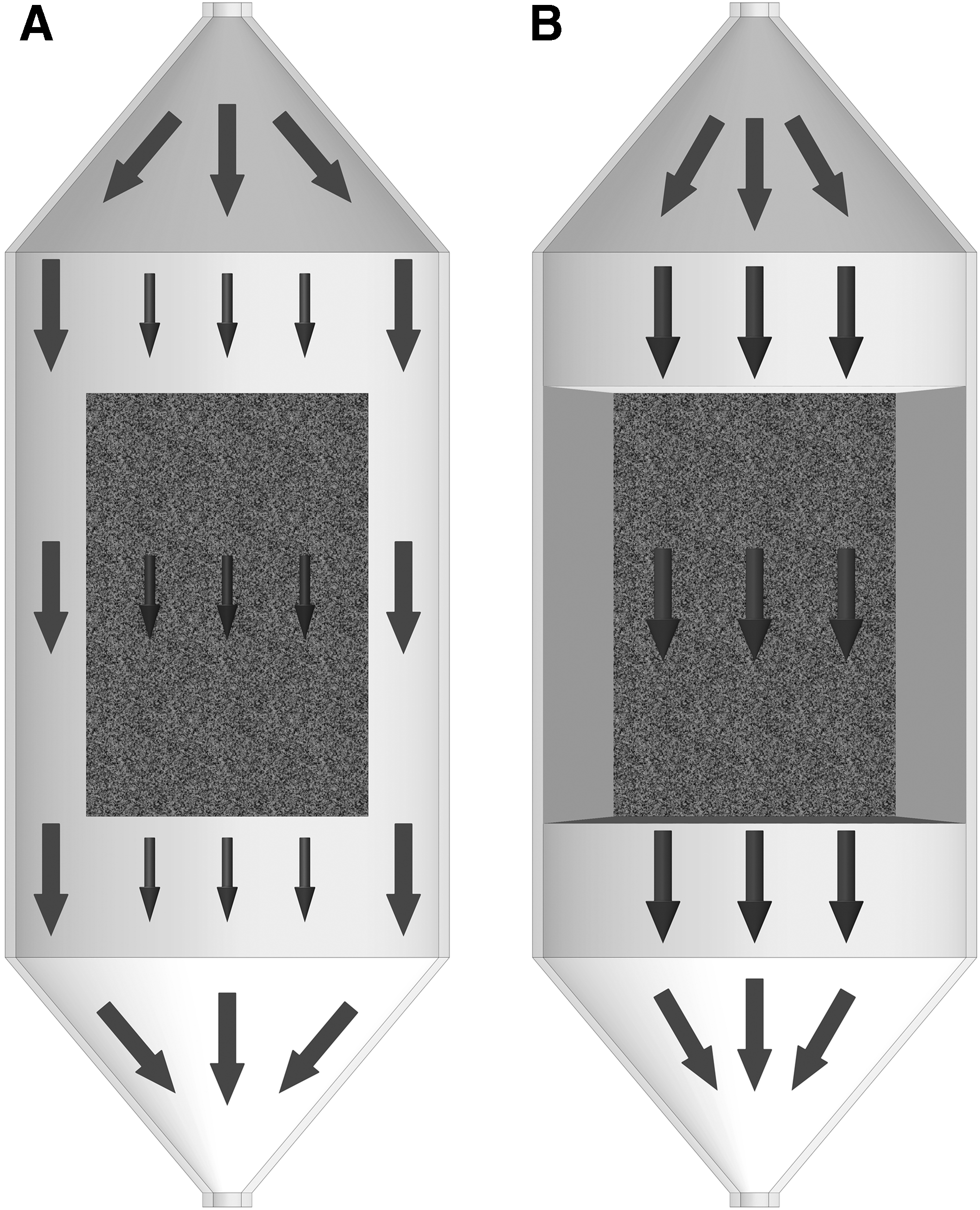
Bioreactors with direct perfusion allow the reduction of internal mass transfer limitations and exert biophysical forces by fluid flow in the interior of the so cultivated cell/scaffold constructs (Fig. 4B). Usually, the scaffolds are fixated in containers or cassettes in a press-fit manner. 101 Systems using direct perfusion have been shown to enhance cell density in the scaffold center, 102 cell proliferation and differentiation of osteoprogenitor cells, as well as the deposition of mineralized ECM.13,19,103,104 Various systems applying direct perfusion method have been described.51,105–108 Janssen et al. introduced a custom-made direct perfusion system equipped with a seeding loop, an oxygenator device, and an online oxygen measurement unit with sensors positioned at the inlet and outlet of the medium flow.50,109 However, in vivo studies in mice using this system showed no statistically significant differences in new bone formation comparing statically and dynamically cultured constructs. The OsteoGen bioreactor (Tissue Growth Technologies; Table 2) allows noninvasive online monitoring of mineralization of 3D cultivated cell/scaffold constructs by use of micro-computed tomography (μCT) technology. 110 The rate of mineralized matrix formation in the perfused constructs increased significantly from 0.69 mm3/week during the first 3 weeks of culture to 1.03 mm3/week over the last 2 weeks. In contrast, the rate of mineral deposition in the static controls was 0.01 and 0.16 mm3/week, respectively. Meinel and colleagues compared the effect of different Col- and silk-based scaffold materials and the influence of hydrodynamic environment (static culture, spinner flask, or perfused cartridge) on the osteogenic differentiation of human MSCs. 76 The authors observed enhanced mineralization on biodegradable Col-based scaffolds in spinner flask cultures compared to perfusion-based bioreactor cultivation. The authors argue that the advanced degradation of the used Col scaffolds by the perfusion bioreactor may be a reason for the unfavorable results obtained with this system. The distribution of mineralization was limited to the outer rim in spinner flask-cultivated constructs, whereas mineralized matrix was more evenly distributed in perfused scaffolds. The authors conclude that osteogenesis in cultured MSCs can be modulated by both scaffold biomaterial properties and flow environment. Fröhlich et al. cultivated bovine cancellous bone cylinders seeded with human adipose-derived stem cells in a novel perfusion bioreactor system. In this bioreactor, medium flowed through a central port at the bottom of the bioreactor vessel from where it was evenly distributed into six channels leading into individual culture wells loaded with the scaffolds. 111 The authors observed enhanced cell distribution, osteogenic differentiation, and bone matrix formation in perfused constructs compared to statically cultivated controls.
In summary, evidence exists that the use of perfusion-based bioreactor systems in bone TE results in improved cellular proliferation, distribution, differentiation, and viability in the interior of scaffolds when compared to static cultivation. Existing devices vary with respect to additional equipment and fluid flow options. Some types of bioreactor systems are suitable for cell seeding, 50 and others can be combined with mechanical stimulation (cyclic compression) 112 or online-monitoring of mineralization. 110 Reported constant perfusion flow rates range from 199 to 600 mL/h. 112 Flow rate levels exceeding a specific range have shown to promote the washing out of cells due to excessive shear stress. 113 It is therefore advisable to determine the optimal medium flow rate for each bioreactor-based setup. In general, upscaling of bioreactor-based engineered bone tissue for clinical use still needs further optimization as the dimensions of cell/scaffold constructs cultivated by use of current perfusion-based systems are comparatively small ranging from 0.04 to 2.7 cm3 (Fig. 5). In general, upscaling of construct dimensions can be either addressed by further upscaling of existing perfusion bioreactor systems to obtain a vascularized construct or by combining smaller tissue-engineered constructs synthesized simultaneously. 50 In an effort to scale up existing perfusion bioreactor systems, Olivier et al. introduced a bioreactor using relatively large porous β-TCP cylinders of 33×14 mm (4.8 cm3) with medium perfused through a dead ending hole. 114 However, this method could lead to an inhomogenous flow and no results were presented about the vitality of the cells in the inner parts of the scaffold.
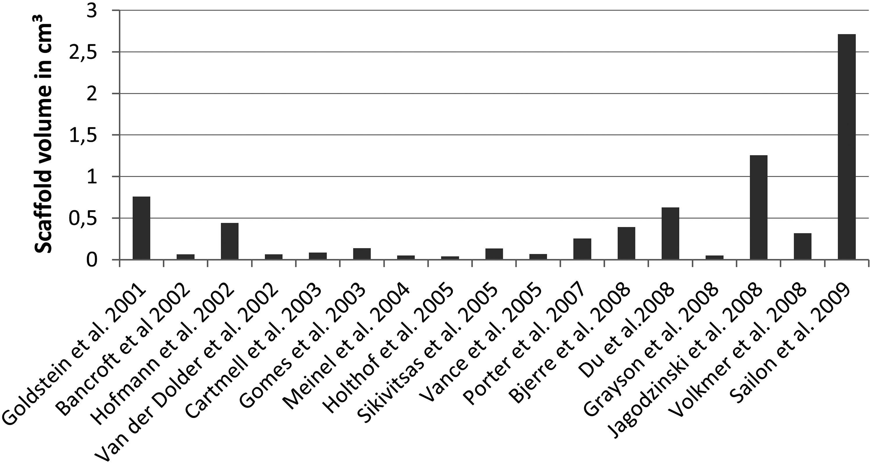
Scaffold volume data of studies using perfusion bioreactor systems.
Systems using direct mechanical strain
Since the German anatomist Julius Wolff in 1892 postulated that bone remodeling depends on mechanical load, numerous scientists have focused on biomechanical effects on the cellular level.115–119 Bone is constantly renewed by bone-forming osteoblasts and bone-resorbing osteoclasts, thereby establishing a homeostasis in healthy humans.120,121 Osteocytes are assumed to sense mechanical stimuli by different means, for example, through the cell body, the dendritic processes, or bending of cilia. 122 The signal transfer is mediated by gap junctions and hemichannels, and the release of signaling molecules into the bone fluid.123,124 However, the distinct pathways involved have not been completely discovered to date. Mechanical unloading that occurs in microgravity during space flights or extended bed rest reduces the number of osteocytes. 125 Loading of bone with strains below 500 μstrains was associated with bone loss, for loading with up to 1000 μstrain the original bone geometry and mass were maintained, and strains between 1000 and 4000 μstrains increased new bone formation progressively. 126 Recent studies identified WNT signaling as an important pathway promoting the early phase of commitment to the osteogenic lineage and subsequent differentiation of C57BL/6J osteoblasts 62 and osteoblastic precursors in general. 127 WNT signaling enhances the expression of osteoprotegerin, but inhibits the expression of high levels of OC, a typical feature of mature, matrix-synthesizing bone cells. 127
Various studies confirmed the principle of mechanical conditioning by the application of direct mechanical strain using, for example, bending, stretching, contraction, and compression (Table 1D). These types of mechanical strain application will be discussed in the following. The mechanical stimulation by a 4-point-bending device (Fig. 6A) resulted in increased levels of ALP activity, mineralized matrix production, and gene expression of ALP and OP in MSCs loaded on demineralized bovine cancellous bone grafts. 48 Interestingly, this effect was only detectable in the presence of dexamethasone at concentrations of 10 nM, but not 100 nM.
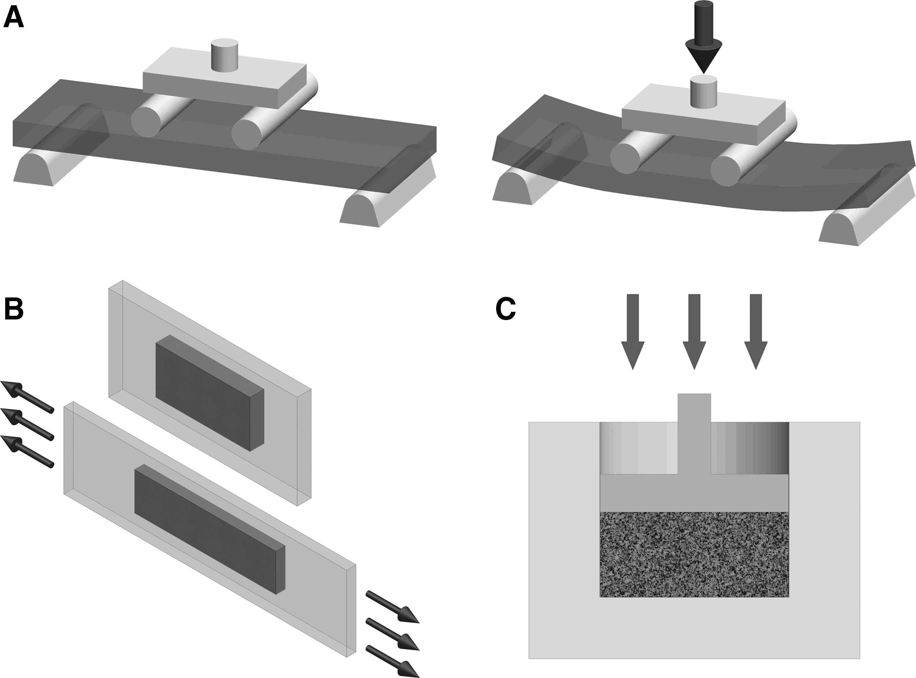
Cyclic stretching of human osteoblasts attached to silicon dishes enhanced proliferation, but did not affect ALP activity. 15 The principle of uniaxial stretching is shown schematically in Figure 6B. Uniaxial stretching of a human osteoblastic cell line in Col1 gels applied for 21 days with a magnitude of 1% (10,000 μstrain) increased both proliferation and gene expression of ALP, OC, OP, and Col1 compared to static controls. Further, the cells and the newly produced type of ECM were strictly oriented according to the direction of the applied mechanical stress. The cell-stretching system consists of rectangular elastic silicone dishes, which were designed for use of a six-station stimulation apparatus driven by an eccentric motor.128,129 Another study supporting the effects of contraction was performed by Akhouayri et al., who observed increased proliferation, ALP activity, as well as Ca2+ and OC protein expression levels by contracted rat osteosarcoma cells cultivated on 3D Col1-matrices. 130
When analyzing effects of compression (Fig. 6C), machines originally fabricated for material testing were used frequently. For example, a study by Lanyon and Rubin from 1984 showed that intermittent dynamic as opposed to continous compression loading induced bone formation in vivo using an avian ulnar defect model. 117 The applied type of stress can influence the effect of tissue response. Using low hydrostatic pressure in a bone organ culture of murine fetal metatarsal and calvariae Burger et al. observed that intermittent stress enhanced mineralization more effectively than continuous stimulation. 131 Wartella and Wayne recently introduced a biaxial bioreactor system for mechanical stimulation of tissue constructs in two perpendicular directions. 131 This type of bioreactor applying both compression and tension forces resulted in elevated proteoglycan production and matrix deposition by human MSCs.
Currently existing bioreactor systems for direct mechanical stimulation have shown beneficial effects on proliferation, osteogenic differentiation, and matrix formation. Several authors used biomechanically instable Col1 gels as matrices for mechanical strain-based cellular stimulation.129,130 This may be disadvantageous in situations where a certain level of initial mechanical stability of an implantable cell/scaffold construct is required for effective bone regeneration and no further stabilization is applied. Further, diffusional limitations occurring in larger constructs in mechanical load-based bioreactor systems may be addressed additionally by other strategies.
EMF-based bioreactor systems
Electric and EMFs have been applied for bone regeneration purposes in patients with, for example, osteoporosis and nonunions as well as supportive therapy during limb lengthening and revision alloarthroplasty procedures for the last three decades.133–137 PEMF has been shown to significantly reduce the loss of bone mass and to accelerate bone formation in vivo.138,139 Endogenous EMF and PEMF arise from muscle movements. 140 The electric potentials generated by mechanical deformation in bone cause piezoelectricity. When bone is fractured, electrons migrate to the injured site, causing a negative potential. Vibrations of human muscles induce mechanical strains and currents of specific frequencies. Frequencies in the ranges of 5–30 Hz and <10 Hz were observed during postural muscle activity and walking, respectively. 141 Interestingly, bone cells exhibit a strong frequency selectivity with EMF effectiveness peaking at 15 Hz. 142 Studies suggest that EMFs affect different subcellular proliferation- and differentiation-related signaling pathways, for example, those including parathyroid hormone and adenosine A2A receptor, resulting in conformation changes or in increase of the receptor density. 143
To utilize these effects for bone TE, EMF-based bioreactors (Fig. 7) were developed. Typically, these systems consist of Helmholtz coils powered by a PEMF generator. The cell/scaffold construct is positioned between two Helmholtz coils and an EMF of a defined intensity is applied. In vitro studies showed that EMFs induce and enhance osteogenesis in human MSCs144,145 and osteoblasts146–148 (Table 1E). Fassina et al. introduced a simple EMF-based bioreactor system with a standard well plate and two parallel Helmholtz coils being kept in a PMMA tube. 149 The applied PEMF frequency used in that study was 75 Hz with an intensity of 2 mT and the magnetic field was measured using a Hall Effect transverse gaussmeter probe. PEMF-stimulated human sarcoma osteogenic-2 cells exhibited increased mineralization and gene expression of decorin, OC, OPN, TGF-β, and Col1. In a study by Schwartz and co-workers, human MSCs cultivated on calcium phosphate discs demonstrated decreased proliferation but enhanced ALP activity and protein levels of OC and TGF-β in the combined presence of BMP-2 and PEMF stimulation. 144 Bodamyali et al. showed superior mineralization and expression of BMP-2 and BMP-4 genes upon PEMF stimulation in rat osteoblasts. 146 The device utilized a saw tooth waveform consisting of 4.5-ms bursts of pulses, repeating at a rate of 15 Hz. Increased proliferation and ALP activity were reported by Tsai et al., who stimulated rat osteoblasts seeded on poly(lactic-co-glycolic acid) scaffolds with PEMFs at a frequency of 7.5 Hz. 150
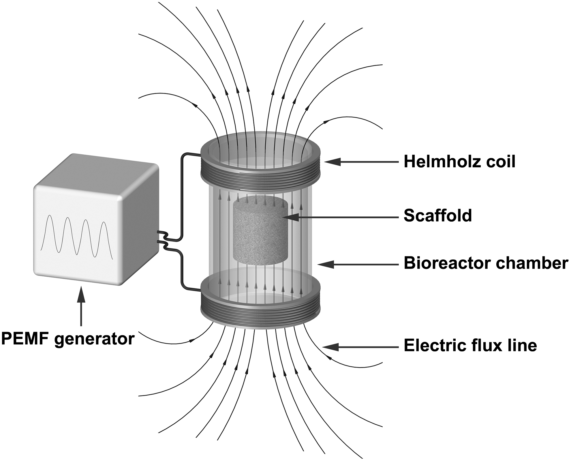
Schematic illustration of a bioreactor system based on pulsed electromagnetic fields (PEMF). The Helmholtz coils are powered by a PEMF generator. The scaffold in the bioreactor chamber is positioned between two Helmholtz coils. The bioreactor applies PEMF stimuli to the cells within the scaffold at a defined frequency, amplitude, intensity, and pulse duration.
CD, cluster of differentiation; DCN, decorin; Hz, hertz; mT, milli tesla; PEMF, pulsed electromagnetic fields; TGF-β, transforming growth factor beta.
In summary, the use of EMF-based bioreactor systems for bone TE resulted in enhanced osteogenic differentiation of cell/scaffold constructs compared to static cultivation. Interestingly, as observed in some studies, PEMF also stimulated proliferation of osteoprogenitor cells. The high initial equipment costs required for PEMF-based bioreactor systems denote a major disadvantage. On the other hand, the noninvasiveness of PEMF-based systems is clearly advantageous with respect to handling and potential GMP approval.
In vivo bioreactor systems
The concept of in vivo bioreactor systems takes advantage of the physiological environment and supply of a cell-loaded scaffold biomaterial with necessary growth factors and nutrients provided by the host organism. Several in vivo bioreactors were developed to generate vascularized bone tissue using different animal models, for example, in mice, 151 rats, 152 rabbits, 153 and miniature pigs, 154 resulting in site-specific de novo bone regeneration. Petite et al. used a combination of a coral scaffold with in vitro-expanded MSCs leading to complete recorticalization and the formation of mature lamellar cortical bone in sheep. 155 Even a man can serve as an in vivo bone bioreactor as described by Warnke et al. 156 A titanium mesh cage filled with bone mineral blocks, infiltrated with 7 mg recombinant human BMP-7 and autologous bone marrow, was implanted in a latissimus dorsi muscle pouch. After 7 weeks the construct was transplanted to repair a mandibular defect. Successful bony reconstruction resulted in improvement in the quality of life of this patient. 157 However, although occasionally applied with success, the application of in vivo bioreactor concepts is currently limited to individual cases.
Commercially available bioreactor systems for bone tissue engineering
Currently, various bioreactor systems for generating mineralized cell/scaffold constructs are commercially available (Table 2). In the following we focus on selected systems representative for the respective type of bioreactor. Besides the systems mentioned, other bioreactor systems are available for bone TE applications.
GMP, Good Manufacturing Practice; RBS, rotating bed system; STLV, slow turning lateral vessel; μCT, micro-computed tomography.
Possibly, the most inexpensive systems are spinner flasks. The Bell-Flow™ spinner flask from Bellco Biotechnology is manufactured from autoclavable borosilicate glass and is available in volumes ranging from 100 mL to 3 L (www.bellcoglass.com). Corning® Lifesciences offers autoclavable spinner flasks as well as disposable systems made of plastic (www.corning.com/index.aspx).
MINUCELLS and MINUTISSUE GmbH offers a variety of perfusion containers, for example, for cultivating cartilage constructs or several types of epithelia in their organo-typical environment (www.minucells.de/index.html). The reactors are referred to the indirect perfusion method. The OsteoGen Bioreactor, a device for direct perfusion, is commercially available from Tissue Growth Technologies. The design is compact and able fit in a standard incubator, and all components of the system are autoclavable. The chambers are designed only for cylindrical scaffolds with 10×10 mm. Besides an optional pulsatile hydrostatic pressure stimulator, the company offers a “GrowthWorks Software and Control platform” (www.tissuegrowth.com/).
Zellwerk GmbH distributes the GMP-conform RBB tissue culturing system BIOSTAT® Bplus RBS (www.zellwerk.biz/). The complete cultivation system comprises a bioreactor, a GMP breeder, and a control unit. Other rotating bioreactor systems are available from for example, Synthecon, Inc. These rotary cell culture microgravity bioreactors originally designed by National Aeronautics and Space Administration are produced as autoclavable and disposable systems. The company also offers a perfused rotating bioreactor allowing the online monitoring of crucial parameters, for example, pH, oxygen, and glucose levels (www.synthecon.com). Another dynamic cultivation system is available from B. Braun Biotech International GmbH offering an RBB meeting GMP standards (www.chemietechnik.de/company).
Systems using tension, compression, and shear stress are available from, for example, Flexcell International Corporation. The Flexcell® FX-5000™ Tension System and Flexcell FX-5000 Compression System apply cyclic or static strain to cells cultured on flexible-bottomed culture plates. Special devices allow to observe signaling responses upon strain stimulation in real-time on a microscope stage (www.flexcellint.com/).
Bioreactors and GMP
GMP is a quality assurance system for medicinal products. Several regulatory requirements, for example, production according to validated standard operating procedures, demonstration of quality control, and in-process controls, have to be applied. In Europe respective directives and guidelines ensure quality and safety standards for donation, procurement, testing, processing, preservation, storage, and the distribution of human tissue and cells. 158 In this context, compliance with the annually updated guidelines for “Current Good Manufacturing Practice” (cGMP) is required in the United States. 159
Protocols were developed to facilitate the adaption of GMP standards for the expansion of human embryonic stem cells in a stirred bioreactor system. 160 In addition, a strategy was described to develop and validate a closed, automated production process to expand stem and progenitor cells in the presence of human bone marrow mononuclear cells. 161 The RBBs Medistat RBS (B. Braun Biotech International GmbH) and Z®RP cell- and tissue culturing systems (Zellwerk GmbH), allowing the cultivation and manufacturing of 3D tissue-engineered transplants91,92 conform to requirements of GMP standards. Successful translation from the laboratory to clinical application can be exemplified in the field of skin TE. The commercially available products TransCyte™ and Dermagraft®40 are generated in a closed perfusion bioreactor system. For this purpose, a scaffold (Biobrane®) is seeded with allogenic dermal fibroblasts and cultivated in a bioreactor system allowing automated cell seeding, media change, in-process monitoring of growth, storage, and delivery simultaneously. To the authors' best knowledge, however, no cell-based, tissue-engineered bone substitute construct cultivated in a bioreactor system has been applied clinically to date. To pave the way for bioreactor-stimulated, tissue-engineered constructs for bone regeneration from bench to bedside, the compliance of potential products with GMP standards will be a basic prerequisite.
Conclusion
Bone graft material is often needed for the treatment of osseous defects. Due to limitations and risks associated with autologous as well as allogenic bone grafting procedures, alternative strategies are needed for skeletal reconstruction. The concept of TE constitutes the framework for the implementation of cell-based bone regeneration strategies. To optimize the cultivation of cell/scaffold constructs, dynamic bioreactor systems, enhancing cellular proliferation and differentiation and resolving mass transport limitations, are appealing components. Bone bioreactor systems include spinner flasks, RWV constructs, perfusion bioreactors, and systems based on mechanical or electromagnetic stimulation of cell/scaffold composites. These systems differ considerably with respect to ease of use, cost-effectiveness, and degree of additional osteogenic stimuli provided, as well as monitoring and manipulation options. Currently available bone bioreactors enable adequate monitoring and controlling of specific biological, physical, and chemical parameters during the process of in vitro bone formation. Further optimization of these systems may be achieved by adapting specific stimuli, for example, shear stress, load, or EMF, and by combining biophysical and biochemical stimuli within one system. A major challenge, however, will be the translation of bioreactor-based concepts into clinically applicable, GMP-conform systems generating newly formed mineralized cell/scaffold constructs.
Footnotes
Acknowledgments
We thank Mike Tipsword, Angela Jacobi, and Corina Vater for proofreading this article. The authors have received financial support from the German Academic Exchange Service/German Federal Ministry of Education and Research (Grant No. D/09/04774) and from the Center for Regenerative Therapies Dresden, Germany.
Disclosure Statement
No competing financial interests exist. We do not claim completeness with respect to the bioreactor systems discussed and do regret if certain products or concepts are not covered in this article.