
Abstract
Osteoarthritis (OA) is a major clinical and scientific challenge. The degradation of articular cartilage in the joints is a common manifestation of painful arthritis. The regeneration of articular cartilage in OA is an unmet clinical need. The assembly of articular cartilage by tissue engineering toward complete regeneration is the goal of most scientists and surgeons. The key ingredients for regeneration are signals, stem cells, and scaffolds. This brief review focuses on the scaffold, with special emphasis on hydrogels and nanomaterials for the assembly of tissue-engineered cartilage, and ultimately leading to the total regeneration of articular cartilage in the joints.
Introduction
Regeneration is, in part, recapitulation of embryonic development and morphogenesis. Morphogenesis is the developmental cascade of pattern formation, establishment of body plan, and the architecture of mirror-image bilateral symmetry of musculoskeletal structures and culminating in the adult form. 2 This concise review will focus on the regeneration of articular cartilage with special emphasis on the necessity of the design criteria for nanomaterials based on the physiology of ECM scaffold in the native tissue and how this can be imitated for optimal regeneration. 3 Several types of cells are currently used for cartilage regeneration including differentiated chondrocytes and undifferentiated mesenchymal stem cells from bone marrow, fat, muscle, or synovium tissue. Also, cells can be implanted in suspension or with different kinds of ECM scaffolds (collagen I, II, III, etc.). It should be emphasized that although one would wish to use tissue-engineered cartilage for OA, at present it is geared mainly for cartilage defect repair.
Articular Cartilage ECM
ECM is the native scaffold in most tissues. 5 In certain tissues such as articular cartilage, the ratio of the volume of ECM to cells is in favor of the matrix. The natural biomaterial in articular cartilage is a composite of collagens, proteoglycan aggrecan, and assortment of glycoproteins including fibronectin, cartilage oligomeric matrix protein, cartilage intermediate layer protein, and matrilins. In addition, there is a distinct zonal organization of surface, middle, deep zones, and a mineralized cartilage with a distinct “tide mark” in articular cartilage at the interface between the unmineralized cartilage and mineralized cartilage ECM. The distinct zones exhibit characteristic biochemical markers of the ECM. The surface zone of the articular cartilage secretes superficial zone protein (SZP), a mucinous glycoprotein with an attached glycosaminoglycan chain, making this protein a novel proteoglycan. 6 SZP is highly homologous to lubricin, a large glycoprotein isolated and purified from synovial fluid.7–10 The middle and deep zones of articular cartilage have distinct fiber organization and have mainly collagen II and proteoglycan aggrecan as the principal constituents. Thus, the structural and chemical composition of articular cartilage has to be taken into account in designing strategies for optimal and complete regeneration of articular cartilage. 11
Although bone has considerable potential for repair and even regeneration, the articular cartilage is feeble in this regard. The inability of spontaneous repair and regeneration of cartilage may be due to lack of vascular elements, endogenous antagonists of morphogens, and other cytokine inhibitors. The optimal regeneration of cartilage continues to be a challenge in view of the known hierarchical organization and the zonal geometry. 12 Despite these challenges, progress continues in articular cartilage regeneration. The initial approaches of autologous chondrocyte implantation (ACI) are being continuously defined. 13 In addition to the use of periosteal membrane, other biomaterial-based approaches are being developed. Composite collagen of types I and III have been used. The second- and third-generation ACI is now in everyday clinical use. In every case, nonintegrated homogeneous cartilage tissue resembling fibrocartilage is formed in the defect. The technique of mosaicplasty is also being used. 14 The relative instability of the chondrocyte phenotype in cell cultures and continuous dedifferentiation remains a challenge. The availability of recombinant bone morphogenetic proteins (BMPs) and transforming growth factor-beta (TGF-β) isoforms permits the redifferentiation of dedifferentiated chondrocytes. In view of the inhibitory effects of endogenous proteoglycans in cartilage on cell migration into the wound and injury site, chondroitinase pretreatment has been investigated with beneficial effects on cell migration. 15 The purity of the implanted chondrocytes remains uncontrolled. Perhaps the presence of fibroblasts drives the synthesis of collagen I inside the defect.
The ECM of the articular cartilage has been systematically studied. 16 Chondrocyte death in articular wound edges 17 and the subsequent lack of matrix producing cells in the interface area are considered major causes of impaired integrative articular cartilage repair. 18 Accordingly, efforts have been made to allow chondrocytes to migrate through their dense matrix toward an interface by combining local enzymatic digestion of the matrix with growth factor stimulation. One approach is to decrease proteoglycans by hyaluronidase treatment, resulting in optimal repair.19,20 This growing information allows one to design a biomimetic scaffold for cartilage regeneration. The design of the scaffold should be based on the physicochemical nature of the ECM and the biomechanical properties of the native articular cartilage. Recent advances in the biology and chemistry of ECM, the cognate receptors, and the downstream targets of transcriptional and regulators will lead to peptidomimetic agonists of the ECM-cell/chondrocyte interface. Such incremental advances will result in synthetic analogs of ECM in articular cartilage. The remaining challenges include the reproduction of the zonal geometry of the articular cartilage, either because the scaffold is different depending on the depth of the defect, or because the undifferentiated cells implanted on the defect are incapable of regional differentiation due to local biomechanical and biochemical constraints.
Modeling Articular Cartilage as a Hydrogel
Native articular cartilage is a durable tissue with a long functional life span unless afflicted by OA and leading to degradation by proteolytic enzymes released by catabolic cytokines such as tumor necrosis factor alpha. The ECM as discussed in the preceding section consists of predominantly proteoglycan, aggrecan that resists compression, and the main collagen component type II responsible for the tensile strength. The aggrecan in the ECM of articular cartilage are negatively charged, inducing both a repelling force due to electronegativity and an osmotic swelling due to water movement into the matrix to ensure electroneutrality. The resultant stress permits the cartilage tissue to withstand large loads due to the tensile strength of collagen II and the water in the tissue that can be exuded. Articular cartilage possesses significant mechanical properties with a compressive modulus of 0.7 to 0.8 MPa, a shear modulus of similar magnitude (0.69 MPa), and a tensile modulus of 0.3–10 MPa.21,22 Thus, the mechanical properties are reminiscent of a hydrogel. During trauma or supra-physiological impact loading, the rate of cartilage failure at both the surface and the subchondral bone increases and results in injury.
The articular cartilage has limited potential for repair. Current surgical options are allografts and autologous osteochondral plugs. The repair response generally results in fibrocartilage and continued pain and discomfort. The ideal design criteria for tissue-engineered articular cartilage require biocompatibility, ease of handling, mechanical properties approaching native tissue, and ease of implantation.
Since articular cartilage has hydrogel-like properties, an attractive scaffold for the native tissue is a poly(ethylene glycol) (PEG) hydrogel. Thus, hydrogels have become a focal approach for cell and morphogen delivery 23 for articular cartilage tissue engineering and regeneration. Hydrogels are hydrated materials in which osmotic swelling is generated entropically by polymers chains and the chemical cross-links that restrain this swelling. It is an idealized model for the swelling characteristics of cartilage ECM. Therefore, considerable research has been focused on hydrogels for cartilage regeneration. One can prepare a solution of hydrogel, incorporate cells and morphogens, and then initiate gelation by changes in temperature, hydrogen ion concentration, and so on. Hydrogels of alginate, agarose, chitosan, and collagen are natural biomaterials. In addition, there are a plethora of synthetic materials where molecular weight, chemistry, and composition can be varied for optimal biological properties. Through novel approaches for structural modifications, the mechanical properties of hydrogels can also be tuned to modulate and direct cellular activity.23–25
A polymeric system based on the hydrophilic copolymer poly(propylene fumarate-co-ethylene glycol) was developed. 26 Solutions of this polymer are in liquid state at temperatures below 25°C and in a gel at temperatures above 35°C. This thermoreversible hydrogel scaffold would appear to be useful for articular cartilage, as one can encapsulate chondrocytes with optimal growth factors and/or morphogens such as BMPs.2,3 Bovine articular chondrocytes were used in this hydrogel, and they exhibited optimal proteoglycan and collagen II synthesis. The addition of BMP-7 to chondrocytes in the hydrogel stimulated cell proliferation but not biosynthesis of proteoglycans. Although thermoreversible hydrogels are a potential delivery system, much additional work is needed.
There is considerable excitement about the use of self-assembling peptide hydrogels for chondrocyte biology and cartilage repair. 27 An ideal self-assembling hydrogel should stimulate both cell proliferation and matrix biosynthesis and assembly, leading to tissue morphogenesis. It is noteworthy that a novel self-assembling peptide hydrogel stimulates cartilage ECM biosynthesis by bovine chondrocytes. The peptide consisted of dodecamer of (K-L-D)4 of lysine-leucine-aspartic acid sequence. This peptide hydrogel has great promise for future applications in cartilage regeneration and basic cell biology of chondrocytes.
The systematic study of the cellular behavior and response of the chondrocytes from various zones is critical in the biology of hydrogels and cartilage. Elisseeff and colleagues have indeed performed such an incisive investigation in PEG hydrogels 28 containing various ECM components such as collagen I, hyaluronan, and chondroitin sulfate seeded with superficial and deep zone chondrocytes. Chondroitin sulfate containing hydrogels stimulated the gene expression and matrix accumulation from superficial and deep zone chondrocytes. 28
Scaffold-less approaches to cartilage tissue engineering have also demonstrated promising results. Athanasiou and colleagues have developed a self-assembling approach where primary chondrocytes are suspended in a nonadherent well at high cell densities. 29 The chondrocytes self-assemble into a nascent tissue and produce large amounts of GAGs and collagens. 30 Through the application of hydrostatic pressure and growth factors, these self-assembled cartilage constructs achieve mechanical properties nearing native tissue levels. 31 Additional use of nanomaterials may further improve the mechanical and biochemical characteristics of these tissue-engineered constructs.
Nanomaterials: Design Criteria for Cartilage Regeneration
The foregoing discussion of hydrogels and the cellular proliferation and biosynthesis of chondrocytes therein reinforces the concept that a natural or synthetic hydrogel mimics the cartilage ECM. The explosive interest and progress in the field of nanomaterials sets the stage for consideration of the potential novel application of nanomaterial hydrogels. Nanomaterials are materials with a dimension of 1–100 nm 32 (Fig. 1). Nanomaterials can be metals, ceramics, and polymers in the form of nanocrystals, nanoclusters, nanoparticles, nanofibers, nanotubes, and nanofilms. Using both top-down or bottom-up technology, a variety of methods have been used to generate nanomaterials. These approaches include electrospinning, self-assembly, phase separation, nano-imprinting, and photolithography. Biomimetic nanomaterials that are cyto-compatible are used for delivering stem cells and chondrocytes. Since hydrogels appear to have the greatest potential and closely approximate the native hydrated cartilage ECM, one can consider articular cartilage as a self-assembled tissue in terms of tissue engineering. After all next to bacteriophage self-assembly, perhaps the self-assembly of the ECM component collagens, including collagen II, has been well investigated. During tissue morphogenesis in cartilage, the basic cellular and molecular mechanisms are dominated by self-assembly and growth. The monomeric constituents of collagens, proteoglycan subunits, and SZP are in the nanoscale and fit into the basic definition of a nanomaterial. The self-assembled tissue structure and the constituent cells are in the higher order meso-, micro-, and macroscales. Thus, the design criteria for the design of a bottom-up hydrogel, whether by self-assembly or directed-assembly, should begin with proto-units of the nanomaterial and self-assemble into higher order structures incorporating chondrocytes, stem/progenitor cells with appropriate signals for morphogenesis of cartilage such as BMPs, and TGF-β family members. 2
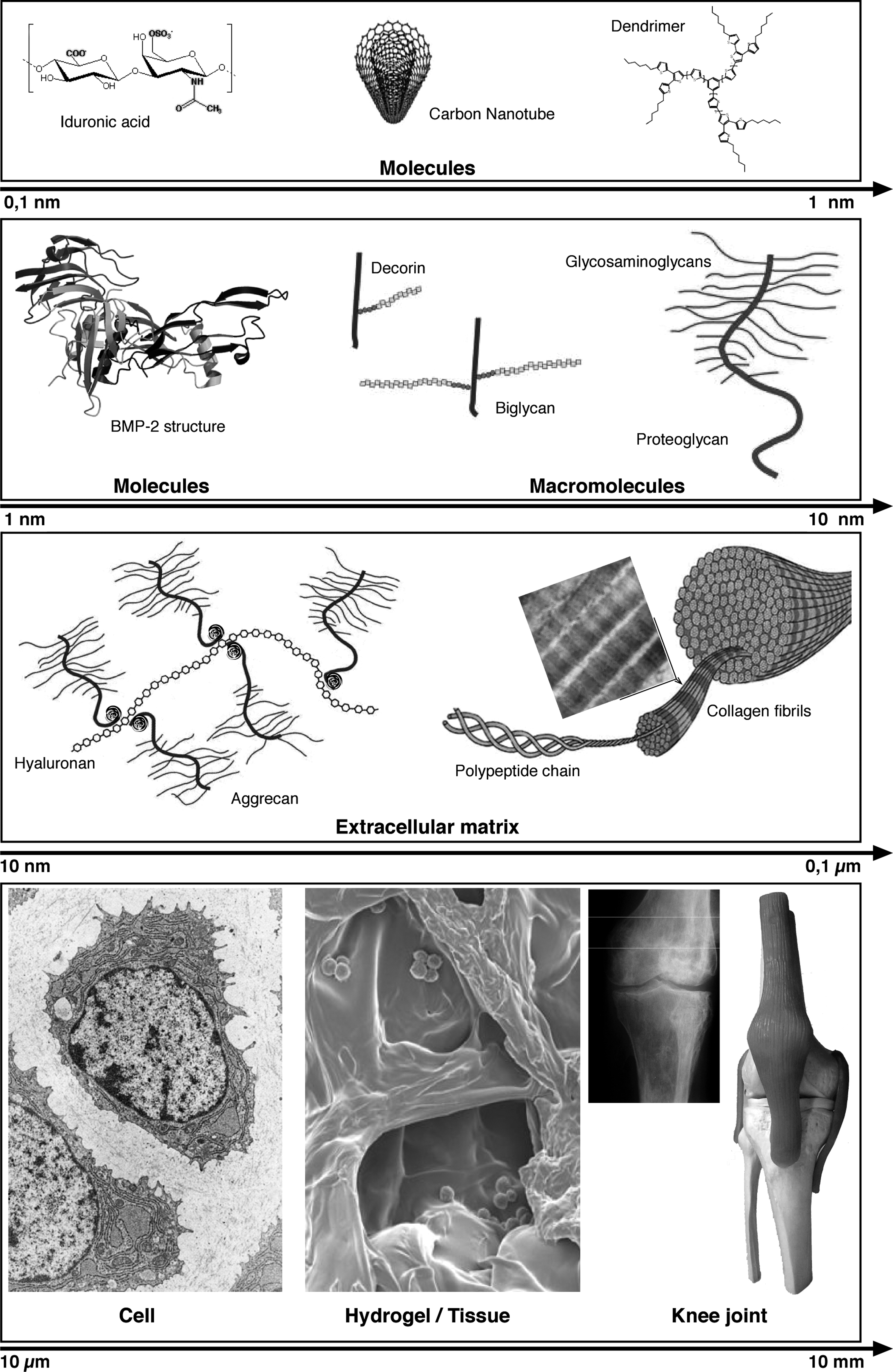
The hierarchical continuum of extracellular matrix molecules, cells, and joint tissue. The continuum consists of disaccharides, dendrimers, bone morphogenetic protein 2 (BMP 2), proteoglycans, collagen fibrils, chondrocytes, cartilage tissue, and, finally, a knee joint. The nanomaterials are in the range of 1 to 100 nm.
The general approaches for regeneration of tissues include injection of cells at the site of injury, the guided tissue engineering of a scaffold such as a hydrogel, and, finally, a scaffold with a growth factor is used. 33 A variety of methods are used to fabricate nanofibrous scaffolds, such as self-assembly, phase separation, and electrospinning. The patterning of cells in a scaffold is aided by contact printing, capillary force, lithography, and other specialized methods. The evolving armamentarium of techniques augurs the development of new hydrogel scaffolds for cartilage regeneration.
Challenges and Opportunities
Degenerative OA requires new, more efficient products for cartilage regeneration. The recent advances in the field of dendrimers are especially exciting in nanomedicine and nanotechnology. 34 In nature, there is a widespread use of branched dendrimer-like structures. The branching pattern of various parenchymal organs is a biological example of dendrimer biology. The basic chemistry of dendrimers include unbranched, branched, dendrons, and dendrimers. In the design of hydrogels for cartilage, one can envisage a classic dendrimer design to mimic the network of molecules in the ECM. The potential utility of dendrimer-based hydrogels incorporating hydrophilic and hydrophobic-domains permits to mimic cartilage matrix. Also, dendrimer chemistry can create a core in a globular structure that provides a protease-protected domain for perhaps growth factors and morphogenetic proteins.
Biodegradable nanofibers and nanomats are made with multiple functional groups for tissue engineering. 35 The form of the nanomaterials can be altered or modified for applications in cartilage regeneration. Carbon nanotubes are increasingly considered for biomechanical applications including tissue engineering.36,37 Due to their strength and material properties, carbon nanotubes may find new uses in various composite biomaterials for cartilage regeneration. Finally, it is encouraging to note that a nanofibrous hydrogel seeded with cells and supplemented with TGF-β3 promoted tissue integration in a cartilage gap model. 38
Footnotes
Acknowledgments
We thank Dr. Jesus A. Santamaria for his collaboration in the design of the figure for this review. This work was supported by grants from the Banco Bilbao-Vizcaya-Argentaria Foundation (FBBVA, Chair in Biomedicine 2007 to A.H. Reddi), the Ministry of Science and Technology (BIO2009-13903-C02-01), the Ministry of Science and Innovation (PLE2009-0163, FIS PI10/02529), and the Andalusian Autonomous Government (P07-CVI-2781, PAIDI BIO-217, PI-0729–2010). Red de Terapia Celular and CIBER-BBN are an initiative funded by the VI National R&D&I Plan 2008–2011, Iniciativa Ingenio 2010, Consolider Program, CIBER Actions, and financed by the Instituto de Salud Carlos III with assistance from the European Regional Development Fund.
Disclosure Statement
No competing financial interests exist.