
Abstract
The reconstruction, repair, and regeneration of the external auricular framework continue to be one of the greatest challenges in the field of tissue engineering. To replace like with like, we should emulate the native structure and composition of auricular cartilage by combining a suitable chondrogenic cell source with an appropriate scaffold under optimal in vitro and in vivo conditions. Due to the fact that a suitable and reliable substitute for auricular cartilage has yet to be engineered, hand-carved autologous costal cartilage grafts and ear-shaped porous polyethylene implants are the current treatment modalities for auricular reconstruction. However, over the last decade, significant advances have been made in the field of regenerative medicine and tissue engineering. A variety of scaffolds and innovative approaches have been investigated as alternatives to using autologous carved costal cartilage or porous polyethylene implants. A review of recent developments and the current state of the art and science is presented, focusing on scaffolds, cell sources, seeding densities, and mechanical characteristics of tissue-engineered auricular cartilage.
Introduction
Ideally, a biopsy of autologous chondrocytes or pluripotent stem cells would be harvested from the patient. If any remnants of auricular cartilage exist, then these tissue sources can be used to avoid harvesting cartilage from the contralateral ear. The cell population would then be expanded in vitro and seeded onto a suitable scaffold to engineer a functional auricle. This approach would avoid the donor site morbidity and other limitations associated with harvesting costal cartilage and manually sculpting an ear-shaped framework. In addition, a tissue-engineered auricle could be custom designed to more closely match the contralateral ear, thus improving the end cosmetic result. Other advantages of utilizing a tissue-engineering approach include the reduction of operative time and avoidance of multiple-stage procedures. Figure 1 illustrates a general methodology that can be potentially applied for the successful production of a tissue-engineered construct.
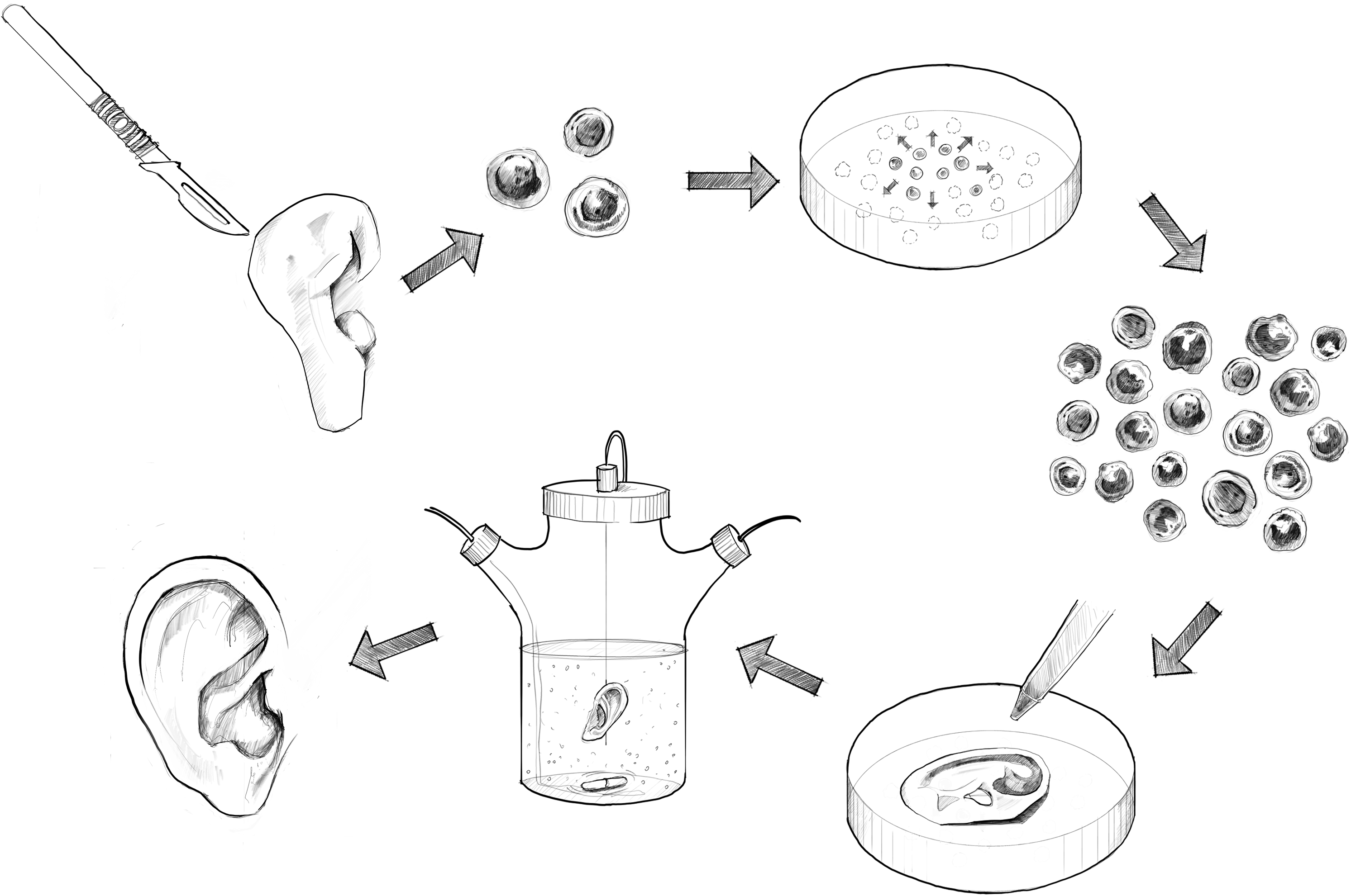
A potential methodology for auricular tissue engineering. Chondrocytes are harvested and isolated from an auricular tissue biopsy or tissue remnants from a damaged or malformed ear. The expanded cells are then seeded onto an ear-shaped scaffold and cultured in vitro to enhance extracellular matrix deposition. The final step would involve implanting the construct in vivo.
The advent of tissue-engineering technologies has prompted researchers to revisit ideas and approaches first outlined over 50 years ago for prefabricated, ear-shaped implants made of perforated external molds and diced cartilage. 5 Cartilage has been successfully engineered in predetermined shapes 6 by seeding cells onto scaffolds or suspending them in hydrogels in both in vitro and in vivo studies, culminating in ear-shaped frameworks in immunocompromised rodent models.7–10
Despite initial success, size and shape retention of such constructs have been unsatisfactory, and efforts to translate the developed methodologies into immunocompetent animal models have been fraught with difficulties. It is crucial to focus on engineering a construct that resembles, as close as possible, the mechanical, biochemical, and histological characteristics of native tissue. A balance between an immunocompetent environment, a biocompatible scaffold, and an optimal cell source and quantity are factors required for successful development of an engineered ear that can be utilized in a clinical scenario. Since the inception of the tissue-engineered ear, there have been a number of developments in this field. Table 1 concisely summarizes research pertaining to cartilaginous ear-shaped frameworks engineered with a variety of scaffolds and cell sources.
Immunocompetent animal model used in study.
PGA, polyglycolic acid; PLGA, polylactic-co-glycolic acid; PLA, polylactic acid; PLLA, poly-L-lactide acid; PCL, poly-ɛ-caprolactone; P-4HB, poly-4-hydroxybutyrate; bFGF, basic fibroblast growth factor; ECM, extracellular matrix; N/A, not applicable.
Scaffolds
The identification of a suitable scaffold is central to the success for engineering an ear replacement. The ideal scaffold should be biocompatible and maintain a structurally stable three-dimensional projection. Candidate scaffolds should be capable of providing a favorable physiological environment for cells as well as temporary mechanical integrity required to create an intricately shaped structure. 11 The scaffold provides a boundary for retention of chondrocytes and a substrate to which the cells can anchor. 12 The rate of scaffold degradation should be balanced by the rate of tissue regeneration to maintain the shape of the construct. Additionally, it should be strong, yet provide a flexible structural support to supplement the neocartilage. Over the last two decades, a number of scaffolds have been used for auricular tissue engineering including synthetic and naturally derived porous and hydrogel-based polymers.
Synthetic polymers, particularly aliphatic polyesters such as polyglycolic acid (PGA) and polylactic-co-glycolic acid, have often been used in auricular cartilage engineering.13–16 A large body of work has focused on the interactions of chondrocytes with these FDA-approved polymers. 12 For example, PGA/polylactic acid (PLA) copolymer scaffolds have been widely used in cartilage tissue engineering17–20 and have demonstrated success in producing human auricle-shaped constructs in vitro. 14 However, the resultant cartilage has proved to be unsatisfactory, brittle, and inflexible. 8 Additionally, the scaffolds have incited unwanted inflammatory reactions in a rabbit model. 10 This foreign body reaction in autologous immunocompetent models has compromised the viability and morphology of the engineered constructs.10,21–23 Failure to produce neocartilage in immunocompetent models has been attributed to cytokine-mediated inflammatory reactions. 24 Specifically, interleukin-1 inhibits the ability of chondrocytes to synthesize extracellular matrix (ECM) and has been shown to degrade intact cartilage.24,25
Shieh et al. evaluated scaffolds made of PGA coated with poly-L-lactide acid (PLLA), poly-ɛ-caprolactone (PCL), and poly-4-hydroxybutyrate and compared the growth of engineered cartilage and ear-like shape maintenance in vivo in both nude mouse and rabbit models. 10 The constructs were assessed over 40 weeks in vivo in the nude mouse, thus observing neocartilage formation in all specimens. However, all constructs suffered variable loss of shape. PCL constructs exhibited the best shape retention—possibly due to the slowest degradation rate of this scaffold material—although they decreased in size over time. In the rabbit model, all constructs made with autologous chondrocytes suffered severe deformation over the 12-week in vivo period irrespective of scaffold material, and a severe inflammatory response and foreign body reaction were observed histologically. These results echo those of previous studies in the rabbit autologous model, using similar synthetic scaffolds. 23
Haisch et al. engineered an ear-shaped construct in vitro by using PGLA-PLLA copolymer and human nasoseptal chondrocytes mixed with fibrin gel. 15 After 6 weeks, only small fragments of the construct were implanted into nude mice, and retention of shape and size was demonstrated in time periods between 6 and 12 weeks. However, no quantitative data analysis or construct dimensions were presented. Isogai et al. later reported several studies, one of which evaluated the maintenance of neocartilage integrity in an auricle-shaped construct in a nude mouse model, by using PLLA/PCL copolymer for a period over 40 weeks in vivo. 26 The results demonstrated that neocartilage had not been homogenously distributed throughout the scaffold; inadequate cell seeding techniques were responsible, according to the authors. Similar to other studies, the biomechanical properties of the final constructs were not measured. In the second study, Isogai et al. used the same PLLA/PCL copolymer impregnated with basic fibroblast growth factor (bFGF) encapsulated into gelatin microspheres. 13 The sustained bFGF release improved chondrogenesis and augmented vascularization of cell-seeded constructs implanted in nude mice; shape retention was improved with addition of FGF due to improved chondrogenesis. In a subsequent study, ear-shaped constructs cultured with autologous serum and bFGF demonstrated better cartilage gene expression and morphological properties versus constructs cultured with autologous serum or FBS. 27 However, gross examination of constructs after 20 weeks in nude mice demonstrated that constructs cultured with autologous serum and no bFGF had better shape retention. More recently, Kusuhara et al. from the same research group demonstrated that retention of the size and shape of the PLLA-PCL scaffolds varied depending on the source (articular, auricular, costal, or nasoseptal) of chondrocytes. 28
Liu et al. described a novel approach of using a CT scan and computer-aided design (CAD) to create precise copies of ear-shaped constructs. 16 The authors demonstrated that coating with PLA increased the mechanical properties of the PGA constructs, and most importantly, demonstrated the feasibility of using CAD to engineer an ear-shaped construct and a three-dimensional laser scan system to evaluate its shape after cell seeding in vitro. Advances in CAD and three-dimensional printing technologies may also aid in scaffold development, and emerging copolymers may be the future scaffolds of choice.
Historically, synthetic scaffolds have demonstrated mixed results depending on the animal model used. The degradation byproducts are an obvious concern that should not be ignored, and efforts should be made to minimize the inflammatory response in immunocompetent species. By using copolymer formulations, the mechanical strength that can be achieved is a definite advantage, particularly when constructs with detailed contours are subcutaneously placed or under a temporoparietal fascial flap using suction to promote construct-tissue integration. Postoperative suction prevents the formation of a seroma or hematoma and enhances the anatomical features and aesthetic appearance.
Hydrogels including sodium alginate,29–31 Pluronics,32,33 and fibrin gel polymer34–37 are commonly used in cartilage engineering and provide a hospitable three-dimensional support matrix, thus permitting phenotypic stability and matrix production. An obvious advantage of hydrogels for auricular engineering is their potential for injectable delivery and ability to mold into three-dimensional structures. 38
Alginate is a polysaccharide extracted from brown seaweed algae that gels in the presence of calcium cations. Previously used clinically in combination with chondrocytes as a potential treatment for vesicoureteral reflux,39,40 this polymer can maintain its shape similarly to fibrin gel. It is possible to manipulate its physical properties by varying the concentration of its constituent parts, alginate and calcium. It has been successfully used to maintain a chondrogenic phenotype of animal41–44 and human30,45 chondrocytes and permit neocartilage formation. Chang et al. used an injection molding technique to produce neocartilage shaped as facial implants by using alginate and articular chondrocytes in nude mice. 46 An extension of this work investigated the production of neocartilage shaped also as facial implants by using an alginate-chondrocyte injection molding system in an autologous ovine model. 29 Unlike their previous study that used articular chondrocytes, this study investigated the use of auricular chondrocytes to engineer neocartilage in vivo. Cartilaginous tissue was engineered, and shape retention was demonstrated over the 30-week implantation period, resembling cartilage on histological, biochemical, and biomechanical evaluation. Neocartilage was found to be uniformly distributed throughout the implant with no evidence of cytokine-mediated degradation of cartilage or areas of central necrosis. Although an actual ear-shaped construct was not engineered, auricular chondrocytes were autologously used to form neocartilage in the subcutaneous ovine model. Other studies have used injectable alginate in combination with human nasoseptal chondrocytes to engineer neocartilage nodules in vivo in nude mice. 47 Further studies, however, are required to examine the biomechanical properties of the engineered cartilage using alginate hydrogels.
Pluronic F-127 is a synthetic bioabsorbable and biocompatible thermosensitive hydrogel. This copolymer, comprised of 70% polyethylene oxide and 30% polypropylene oxide, 21 is distinguished by its ability to successfully support engineered neocartilage in the immunocompetent autologous animal model.21,22,32 Cao et al. found that Pluronic mixed with autologous auricular chondrocytes caused a minimal inflammatory response compared with both calcium alginate and PGA when subcutaneously implanted in a swine model for a 6-week period. 21 Despite the ability to generate contiguous neocartilage in its host, the clinical application of this copolymer is limited by its inferior biomechanical properties and the inability to maintain a predetermined shape. 32 In an effort to overcome this limitation, Saim et al. injected Pluronic F-127 with freshly isolated autologous auricular chondrocytes into a surgically created subcutaneous skin fold channel in the shape of a human auricular helix in a swine model. 32 After 10 weeks in vivo, histological and biochemical analysis revealed that some of the engineered tissue was consistent with elastic cartilage. However, the core of the neocartilage contained fibro-vascular tissue. Despite engineering a construct with the size and shape approximate to the human auricular helix, the skin channel failed to resemble a complete auricular-shaped structure. Encouragingly, the authors reported maintenance of construct volume and mass over the in vivo period. Further studies would be necessary to validate the capability of Pluronic polymer to produce and retain a complex three-dimensional structure.
Fibrin gel is a naturally derived hydrogel formed through the combination of thrombin and fibrinogen. It offers the advantage of being resorbable, readily available, and biocompatible. It can be prepared from autologous plasma and is commercially available as glue. The malleability of this polymer affords versatility that facilitates complex construct design. 34 Early studies provide evidence that neocartilage could be successfully engineered from a variety of chondrocyte sources by using a fibrin glue polymer in the nude mouse model.35,36,48,49 It can be used as an injectable vehicle for cell delivery. 36 Ting et al. were the first to generate cartilage in the form of a nose tip from a fibrin gel polymer and human costal chondrocytes in nude mice. 34 In the same study, they also synthesized an ear-shaped construct from bovine articular chondrocytes suspended in fibrin gel. It was maintained in vitro for 4 weeks, demonstrating the feasibility of engineering a complex contoured construct using fibrin. Fibrin gel was then employed to engineer a composite flexible human ear-shaped construct in a nude rat model by Xu et al. 9 They demonstrated that the construct maintained both size and shape over a 12-week period. The adhesive properties of the fibrin gel were responsible for the successful integration with pseudoperichondrium, which was added to improve flexibility. More recently, Neumeister et al. created an ear-shaped construct by injecting fibrin gel mixed with expanded autologous auricular chondrocytes into a surgically created fibrous vascularized capsule. 50 The capsule was formed by subcutaneously implanting an ear-shaped silicone block over the transposed femoral vascular pedicle in the abdomen of rats. The fibrin-based ear-shaped construct was explanted after 8 weeks, and the histological analysis demonstrated viable neocartilage inside the vascularized capsule. Although in both of the previously mentioned studies, cartilage had been successfully engineered, a well-maintained construct contour was achieved only when an external mold support was used.9,50
Recently, there has been a shift in the approach that tissue-engineered auricles are assembled by combining degradable and nondegradable materials due to the fact that maintenance of the complex auricular contours is a major issue. A recent study investigated using auricular chondrocytes suspended in fibrin gel to cover commercially available porous polyethylene implant material (Medpor). 51 The material surface was oxidized to improve surface hydrophilicity. Small square-shaped constructs were implanted into nude mice for 12 and 24 weeks, thus demonstrating adequate ECM deposition. This concept of creating a “cartilage bioshell” on porous polyethylene was introduced by Monroy and colleagues by using Pluronic F-127 as a hydrogel carrier for chondrocytes. 52 Using human nasoseptal chondrocytes, fibroblasts, keratinocytes, and FG, a subsequent study also utilized porous polyethylene to form an ear helix. 53 Improving surface chemistry and achieving cartilage formation on porous polyethylene may overcome limitations of rigid implants, including construct extrusion and skin erosion. Bichara et al. reported using alginate hydrogel in combination with porous poly(vinyl alcohol) (PVA). 30 This study demonstrated the feasibility of combining a degradable (alginate) and a nondegradable, flexible, porous PVA hydrogel for cartilage engineering. The authors also demonstrated that the PVA-based flexible polymer could be shaped into a human ear. Nondegradable scaffolds should assist in maintaining the original dimensions of the construct over time.
With regard to shape retention, only two studies to date demonstrate maintenance of cell viability and construct structure for up to 40 weeks26,28 (see Table 1). Further studies should seek a scaffold of superior biocompatibility that will reduce foreign body reaction and thereby result in minimized remodeling in situ. 54 Minimizing remodeling and construct shrinkage or distortion in vitro can be achieved by using composite scaffolds. For example, a recent study from our group reported using fibrous collagen in combination with an embedded coiled titanium wire framework to engineer ear-shaped constructs with preserved shape in nude mice. 55 This is another example of using both a degradable and nondegradable material, demonstrating the feasibility of combining a naturally derived scaffold with a flexible wire to support the ear contour and prevent construct shrinkage. Novel scaffold combinations are essential for the successful engineering of an ear, and efforts should be made to maintain biocompatibility and structural integrity.
Cell Sources and Seeding Densities
One of the major obstacles regarding auricular engineering is obtaining a sufficient number of chondrogenic cells to generate a cartilaginous framework. Based on our literature search, 100–150 million cells are required to engineer an adult human ear-shaped cartilage depending on the type and porosity of the scaffold material. If available, chondrocytes could be isolated from a biopsy of cartilage tissues of a patient. Otherwise, bone marrow or adipose tissue-derived pluripotent mesenchymal cells can be harvested and expanded in vitro and differentiated toward chondrogenic lineage. However, despite considerable success in engineering auricular cartilage from stem cell sources, 56 only small cartilage constructs have been engineered, and long-term consequences of stem cell-based implants in humans remain unknown. To the best of our knowledge, no studies using stem cells for ear-shaped construct engineering have been reported to date, and chondrocytes remain the only source of engineered cartilage.
Chondrocytes have been historically and easily isolated from cartilage by enzymatic digestion. 57 To obtain the cell numbers required for the production of an ear-shaped construct, the cells should undergo in vitro expansion. However, chondrocytes de-differentiate after being passaged multiple times in two-dimensional culture, whereby cells lose their chondrocyte phenotype and become fibroblastic. 58 The resultant engineered tissue from the de-differentiated cells lacks the histological, biochemical, and biomechanical characteristics of native cartilage. 59 A large body of work has found that the modification of culture methods, scaffolds, and supplementation of culture medium with growth factors60,61 can promote successful re-differentiation of chondrocytes after expansion in monolayer culture. Suspension of chondrocytes in three-dimensional matrices 3 or culture systems62–66 that mimic their native environment has been shown to permit re-differentiation, whereby cells recover their chondrogenic phenotype.67,68 However, the ability of extensively expanded cells to produce adequate cartilage in vivo remains to be demonstrated. 3
Two additional potential drawbacks associated with using autologous chondrocytes involve the difficulty of expanding cells in a short period of time, as well as possibility of complications associated with prolonged in vitro expansion including cell culture contamination and chondrocyte de-differentiation. A number of advances have sought to optimize chondrocyte growth and maximize cell yield: growth factors, bioreactors, and optimization of cell concentrations and seeding densities. Bioreactors could also represent a useful adjunct to the manufacturing of tissue-engineered products. They have been shown to produce large amounts of tissue in shorter time periods in a controlled environment that can be modified in accordance with chondrocyte development to optimize the growth environment. 69
Growth factors including bFGF, FGF-2, transforming growth factor-beta (TGF-β), and insulin-like growth factor I (IGF-I) positively impact the in vitro and in vivo growth of the engineered cartilage and could offer an important contribution to its clinical application. By stimulating chondrocyte expansion and enhancing their ability to produce matrix, growth factors increase the yield of neocartilage production. Basic FGF prevents chondrocyte de-differentiation, whereas TGF-β and IGF-I promote re-differentiation of rabbit and human chondrocytes cultured in vitro, 61 respectively, and have been shown to stimulate the production of glycosaminoglycans (GAG) and collagen type II 70 when combined with serum-free medium. Terada et al. demonstrated that chondrocytes from different species require different growth factors for improved matrix formation. 71 Researchers have shown variability in the re-differentiation response of several cartilage sources to scaffolds and growth factors. 59 In a case-series report, Yanaga et al. utilized FGF-2 to expand autologous chondrocytes harvested from patients with microtic auricles. 72 The cells were subsequently implanted into a subcutaneous pocket on the fascia of the patient's lower abdomen. A block of cartilage was successfully harvested after 6 months, and an ear-like framework resembling the one usually made from costal cartilage was carved and implanted. Although successful in this circumstance, the ideal combination of growth factors necessary for proliferation and preservation of the chondrogenic phenotype remains unknown. Future studies should focus on understanding the influence of growth factors on in vivo neocartilage formation. Advances in polymer technology will permit scaffold modification to facilitate the controlled release of growth factors concurrent with scaffold degradation.
With regard to the seeding density of chondrogenic cells in or onto a scaffold, a sufficient number of cells are essential for the formation of functional cartilage. Insufficient cell density or nonuniform distribution of cells within the scaffold permits fibrous tissue ingrowth that can compromise the characteristics of the engineered tissue. 73 Several investigators have optimized seeding densities for a variety of scaffolds with the aim of maximizing neocartilage formation and construct coverage. 10 The pretreatment of scaffolds with ethanol, sodium hydroxide, poly(l-lysine), or type II collagen, depending on the type of scaffold material, has been shown to improve cell infiltration and attachment.20,74–76 Further, seeding uniformity has been aided by using bioreactors. 69
A variety of chondrocyte sources have been investigated in both immunodeficient and immunocompetent animal models for auricular cartilage engineering with varying degrees of success. Reproducible neocartilage formation has successfully been demonstrated in immunodeficient mice by using chondrocytes from swine, ovine, bovine, canine, and lapine species.35,77 Kusuhara et al. compared the performance of four chondrocyte sources—articular, auricular, costal, and nasoseptal—and how they would perform with regard to engineering ear-shaped constructs in nude mice. 28 Cells were seeded onto PLLA-PCL copolymer human ear-shaped scaffolds and implanted for up to 40 weeks. Constructs seeded with auricular chondrocytes retained an acceptable contour, whereas those seeded with articular chondrocytes decreased in size. Constructs engineered using costal chondrocytes demonstrated a high relative gene expression of bone sialoprotein and rigid calcified protrusions were also identified, thus suggesting ossification. Scaffolds seeded with nasoseptal cells were thicker and demonstrated prominent Safranin-O staining. In another comparative study, Xu et al. used fibrin gel and swine auricular, costal, and articular chondrocytes to engineer neocartilage in nude mice and demonstrated that auricular cartilage constructs increased in dimensions by 20%–30%. Further, auricular constructs had the highest modulus and GAG content. 35
In comparison to published studies using animal chondrocyte sources, a limited number of studies using human chondrocytes have been carried out. Human costal and auricular chondrocytes have been investigated with varying degrees of success.34,78 Park et al. found that auricular neocartilage engineered in nude mice using porous PGA/PLLA scaffolds had mechanical properties similar to native auricular cartilage, but had an irregular distribution of chondrocytes. 78 Studies on engineering neocartilage constructs using human nasoseptal chondrocytes have also been reported. However, these studies engineered small constructs in nude mice and did not demonstrate cartilage formation and shape of an intricately shaped structure.30,47,79 Additionally, cells from microtic58,80 and pediatric 81 auricles have been used to generate tissue resembling cartilage. Most studies have focused on isolation techniques and characterization of the cells and neocartilage.45,58–60,79 None of the previously cited studies demonstrate that human chondrocytes can be successfully used to engineer a human-shaped and sized auricle, but the obtained results lay a foundation for future work.
As previously mentioned, insufficient research has been carried out with human chondrocytes for auricular engineering. There has been partial success in producing cartilage in athymic models with fibrin gel 34 and the combination of PGA/PLLA and fibrin gel. 82 Ting et al. was the first to combine human costal chondrocytes with fibrin gel to form three-dimensional ear-shaped cartilage in the nude mouse model. 34 However, over the 8 week in vivo period, constructs underwent severe volume loss and shape deformation. These changes may have been due to the use of abnormal costal cartilage harvested from a patient with Marfan's syndrome and pectus excavatum. Further work by Haisch et al. investigated the feasibility of engineering an ear-shaped construct from a biopsy of human nasoseptal chondrocytes. 15 After cell expansion, the cells were seeded onto PGLA-PLLA scaffolds and cultured for up to 6 weeks in a bioreactor. The ear-shaped constructs were then cut into small fragments and implanted in nude mice for 6 and 12 weeks. The implants maintained their original shape and demonstrated only a slight decrease in size. This is an example of a combined in vitro-in vivo approach that permitted the preservation of specifically shaped structures.
Mechanical Properties and Biochemical Composition
Engineered cartilage should ideally have the histological, biochemical, and biomechanical characteristics similar to native auricular cartilage. It is crucial to generate a functional replacement tissue that is capable of withstanding the in situ forces of the skin on implantation. Determination of the biomechanical properties of both native and engineered cartilage is also important for establishing benchmarks for the engineered auricle. The biochemical composition of auricular cartilage should greatly influence and determine its mechanical properties. Specific emphasis should be placed on the flexibility and elasticity provided by the elastin fibers embedded within the ECM. Researchers have previously characterized native nasoseptal and costal cartilage79,82,83; however, the specific biomechanical characteristics of auricular cartilage remain unknown. Flexibility is a key to the success of an engineered auricle. Indeed, a flexible auricle could result in a functionally and cosmetically superior reconstruction, which would likely be more comfortable for the patient. It is the flexibility of elastic auricular cartilage that allows it to withstand structural deformation without failure.
Cartilage has been successfully engineered in the shape of a human ear7,13; however, it was brittle and inflexible. Saim et al. demonstrated flexibility of engineered cartilage, but failed to create an entire auricle, producing only a helical framework and demonstrating flexibility only in one plane. 32 Xu et al. investigated the role of native swine perichondrium and found that it conferred flexibility and structural integrity to both native and engineered auricles. 9 To date, only one study has described—utilizing a large-deflection elasticity model—the mechanical characteristics of auricular and costal neocartilage tissue during a three-point bending test. 84 Most of the literature concentrates on studying the biomechanical properties of articular neocartilage comparing native and engineered cartilage.46,85 Further studies should aim at improving the material properties of tissue-engineered cartilage and at identifying the biomechanical benchmarks for withstanding the biomechanical forces overlying the construct produced in situ in the human.
A substantial effort has been allocated to evaluating the composition of engineered neocartilage through the extraction of proteins and GAGs. By extracting matrix molecules from a sample (20–40 mg) of both engineered and native tissue, researchers are able to determine the contents of GAGs, DNA, and hydroxyproline. Despite broad similarities between native and engineered cartilage, a number of differences have been identified, namely, tissue-engineered cartilage contains smaller chondrocytes that are less uniformly distributed. 79 Biochemical analysis of engineered auricular cartilage have found it to contain significantly less elastin and collagen than native cartilage, 35%–48% and 40% of native values, respectively.10,86 The significance of these data is unknown, and the relationship between the histological appearance, biochemical content, and mechanical properties of engineered cartilage remains controversial.10,78 It is important to note that the biochemical composition of tissue will vary within species and age, and that engineered tissue should always be compared with that of native tissue. Currently, the specific biochemical values remain unknown as to which are required to create an auricle that is strong enough to withstand the remodeling forces in vivo while maintaining its flexibility. Although it may not be possible to precisely replicate native cartilage, a realistic goal is to achieve a functional and durable tissue substitute for auricular cartilage.
Concluding Remarks and Future Directions
Despite worldwide efforts and multiple descriptive articles regarding the tissue engineering of auricular cartilage and ear-shaped frameworks, a functional tissue replacement for auricular repair and reconstruction remains to be found. The complex interactions between cellular biochemistry, immunology, and biomechanics of native and engineered auricular cartilage require further studies before orchestration of the optimal characteristics for the engineered ear and its subsequent clinical application. Table 2 compares the characteristics of current clinical approaches (carved costal cartilage and porous polyethylene ear implants) and those of an ideal engineered ear. Future studies should focus on the advances regarding chondrogenic cell sources and biocompatible, biomimetic scaffolds to facilitate the realization of the tissue-engineered ear. Further, standardized and cost-effective tissue engineering methodologies need to be established.
Ideal circumstances.
Footnotes
Acknowledgments
The authors of this study would like to thank illustrator Mariano Recalde for the figure in this article. This research was sponsored by the Armed Forces Institute of Regenerative Medicine award number W81XWH-08-2-0034. The U.S. Army Medical Research Acquisition Activity, 820 Chandler Street, Fort Detrick MD 21702-5014, is the awarding and administering acquisition office. The content of the article does not necessarily reflect the position or the policy of the Government, and no official endorsement should be inferred.
Disclosure Statement
No competing financial interest exists.