
Abstract
Within the field tissue engineering, new and novel approaches to bone and cartilage regeneration have been explored. These novel approaches are being developed in order to mediate and expedite the natural healing process. Electrospinning is a well-established nano-/microtechnique for the manufacture of biomimetic fibrous constructs for various tissue growth and healing. This review will focus on recent advancements in the area of tissue-engineered construct fabrication via electrospinning-based technologies with emphasis on multimaterial electrospinning, incorporation of morphogenetic/bioactive factors, as well as the combination of electrospraying for bone and cartilage regeneration. The review will also place special emphasis on novel biologically inspired nanomaterials for electrospun nanocomposites.
Introduction
Cartilage defects and injury
Cartilage is a connective tissue found in articulating joints between areas of bone contact and other semi-rigid structures in the body. Specifically, articular cartilage is highly acellular tissue composed of a small percentage of mature chondrocytes within a dense ECM of collagen, proteoglycans, and other noncollagenous proteins.4,6–8 Damage to the tissue may occur via traumatic injury or gradually as in degenerative joint disease over a person's lifetime. Progressive wear and tear of articular cartilage may lead to gradual tissue loss, resulting in direct bone–bone contact leading to osteoarthritis. Traumatic injuries resulting in cartilage damage include, but are not limited to, joint dislocation, ligament/meniscal tears, impact, infection, and inflammation. 9 Many other tissues are able to repair defects through regenerative healing cascades, but, due to the acellular nature of mature cartilage in addition to the lack of vasculature, neural and lymphatic networks, it is unable to self-heal.4,6,7,9 Several surgical options to treat cartilage defects are currently available to include microfracture surgery for small cartilage defects where the damaged cartilage is debrided and the underlying bone is induced to bleed forming a clot from the exudative bone marrow in order to allow blood and mesenchymal stem cells (MSCs) to penetrate and repair the damaged cartilage. A graft may also be taken from the patient (autograft), a cadaver (allograft), or an animal (xenograft) and used to replace the damaged cartilage tissue.7,9,10 However, these methods have a number of issues associated with them such as eliciting an immune response, insufficient donor tissue, donor-site morbidity, and formation of incomplete or weak fibrocartilage tissue.4,9
Bone defects and injury
Bone is a multifunctional nanocomposite tissue that provides the rigid support of the human skeleton. There are two types of bone: a densely organized cortical tissue that comprises the outer layers and a spongy cancellous tissue that makes up the interior architecture.3,11,12 The most common type of bone defect is a fracture wherein the cortical bone is cracked or broken, leading to considerable pain and discomfort, as well as patient immobilization. Fractures may also lead to excessive internal bleeding due to the highly vascularized nature of cancellous tissue resulting in internal damage to surrounding tissue. 11 Currently, bone fractures are treated with a hard cast or splint, which immobilizes the fracture site allowing the body to naturally heal.3,4,11,13 In more severe cases where bone discontinuity exists, metal screws and plates are internally fixed to bone fragments for stability and coordinated new tissue formation. Autografts and allografts are also clinically used for large bone defect repair. In addition, bone cements containing hydroxyapatite (HA) (the inorganic mineral component in bone) particles within a hydrogel matrix have been implanted into bone defects as a viable treatment option.3,4,13–15 However, similar to cartilage repair, traditional surgeries for implantation of fixation devices still have many shortcomings such as being highly invasive and/or requiring extended healing time.14,16–18 In addition, infection and implant loosening frequently occur potentially leading to implant failure.
Electrospinning
Considering all of the aforementioned issues related to cartilage and bone regeneration, it is desirable to develop a new generation of easy-to-use cartilage and bone substitutes with biomimetic architecture and controllable properties. Since natural bone and cartilage ECM are multi-scale, hierarchically structured composites with a rich amount of fibrous collagen, it is postulated that electrospun nano-/microfibrous scaffolds could provide an advantageous new approach toward the treatment of bone and cartilage defects. In addition, it is a promising method owing to the ease with which one can design and fabricate a scaffold of desired physical and mechanical properties through the incorporation of nano- and micro-sized materials within a tissue-engineered composite scaffold for enhanced cell adhesion, proliferation, differentiation, and resultant de novo tissue formation. 19
Electrospinning parameters such as voltage, working distance, and polymer concentration have been investigated as they relate to scaffold characteristics and fiber morphology of fabricated natural and synthetic polymers. This being the case, electrospinning has been used in a wide variety of practical and experimental applications. Because of the ability of electrospinning to produce ECM-like structures, it is often used to create coatings on implants and devices in order to promote adhesion of host tissue to the implant surface 20 through the deposition of natural polymers, proteins, and peptides, as well as onto larger diameter electrospun polymer scaffolds to promote cell growth.7,14,20 The same principles that render the surface coating of electrospun materials useful for promoting tissue regeneration also make electrospinning advantageous for scaffold fabrication. Unlike coating an implant, where the goal is to induce existing tissue to adhere strongly to the implant surface,3,4,21 electrospinning has been employed in the manufacture of two-dimensional (2D) and three-dimensional (3D) scaffolds. Tissue-engineered scaffolds provide the structural support, as well as a tunable microenvironment for enhanced cell adhesion, proliferation, directed cell differentiation, and tissue growth. In regard to hard tissue scaffolds, it is important that a scaffold exhibits similar mechanical properties and fiber dimensions closely matching that of native bone. Once an appropriate scaffold is fabricated, cells are seeded onto sterile fabricated scaffolds and cultured for a designated period to induce mature phenotypic expression. In recent years, research has been moving in the direction of seeding bioactive, 3D electrospun scaffolds with pluripotent stem cells in an attempt to exploit their proliferative capacity and direct their differentiation toward a desired tissue type.17,22 A highly investigated and promising stem cell source, adult MSCs originating from a variety of human tissues, including bone marrow, fat, and skin, have been widely employed in bone and cartilage regeneration.8,17,23,24
Current Advancements and Applications of Electrospinning in Orthopedic Tissue Engineering
Several parameters have been extensively studied in modulating properties of electrospun scaffolds to include the choice of polymer, polymer concentration, working distance, and voltage. The effects of altering the physical and chemical properties of electrospun scaffolds on cell behavior have been extensively studied on osteoblasts and chondrocytes. The current landscape of bone and cartilage regeneration research via electrospun scaffolds has focused on novel electrospinning techniques, and the employment of biomimetic composite materials for enhanced cell function, as well as directed stem cell proliferation and differentiation through chemical modification of fabricated scaffolds. With regard to hard tissue scaffolds, modification of scaffold physical properties without compromising mechanical integrity is of great concern. In the following sections, recent progress in novel electrospinning techniques for cartilage and bone regeneration will be discussed.
Electrospun scaffolds for cartilage regeneration
Although relatively limited research has focused on the regeneration of cartilage via electrospun scaffolds, some important findings have been reported with emphasis on increasing scaffold porosity, as well as enhancing cell function via fiber surface modification. Early work explored techniques for electrospinning nanoscale fibers onto microscale fibrous structures. 7 Microfibrous poly(lactic) acid scaffolds served as the base material that was placed within a nonconducting enclosure and subsequently deposited with a nanofibrous material (natural polymer or protein). 7 This approach allows for the enhancement of a very structurally sound biodegradable microfibrous scaffold via nanoscaled bioactive surface topography leading to improved bioactivity.7,20,25 Additionally, the incorporation of various morphogenetic factors or biomimetic tissue components such as HA into electrospun collagen scaffolds have been extensively explored.26–28 Bioactive molecules have been incorporated and spun within the polymer solution. Currently, research has focused on the development of electrospun constructs with greater spatial control of incorporated constituent materials in an effort to mimic the natural stratified structure and varying mechanical characteristics of cartilage. Stratified scaffold approaches have focused on repairing meniscal defects and the interface between bone and ligaments.6,27–29 The scaffold is spun in a step-wise fashion with each layer separately spun and coated or embedded with the desired morphogenetic factor for that particular layer. However, this approach can lead to mechanical and physiochemical scaffold environments that are not strong enough at the tissue-type interfaces to withstand natural loading conditions.27,28 Samavedi et al. employed co-electrospinning in order to create a biphasic scaffold for the bone–ligament interface that would be continuously connected, and withstand some of the stress concentrations present. 28 In this study, nanohydroxyapatite/poly(caprolactone) (nHAP-PCL) and poly(ester urethane) urea elastomer solutions were co-spun from offset spinnerets, and mineral crystallites were selectively deposited on the nHAP-PCL fibers by treatment with a simulated body fluid. Tensile testing demonstrated the presence of a mechanical gradient, and biocompatibility of the graded meshes was verified using an MC3T3-E1 osteoprogenitor cell line, yielding improved osseointegration over traditional step-spun biphasic scaffolds. 28 In all of the cases described above, scaffolds must be fabricated, seeded with cells, and subsequently cultured to allow adequate tissue growth in order for the scaffold-assisted tissue to be implanted in the defect site. Some work has been done into fabricating a scaffold that could be implanted directly into a defect without necessitating preimplant culturing. Toyokawa et al. fabricated two types of poly(D,L-lactide-co-glycolide) scaffolds, a solid cylindrical type and a cannulated tubular type, using electrospinning. Osteochondral defects were made on the femoral condyles of rabbits and filled with these scaffolds, and the repair process was investigated. Ingrowth of cartilage tissue in vivo was seen, and was very uniform compared to the surface of the tissue regenerated in untreated control defects, which was not regular and not well organized. 10
Current electrospinning work for bone regeneration
More focused research efforts have been concentrated on electrospinning biomimetic constructs for bone regeneration similarly to that previously discussed for cartilage. This has been true for a number of reasons, including the fact that bone tissue has a higher cell density and more vasculature, making it more conducive for scaffold-assisted tissue regeneration. The highly porous cancellous tissue of bone can be replicated with an electrospun scaffold resulting in a more biomimetic environment.5,23 Modification of electrospun polymeric scaffolds for bone regeneration with desired physical, mechanical, and chemical properties has been explored through the fabrication of novel electrospun nanocomposite scaffolds with varying biomimetic and osteogenic factors. The current focus of bone regeneration research can be divided into three distinct focus areas: (1) drug delivery, (2) surface modification of fabricated scaffolds, and (3) fabrication of biomimetic composite scaffolds.
Drug delivery
Tissue-specific bioactive growth factors such as bone morphogenetic protein (BMP-2 or BMP-7) and transforming growth factor-β (TGF-β1) play an integral role in regulating bone cell behavior and tissue deposition. Thus, the main focus of drug delivery in tissue-engineered bone scaffolds is the incorporation of various osteogenic factors (growth factors and other signaling chemicals/proteins) within a biodegradable scaffold for guided cell adhesion, proliferation, and directed differentiation. 30 Because most osteogenic factors have short-term retention, quick half-life in circulation, and rapid loss of biological activity in vivo even when given at high doses, a secondary design consideration is the efficient, gradual controlled release of osteogenic factors.13,22 The traditional method of creating a gradual drug release gradient is to embed the desired drug to be delivered in a polymer that will naturally degrade and release the drug. In a bulk polymeric structure, the drug release profile begins with an initial burst, which quickly decreases and equilibrates as the bulk of the material has degraded. This issue can be addressed by increasing the surface area of a material such as electrospinning smaller diameter fibers (nanoscale as opposed to microscale); the increased surface area of a material composed of many small fibers greatly lessens the initial burst release.22,24,30 Release rates may further be modulated by altering the fiber dimension, as well as by using a material with a faster degradation rate; however, this presents a potential problem with regard to structural integrity, something that is especially important for hard tissue regeneration. Scaffolds fabricated of thin fibers and/or fast-degrading polymers may lead to compromised tissue ingrowth where the regenerated tissue is unable to withstand mechanical loads leading to inhibited growth and implant failure. A novel technique has been developed to address this by co-spinning two polymers creating core-shell fiber scaffolds where a stiff polymer shell surrounds a softer polymer core. The core polymer is doped with the desired drug for delivery while the shell is made to be porous as to allow the core to degrade and release the drug.24,28 Srouji et al. developed a core-shell fiber scaffold for BMP-2 release to support bone regeneration. 24 BMP-2 was incorporated in an aqueous core solution of poly(ethylene oxide) (PEO), whereas the shell solution was fabricated of a poly(caprolactone) blended with poly(ethylene glycol) (PEG). The blending of PEO and PEG induced pores in the shell, which dramatically affected the diffusion of BMP-2 and other proteins out of the fiber core. 24 New tissue formation was seen in a rat cranial defect model.
Surface and chemical modifications of electrospun TE scaffold
Modifications to the surface of polymeric electrospun TE scaffolds, as well as chemical modifications to the polymeric material itself are two avenues by which scaffold properties can be modulated. In native tissue, the ECM forms a naturally porous nanostructured microenvironment that promotes and modulates tissue-specific cell function.3,4,15 Therefore, it is important to construct a scaffold that closely resembles the structure and architecture of native ECM. 23 However, it is difficult to electrospin polymer fibers on the truly nanoscale due to limitations of the electrospinning processes and the loss of structural and mechanical integrity of a scaffold with small fiber dimensions.29,31,32 Owing to these considerations, more and more research has been done to modify the surface characteristics of microscale and submicron scaffold fibers. As previously discussed, one potential technique involves depositing nanoscale fibers onto larger fibers in order to achieve a nanotextured surface morphology. This technique creates nanostructures on microscale fibers promoting desirable cell behavior. 7 A novel method for creating more 3D porous scaffolds is through a process known as wet electrospinning. In wet electrospinning, fibers are collected in a coagulation solvent bath (e.g., methanol) as opposed to a collector plate in open air. 33 This method yields a highly porous structure, which greatly improves cell adhesion. Fang et al. reported that cellular growth was four times greater than a control group after a 28-day culture period. 34 Another method for generating highly porous fibers is the fabrication of a composite scaffold that combines a chosen electrospun polymer as the matrix and an inherently porous material. In one experiment, a scaffold was co-spun with PCL and mesoporous bioactive glass (MBG), which is a commonly used material in bone regeneration due to its high bioactivity 35 ; the scaffold was subsequently coated with HA and collagen to further enhance cell adhesion and tissue growth. The initial incorporation of MBG into the scaffold not only greatly enhanced osteoconductivity, biocompatibility, and cell adhesion, but allowed for a more effective and efficient coating of HA and collagen. 35
Fabrication of biomimetic composite materials
Recent investigations have focused to the fabrication of biomimetic electrospun composite scaffolds with promising results (see Table 1). As with the previous methods discussed, tissue-engineered scaffolds containing nanostructured materials have been employed to enhance the structural and mechanical characteristics of bulk materials, modify the surface chemistry, as well as increase the porosity and surface roughness for enhanced cellular differentiation and resultant osteogenesis. Enhanced osteogenesis is frequently investigated through the incorporation of HA. 20 Typically, HA is dispersed within a polymer solution at a desired concentration and electrospun into a scaffold. 18 HA is widely used because it serves to enhance several important scaffold design parameters, including mechanical strength and cell behavior.18,34,36 In addition to mechanically fortifying a scaffold, an important consideration for hard tissue scaffolds, HA has been shown to increase osteoblasts and osteoblast-like cell proliferation, as well as MSC directed differentiation.23,25,37,38 In addition to stimulated cell growth, HA also greatly increases the surface roughness of electrospun fibers creating a nanotextured surface14,36 onto which cells preferentially adhere. Alterations in the surface morphology of electrospun fibers provided by the addition of HA greatly enhance cell adhesion and growth,23,33,34,39 increase tensile strength.25,36 Inclusion of HA within a scaffold matrix increases the yield strength, as well as Young's modulus resulting in a more robust and functional scaffold while directing favorable MSC differentiation. 25
HA, hydroxyapatite; TCP, tricalcium phosphate.
Other novel materials and methods have been investigated in an effort to increase porosity and surface roughness by incorporating bioactive glass, gelatin, and the blending of multiple polymers.14,23,34,38,40 One particularly interesting application of a blended polymer was the inclusion of a sacrificial PEO fibrous element to an electrospun PCL/collagen scaffold. In an aqueous and/or cellular active environment, the PEO fibers degraded much more rapidly, leaving a highly porous, complex PCL/collagen scaffold that greatly enhanced osteogenesis of MSCs. 23 This example serves to represent a recent trend in composite scaffolds where materials are utilized in novel approaches where more than just the diversity and concentration of constituents is being investigated.
While the approaches to electrospinning scaffolds for bone defect repair described herein are well investigated and generally deemed successful, there is still much room for improvement. The creation of physically biomimetic electrospun scaffolds has been extensively investigated and the benefits of the addition of biological materials such as HA and growth factors have also been established. The challenge now is how to include these constituent materials more effectively, in order to controllably manipulate the material properties of a scaffold or to incite desired tissue growth.
Future Directions of Electrospun Scaffolds for Bone and Cartilage Tissue Regeneration
As has been shown, there are many newly developed approaches in the manufacture of electrospun TE scaffolds for cartilage and bone regeneration. Novel methods have been developed and evaluated to improve characteristics of scaffolds, ranging from fiber dimension to yield strength to directed stem cell tissue growth. While the direct correlation of these modifications on tissue growth has yet to be fully understood, promising results have lead researchers towards novel ways of modulating the properties of electrospun TE scaffolds (see Table 2). Novel methods largely consist of new modifications and manipulations of the electrospinning fabrication process and the use of novel biomaterials as discussed below.
Co-spun scaffolds and co-deposited materials
One of the most heavily researched experimental methods for electrospun scaffolds has been the fabrication of co-electrospun scaffolds. Co-spinning refers to the simultaneous deposition of two or more different materials onto a single collection agent in an effort to fabricate novel spatially controlled composite materials, and geometries. This method has been shown to produce scaffolds of desired spatially distributed morphogenetics, create novel core-shell fiber structures for drug delivery, and used to deposit nanostructured materials creating nanotextured surface features on larger, structural elements.7,10,24,28,30 Figure 1 illustrates a co-spun scaffold with core-shell fibers for the controlled release of BMP-2 to improve bone regeneration in vitro and in vivo. In addition, this scaffold enables the separation of organic and aqueous phases allowing the incorporation of biologically active components such as growth factors into the aqueous phase without exposing them to harmful organic solvents. This method is also being investigated in relation to combining electrospinning and electrospraying (a process similar to electrospinning where the material is deposited in micro- or nano-sized beads).20,41,42 The unique combination of electrospinning and electrospraying has begun to attract greater attention and has been utilized in the fabrication of TE constructs.
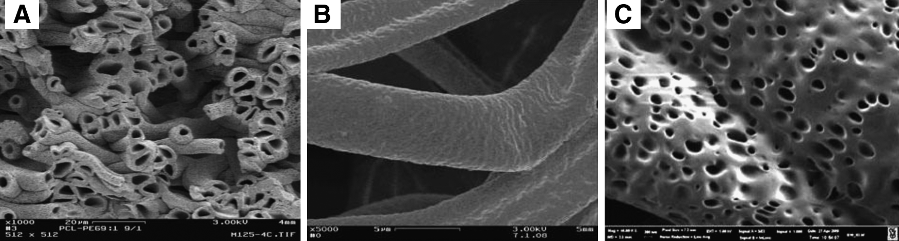
SEM images of
Combinations of electrospinning/electrospraying have been employed in the fabrication of composite nanocomposite materials. Francis et al. used the process to create composite scaffolds consisting of electrospun gelatin (Gel) and electrosprayed nanohydroxyapetite (nHA). Scaffolds were fabricated in a 4:1 and 2:1 Gel:nHA compositional ratio, and evaluated for surface topography alterations, material distribution, and mechanical properties. Biocompatibility was investigated in vitro by culturing human fetal osteoblasts on the fabricated scaffolds yielding enhanced cell proliferation and biomineralization. 41
The novel application of combining electrospinning/electrospraying has not been limited to the fabrication of composite scaffolds. Paletta et al. published a recent study where osteoblasts suspended in medium and electrosprayed onto scaffolds made of poly-l-lactic acid (PLLA) and PLLA/collagen displayed no adverse or inhibition to cellular growth or scaffold degradation providing positive preliminary results of this method being suitable for cell seeding of tissue-engineered scaffolds. 42
Wet electrospinning
The process of wet electrospinning involves a standard electrospinning setup with a modified collector plate submerged in a collecting bath. Polymer fibers are spun and collected into this bath with physical fiber bridging resulting in the formation of a highly porous polymeric structure. Shin et al. wet electrospun a 3D poly(trimethylenecarbonate-co-epsilon-caprolactone)-block-co-poly(p-dioxanone) scaffold for bone regeneration with 90% porosity with good pore interconnectivity. This highly porous scaffold showed good osteoblast cell adhesion at the center of the scaffold after 4 days of in vitro cell culture and cells proliferated 1.5 times faster than the control after 7 days. In addition, alkaline phosphatase activity was four times faster than the control at 28 days. 33 These results illustrate the effectiveness of wet electrospun scaffolds in the fabrication of biomimetic scaffolds for bone regeneration.
Novel nanocomposites
With increased attention on the use of natural materials native to bone and cartilage tissue, a novel area of research area has begun to focus on the incorporation of non-natural or unconventional, yet novel biologically inspired nanomaterials into scaffolds. One such novel material is the DNA-based rosette nanotube (RNT), which is a supramolecular nanomaterial obtained through the self-assembly of DNA base pair motifs in water (Fig. 2). Tailorable amino acid and peptide side chains (such as lysine and RGD) make them intriguing nanomaterials for bone and cartilage regeneration.43–48 In a recent study, electrospun RNTs with a lysine side chain in a hydrogel scaffold has been shown to significantly enhance chondrocyte and synovial cell viability and chondrogenic differentiation. 48
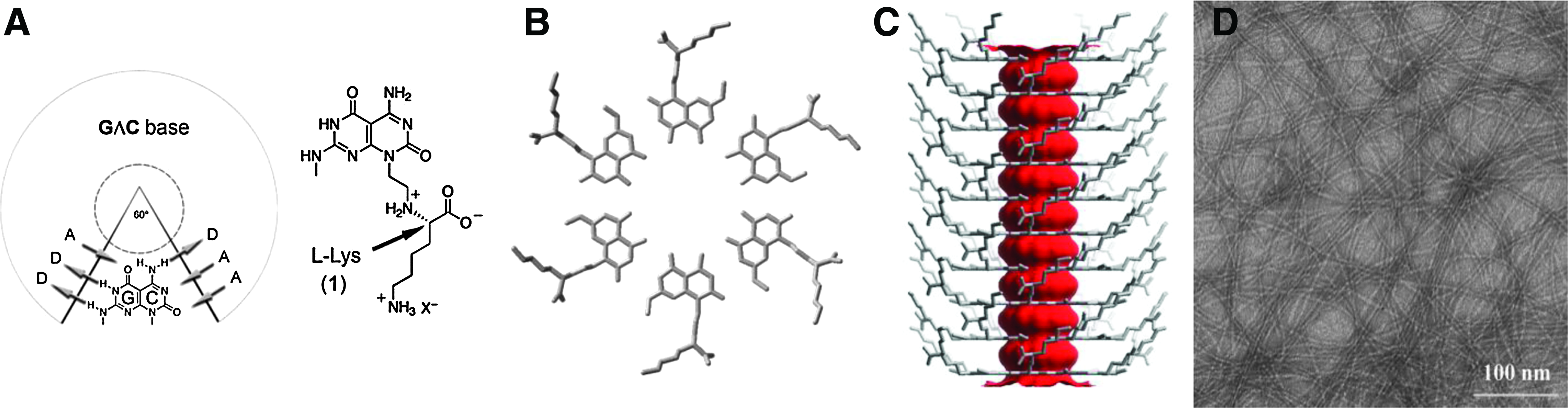
Self-assembly of rosette nanotubes with lysine side chain (RNT-K).
Nano diamonds (NDs) are another example of novel nanocomposite materials that have been employed in the regeneration of orthopedic tissue. NDs are 5-nm diamond particles surrounded by amorphous and graphitic carbon, thus providing a mechanism for functionalization. Because of this, NDs have chemically complex surfaces with potential for combination with a variety of chemicals. 49 In a recent study, Zhang et al. synthesized octadecylamine-functionalized nano diamonds (ND-OCT) 50 to serve as a fluorescent mechanically enhancing constituent for electrospun PLLA scaffolds. OCT was chosen as a functional group because it renders the NDs immiscible in water and hydrophilic organic solvents, but highly miscible in hydrophobic solvents, making them more easily dispersed within a polymer while also being resilient in a biological environment.49,50 At 10% (wt/wt) concentration, PLLA-ND-OCT scaffolds exhibited a 200% increase in Young's modulus and an 800% increase in hardness closely mirroring the mechanical properties of natural bone. 50 Unlike other fluorophores, ND-OCTs are nontoxic and very stable, which could make them a potentially powerful tool for in vivo analysis of polymer migration.49,50
Vascularization of electrospun tissue-engineered scaffold
Adequate vascularization is an essential prerequisite for implanted electrospun tissue-engineered constructs for successful integration with host tissue. Without the direct and immediate availability of a vascular system and potential for angiogenesis, cell viability within tissue-engineered constructs is impeded, thus limiting their therapeutic capability. Furthermore, a successful tissue-engineered construct should be evaluated not only for de novo blood vessel formation, but also for functionality shown within the structure. Native tissue can heal up to 100–200 μm without the need of blood infusion since oxygen and nutrients can reach cells by diffusion. 51 However, various studies have indicated that blood perfusion/infusion is still a major limitation in the development of various tissue-engineered structures.51–53 In general, none of the approaches has so far proven successful in maintaining viability of cells in large tissue-engineered constructs. In most studies performed to date, nutrients failed to reach the center of the tissue-engineered construct leading to cell death and tissue necrosis. This major drawback is because the scaffolds are designed in a way that does not allow for successful functional vascular ingrowth. Therefore, designing novel electrospun scaffolds with increased vascular ingrowth and blood perfusion holds great promise for future regenerative applications.
Conclusions on Electrospun Scaffolds for Bone and Cartilage Tissue Generation
A number of innovative methods for enhancing electrospun scaffold characteristics have been investigated. One of the most widely researched areas being co-electrospinning. It has been shown to be a highly versatile option when attempting to fabricate a scaffold with desirable characteristics. Another advanced method for electrospun scaffold fabrication is wet electrospinning, where fibers are collected in a solvent bath. This method has been shown to create highly porous materials with complex, interconnected pore structure rendering it ideal for bone regeneration. Aside from variations in fabrication techniques, novel nanomaterials such as DNA-based self-assembled nanotubes have also been used in electrospun nanocomposites.
Traditional materials, such as natural polymers, polysaccharides, and inorganic ECM components, have been used extensively via incorporation within polymeric scaffolds in an effort to enhance the mechanical characteristics of scaffolds, as well as improve cell behavior. Although progress has been made, the improvements made are still not ideal, prompting researchers to investigate novel materials, such as ND-OCTs and RNTs, with attractive and unique qualities. Increasingly, researchers have begun to turn to unconventional, unique materials to improve the functionality of electrospun scaffolds beyond what has been capable with conventionally applied materials for electrospun biomimetic nanocomposite scaffolds. In addition to the use of new materials, the methods for which scaffolds are fabricated will also be modified. The field has virtually exhausted all of the ways in which electrospinning parameters can be modulated. However, research is moving in the direction of developing new, more complex electrospinning methods which have the potential to yield more complicated and characteristic architectures (i.e., vascularized electrospun scaffold). With the combination of both novel methods and novel materials, electrospinning has never looked more promising as a method for tissue engineering of electrospun fibrous scaffolds for bone and cartilage regeneration.
Footnotes
Acknowledgments
The authors would like to thank the financial support from the George Washington University Facilitating Fund (UFF).
Disclosure Statement
No competing financial interests exist.