
Abstract
Articular cartilage is a complex, multilayered biological composite material, comprised of chondrocytes encapsulated in a water-based glycosaminoglycan matrix reinforced with collagen fibers. Once damaged by osteoarthritis or traumatic injury, this aneural, avascular tissue has little self-repair capacity. Over the last 20 years, cell therapies and tissue-engineering strategies have shown significant promise for the repair or regeneration of damaged cartilage. In particular, mesenchymal stem cells (MSCs) have great potential owing to their ability to create a reparative environment. Despite the fact that there have been great strides in the design and development of three-dimensional scaffolds, there is an upper limit to the number of viable cells that can be delivered using current approaches. To this end, this review examines current strategies for optimizing MSC localization, evaluates their limitations, and looks to other technologies to devise a combinatorial strategy for the creation of an MSC-seeded composite structure that addresses both the mechanical and biological property requirements for enhanced cartilage repair.
Introduction
With respect to MSC transplantation, early studies into the arthritic knee in a caprine model revealed that bone marrow-derived MSCs slowed degradation of damaged cartilage tissue and contributed to repair.1–3 However, the injected cells did not engraft to the articular cartilage, but instead to the meniscus, indicating an indirect influence on repair. Supporting this hypothesis, MSCs have previously demonstrated a wide spectrum of therapeutic properties other than direct chondrogenic differentiation, including the secretion of antiapoptotic factors,4,5 immunomodulatory molecules,5–9 and chemoattractants.5,10–12 Despite recent claims, 13 MSC transplantation studies for cartilage repair have to date demonstrated the retention of only a small percentage of injected cells.1,2 The exact number has yet to be elucidated, but the general consensus remains that more cells are required at the site of damage to support repair. Caplan suggested the potential use of MSCs acting as multidrug dispensers through site-directed delivery. 14 By increasing reparative cell retention at the site of tissue damage, the potential of retained cells to either contribute through engraftment or through conditioning the local environment with restoring factors increases. Further, the conditioned local environment will attract native progenitor cells and sustain the survival of surrounding tissue while providing immunomodulatory support to the microenvironment.5,14,15
To this end, this review discusses current cell retention and encapsulation strategies as well as future combinatorial approaches, enabling the field of cartilage repair to retain larger numbers of regenerative cells at the site of injury. A review of all three-dimensional (3D) biomechanical scaffold structures is outside the range of this review; many have been reviewed comprehensively by others.16–18 In particular, the main focus of this article lies in examining the current state of the art in hydrogels, peptides, antibodies, and nanostructured guidance cues (Fig. 1) and in looking to the future on how these approaches can or may be combined to create an effective biomimetic strategy for cartilage repair (Table 1).

Current and future strategies targeting mesenchymal stem cells (MSCs) to articular cartilage. MSCs (in blue) may be directly administered to the articulating joint, resulting in the adhesion of a small percentage of delivered cells to the articular cartilage (purple). Targeting strategies, such as hydrogels (yellow), antibody- or protein-/peptide-based systems (orange), nanomaterials (red), or microcarriers (green), may improve the attachment and/or retention of therapeutic MSCs to damaged cartilage, thereby resulting in enhanced clinical effect. Color images available online at www.liebertpub.com/teb
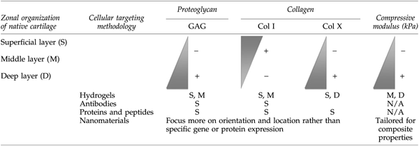
Current approaches focus on MSC hydrogel delivery vehicles with homogenous mechanical properties, targeting antibodies, proteins, peptides, or nanotopographical cues that address surface and middle-layer cell requirements. The future of biomimetic repair may lie in a combinatorial approach to produce a composite mechanobiological substrate for enhanced repair, regeneration, integration, and neotissue formation.
GAG, glycosaminoglycan.
Over the past 20 years, a tremendous range of scaffold structures and materials have been employed to deliver cells in vivo and to provide mechanical support for neotissue formation. However, previous research has also shown that there is an upper limit to the number of cells that can be successfully delivered using 3D scaffolds, and in fact, higher cell-seeding densities can have an adverse effect on nutrient availability, cell metabolism, and cell viability.19,20 Moreover, Hansen et al. have shown that glycosaminoglycan (GAG) accumulation is reduced when porous methoxy-polyethylene glycol–polylactic-co-glycolic acid scaffolds are seeded with cell-seeding densities up to 1.2×10⁶ cells per scaffold. 21 In contrast, hydrogels have been used successfully for cartilage repair 19 with seeding densities up to 60×106 cells, 22 demonstrating potential for localizing large number of cells in cartilaginous defects.
Hydrogel-Based Localization
Hydrogels, a network of water-insoluble super-absorbent polymer chains, are optimal for cell encapsulation. The high water content of hydrogels facilitates the transport of nutrients, mimicking the fluidic properties of native tissue. 23 Hydrogels were one of the first systems to be utilized as a vehicle for cell delivery and encapsulation for cartilage repair, primarily those composed of hyaluronic acid (HA), fibrin, or alginate. 24
HA, a major component of the cartilaginous extracellular matrix (ECM), is often desired as a scaffold for cartilage repair as it supports chondrogenic differentiation over inert matrix molecules such as polyethylene glycol (PEG). 25 In vivo, the assessment of rabbit progenitor cells encapsulated in an HA-based scaffold demonstrated the ability for a combined HA-MSC therapy to generate a repair tissue in situ. 26 More so, cartilage repair with MSC-containing HA-based hydrogels supplemented with growth factors, such as transforming growth factor (TGF) β3, resulted in increased deposition of collagen type II, aggrecan, and Sox 9, all markers of a cartilage-like tissue.27,28 Alternatively, a combination with additional strata, such as MSCs in HA–gelatin scaffolds, 29 generates firm, translucent cartilage seamlessly integrated with host tissue, a clear advantage over cell or matrix treatment alone.
Fibrin, a fibrous protein involved in the clotting of blood, can be used as an injectable biomaterial in combination with MSCs, providing the opportunity for gelation in situ through the addition of thrombin.27,30 The encapsulated cells remain viable and biologically active and are able to produce a cartilaginous-like matrix. 31 The efficacy of progenitor cell-containing fibrin hydrogels to repair cartilage has been investigated using several animal models and bone marrow-derived progenitors. 32 Most interestingly, the repair of equine full-thickness cartilage defects using MSCs in fibrin hydrogels demonstrated the support of chondral repair, yet poor cell retention with a time-reduced efficacy when compared to controls. 33 More importantly, the combination of bone marrow-derived MSCs with fibrin glue has been investigated clinically in humans. All patients demonstrated an improvement over the 1-year study, but only three of five resulted in complete defect fill. 34 A limitation of fibrin-based hydrogels is their suboptimal mechanical properties, which make them unable to bear high mechanical loads as experienced in vivo, 35 regularly resulting in delamination. Using similar technologies, such as chondrocyte-containing fibrin gels in combination with a polyurethane macroporous matrix to enhance the mechanical integrity of the construct, 36 novel mechanically sound progenitor-containing hydrogels may be developed, allowing for greater cell viability, cell distribution, and enhanced ECM retention.
Alginate, a natural derivative of algae, is regularly used as a scaffold in combination with chondrocytes to support in vitro chondrogenesis. In vitro encapsulation of progenitor cells in alginate has demonstrated the potential for this hydrogel to support MSC chondrogenic differentiation.37–39 Upon encapsulation and differentiation, the signature upregulation of collagen type II, aggrecan, and Sox 5, 6, and 9 was observed, 36 coordinating with the suppression of collagen type 1 expression. Ahmed et al. have additionally shown that alginate-encapsulated MSCs communicate with adjacent mature cartilage tissue via soluble factors, resulting in the upregulation of Sox 9 expression and suppression of collagen type X during differentiation.40,41
Agarose, a material well established to support chondrocytic development of cartilaginous constructs, 42 has more recently been employed in conjunction with bone marrow-derived progenitors to regenerate cartilage. Bovine MSCs seeded in agarose constructs and induced to undergo chondrogenesis resulted in the deposition of cartilaginous ECM molecules. However, the quantity of matrix and its mechanical properties were reduced as compared to that produced by chondrocytes cultured similarly. 43 Intensive transcriptional studies comparing matched chondrocytes and MSCs in agarose have identified 324 dysregulated genes between the two cell types, highlighting an inherent difference in chondrocytes and MSCs in this system even when both are able to generate engineered cartilage. 44 These results highlight the need to better understand the difference in these cell types and their process for generating de novo cartilage tissue as a means to enhance their application in regenerative medicine.
Hydrogels have therefore shown the potential to retain therapeutic progenitor cells on the damaged cartilage surface. They support chondrogenic differentiation, maintain cell viability, and enable uniform cellular distribution, attributes that are required for adequate tissue regeneration 45 (Fig. 2). At the macrostructure level, the mechanical properties of hydrogels in the kPa range regularly lack the physical strength of native articular cartilage tissue in the MPa range 46 ; however, at the microstructural level, they have been shown to provide support for MSC differentiation. Using a composite engineering approach, the potential of hydrogels to support chondral repair is possible. In combination with antibodies or peptides, hydrogels can provide cell or differential specificity by enhancing the reparative milieu.28,47 Alternatively, just as cells can be encapsulated within hydrogels, growth factors such as insulin growth factor-1 or TGF-β family members can be incorporated for sustained release over time.48,49 With optimization, this technique can be extrapolated to control progenitor cell differentiation in situ, demonstrating the potential for improving and tailoring these materials for specific needs. To develop a deeper understating of what is required for the generation of such a biocombinatorial approach, protein-, peptide-, and antibody-based cell localization technologies are discussed in detail in the following sections.
Repair of chondral defects with hydrogels containing bone marrow progenitor cells. MSCs delivered in a modified hyaluronic acid gel to a rabbit chondral defect model resulted in neocartilage formation after
Protein- and Peptide-Based Cell Localization
In the cartilage field, arginine-glycine-aspartic acid (RGD) peptides, the integrin-binding sequence in fibronectin, have been investigated in cell localization technologies. Alginate gels modified with RGD demonstrated an upregulation of Sox9 and collagen type II in MSCs undergoing chondrogenic culture in vitro. 50 Similarly, You et al. demonstrated upregulation of Sox 9, aggrecan, and collagen type II and a downregulation of collagen type I on MSC-seeded polyhydroxyalkanoate scaffolds combined with RGD peptides, suggesting that RGD may provide a biological stimulus for MSC differentiation in addition to enhancing cellular retention within the scaffold. Concurrently RGD peptides retained local native cells at the tissue surface. 51 Interestingly, increased cellular adhesion at higher RGD concentrations resulted in a decrease in matrix production, as demonstrated by the inhibition of GAG synthesis. 52 Re'em et al. hypothesize that further differences in the scaffold type, cell source, cellular environment, and peptide sequences may account for contrasting results in this field of research. 50
Naturally, fibronectin protein expression is regulated during embryonic chondrogenesis: increased at times of condensation and decreased in quantity during maturation. Similarly, the presence of fibronectin, or the RGD sequence, enhances early chondrogenesis and maintains MSC viability in vitro, while its continued expression inhibits chondrogenic differentiation.47,53 In an attempt to improve the temporal regulation of RGD expression on scaffolds, Salinas and Anseth developed an RGD-releasing PEG gel to allow for timely exposure of MSCs to the peptide. As a result, encapsulated MSCs produced 10-fold more GAG and greater quantities of collagen type II as compared to cells exposed to an uncleavable RGD gel. 47
An alternative, generic approach to selectively retain progenitor cells is modifying the cell's natural behavior, thereby homing cells in vivo to a site of tissue damage. Sarkar et al. demonstrated that the surfaces of MSCs could be chemically modified in vitro to possess leukocyte-like adhesion properties, promoting cell rolling. For example, the sialyl Lewis X (SLeX) moiety was covalently coupled to the surface of MSCs through biotin–streptavidin modifications, or transiently with biotinylated lipid vesicles with SLeX, in an attempt to control the adhesion time.54–56 Engineered MSCs exhibited an increased homing response over native MSCs to inflamed endothelium in vivo 57 (Fig. 3). In a different proof-of-concept approach, altering the native MSC CD44 glycoform to a hematopoietic stem cell (HSC) E-selectin/L-selectin ligand resulted in enhanced HSC-like homing of MSCs to bone marrow in vivo. 58 Such technologies could be specifically tailored, so that MSCs might overexpress a peptide moiety to an epitope found specifically on the surface of degraded cartilage, thereby enhancing their binding to a site of interest.
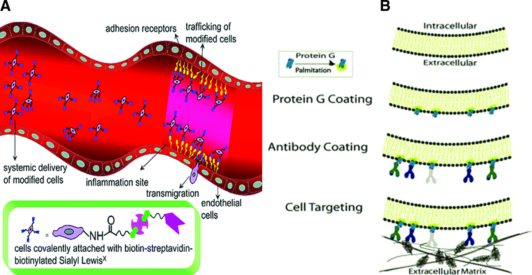
Current state-of-the-art protein and antibody cell localization technologies.
Despite initial successes, peptides and proteins for localization or the modification of cell surfaces will heighten regulatory costs. Specifically, the context of application and desired outcome needs to be considered.47,53,59 The RGD peptide shows a desirable retention capability,34,35 but a potential risk may be the localization of undesirable cells due to its generic nature. This problem has not yet been documented in published in vivo studies. Additional preclinical studies will further elucidate undesirable peptide effects or limitations that these altered cell properties may have and secondly explore the possibility of requirement for a biotailored vehicle for improved cell localization. 49
Antibody-Based Cell Localization
Antibodies are one of the strongest candidates for cell localization and enhancing cellular retention since they are natural molecular tags that may be employed to specifically direct cell engraftment. Pioneering work in the use of antibodies for cancer and immunology therapeutics has demonstrated the exciting potential of bispecific antibodies (BiAb), antibodies that can bind two different antigens, in disease treatment. Directing the body's self-defense system to specifically target a cancerous tissue was the earliest example of utilizing the natural tagging system of antibodies for delivering reparative cells to a disease site.60,61 More recently, BiAb have been considered for cell localization in tissue engineering, demonstrating positive preclinical evidence for increasing cell retention to a site of tissue injury.62,63
Coating cells with antibodies, or cell painting, was first introduced by Chen et al. 64 and further developed by Dennis et al., 65 for the cartilage repair field, by which lipidated protein G was intercalated into the cell membrane of cultured chondrocytes (Fig. 3). Coated cells were subsequently incubated with antibodies specific to cartilage matrix antigens, allowing the binding of antibodies to protein G on the surface of the chondrocyte. Antibody-coated cells were added to cartilage explants ex vivo, successfully resulting in enhanced binding to the cartilage surface.64,65 Similar methods have been employed more recently to successfully deliver MSCs to activated human vascular endothelial cells.64–66 Cell painting has been successfully investigated in vivo in acute colitis, where indeed increased MSC numbers were observed; however, the efficacy of retained cells was associated with not only increased numbers, but also the specificity and functionality of the labeled cells. 67 Significant potential lies in the technologies developed in these nonorthopedic applications; they hold great promise if tailored to localize MSCs at the articular surface.
It is important to consider the specific requirements for antibody technology in the context of tissue engineering and regenerative medicine. The design of an antibody construct needs to be carefully chosen depending on the specific function and application to avoid misdirection of administered therapeutics. 63 However, the redirection of targeted cells might be reduced by local delivery of cells and construct into the site of injury. Alternatively, the development of a less tissue-specific construct may provide a therapeutic with a larger variety of tissue-engineering applications, providing that there is no undesirable retention in other tissues.
Few studies have been reported using systemic administration of antibodies and peptides to direct progenitor cell localization to cartilage. This is likely due to the zonal complexity of cartilage, the production costs of such therapeutics, and the absence of a blood supply within cartilage. In addition, the fluidic state of the joint presents further challenges for cellular retention with proteins alone. Future studies may demonstrate the potential of antibodies and peptides in a combinational approach with biomaterials providing both structure and further biological cues for advancement in this field.
Nanostructured Cues
We have discussed the repair potential of MSCs, the possibility of increasing the number of functional cells delivered to the site of injury using hydrogels, proteins, peptides, and antibodies, but the question of mechanical support at a macroscopic or tissue level remains to be addressed. Again, looking at composite engineering for inspiration, the structural strength of polymers has been enhanced by the addition of particles and fibers in the order of micrometers and nanometers. Indeed, many of the nanostructured scaffolds for tissue-engineering applications are inspired by the supramolecular structures found in the natural world. Certainly, many studies have focused on developing artificial ECM-like scaffolds that replicate the 3D environment of native tissues. Articular cartilage is a complex composite structure, with features ranging from the nanoscale for the collagen fibers to the micron scale for the chondrocyte cells and their lacunae. In an effort to recreate this biomechanical environment, recent studies have focused on developing nanotopographical cues to direct the differentiation of cells without the addition of growth factors.68,69 Alternatively, nanostructures can be employed to deliver and retain cells at the site of injury, 70 can be tethered with antibodies and peptides to target specific cells to improve integration and neotissue formation, 71 or internalized within cells to promote repair without the use of genetic alteration or growth factor manipulation. 72
Recently, electrospinning has been used to create cartilaginous ECM-like features in the nanoscale range. Li et al. demonstrated the feasibility of electrospun biodegradable polycaprolactone (PCL) with fiber diameters of 700-nm nanofiber scaffolds to maintain and support a range of chondroprogenitor cells both in vitro and in vivo.70,73–75 Although electrospinning offers great potential for replicating the ECM environment of articular cartilage, further improvements are required to optimize the 3D architecture and biological function of electrospun nanofibers. As with other biomaterial techniques, the complex multilayer architecture of native cartilage has yet to be successfully replicated by electrospinning.
Naturally, complex molecular suprastructures are created by molecular self-assembly. Indeed, proteins and peptides self-organize to create natural scaffolds such as collagen, keratin, coral, and pearl.76–78 Recently, many researchers have been taking the lead from nature and using the self-assembly ability of proteins and peptides to create artificial ECM environments for the delivery of cells and de novo tissue repair. It has been shown that peptides with alternating hydrophilic and hydrophobic amino acid groups form stable β-sheet structures when dissolved in deionized water.76–79 Furthermore, peptide sequences have been tailored to promote specific cell responses, such as proliferation, differentiation, and tissue formation. 80 Using this bottom-up method of assembly, porous 3D woven scaffolds with nanofibers ∼10 nm in diameter have been developed for a range of tissue-engineering applications. Kisiday et al., developed chondrocyte-seeded self-assembling peptide scaffolds for cartilage repair, made from the self-assembling peptide containing lysine-leucine-aspartic acid (KLD) amino acid repeats. 81 Over a 4-week culture period in vitro, the mechanical properties and GAG accumulation of the 3D construct began to replicate that of native cartilage. 82 Although, recent studies by Petrie et al. have shown great promise for osteointegration, the full potential of 3D self-assembled constructs has yet to be demonstrated in vivo for cartilage applications. 72
As previously mentioned, nanomaterials are generating great excitement for biomedical applications, since they can be designed to create nanostructures with dimensions up to 3 orders-of-magnitude smaller than those created by conventional processing techniques. 81 By nanoengineering substrate surfaces, features such as nanotube diameter, lateral spacing, surface chemistry, and geometry can be tailored to create biomimetic architectures for enhanced cell interaction and delivery. 83 Taking this approach, researchers have adapted a similar top-down methodology to that used in the electronics industry to create nanowires and nanorods with similar dimensions (∼15 nm) to the spacing of integrin receptors in focal contacts of native ECM. 69 One such technique that has attracted much attention for the creation of nanostructured scaffolds structures is template synthesis via a layer-by-layer (LbL) assembly.
The LbL fabrication process involves depositing layers of oppositely charged species, such as polymers, proteins, and nanoparticles, layer-upon-layer to create multilayer structures such as films and more recently colloid particles. 84 By combining the LbL assembly with template synthesis, nanotubes with controlled wall thickness of nanometer resolution, diameters ranging from 30 nm to a few micrometers, and lengths of the order 5–50 mm have been created.84–86 Indeed, studies have shown that MSC adhesion, growth, and spreading can be manipulated at the nanoscale and are dependent on tube diameter and lateral spacing. Park et al. demonstrated that a maximum stimulated cell response was achieved with lateral spacing in the range 15–30 nm, which in turn approximates the lateral spacing of integrin receptors in focal contacts in the ECM. 69 Using this approach, Landoulsi et al. recently demonstrated the feasibility of template synthesis combined with the LbL assemble for creating collagen nanotubes with highly regular dimensions. 85 In parallel, Porter et al. created PCL-based nanowire scaffolds where, in addition to showing enhanced MSC performance, through retained viability and morphology on the PCL nanowires, the possibility of solvent-free encapsulation of bioactive molecules was also demonstrated.87,88 These studies demonstrated the potential of nanostructures for promoting cell-specific responses and manipulating MSC differentiation along different lineages in vitro; however, cell localization and cell retention have yet to be demonstrated in vivo.
Over the last 5 years, carbon nanotubes (CNT) have been attracting attention for tissue engineering and regenerative medicine applications. CNT, rolled sheets of graphene, display magnetic or electrical properties depending on their chirality. 89 In terms of biological applications, CNT are currently being investigated as potential nanovehicles for drug and gene delivery, cellular labeling and tracking particles, and scaffold matrix enhancers for tissue-engineering applications.90–96 Furthermore, cellular uptake of CNT has been demonstrated for a number of cell types, including HeLa cells, Jurkat cells, and MSCs.93,97 Despite the controversy about CNT cytotoxicity, 98 it has been revealed that low concentrations of CNT have no adverse effect on MSC viability, proliferation, or differentiation toward a chondrogenic lineage. 97 As it is possible for CNT to be internalized within cells, offering an opportunity to deliver bioagents such as DNA, peptides, and fluorescently labeled biotin–streptavidin moieties into stem cells to promote MSC differentiation or target chemokines for cartilage repair. Despite the fact that many of these studies have shown great potential in vitro, the functional effect of progenitor cells in combination with nanostructured cues for cell retention, localization, and support has yet to be demonstrated in cartilage repair models in vivo.
Microcarriers
Microcarrier culture systems were first introduced by van Wezel for the large-scale manufacture of viral vaccines and biological cell products.99–101 Today, microcarriers are regularly used in tissue engineering and regenerative medicine applications to produce large numbers of therapeutic cells and deliver them to the site of injury.97,102–105 Microcarrier culture systems consist of cell-seeded microbeads suspended in tissue culture vessels such as spinner flasks or bioreactors. One of the advantages of using microcarriers is the large surface area available for the generation of significant cell numbers where, according to Malda et al., 1 g of microcarrier beads provides a surface area equivalent to fifteen 75-cm2 tissue culture flasks.100,104 By manipulating the chemical composition, charge density, porosity, particle diameter, and bioreactor conditions, cell attachment and propagation can be controlled. 100 In terms of chemical composition, a range of materials, including polymers, glass, ceramic, dextran, collagen, and gelatin microcarrier beads, are commercially available. Furthermore, a number of biodegradable polymer microcarriers, microcarriers modified with growth factors and ECM protein coatings, are being developed to direct or improve differentiation. 106
Microcarrier cultures have been employed as in vitro model systems to increase cell numbers and maintain cell phenotype for cartilage repair applications.102,103 Furthermore, the size of chondrocytes and lacunae is of the order of microns in scale, so cell localization at the micron level can be examined using microcarriers. In terms of delivery after culture on the microcarriers, cells can be delivered arthroscopically, directly to the site of injury, or reseeded on scaffolds for improved mechanical stability. Indeed, chondrocytes retrieved from microcarriers, reseeded on PEG-terephthalate/polybutylene terephthalate scaffolds, and implanted in a mouse model were found to produce twice the amount of proteoglycans in vivo as cells grown in a monolayer. 104 In a study by Yang et al., 105 MSCs were cultured on CultiSpher-S gelatin microcarrier beads, expanded in spin culture, and directly implanted in a rat model. Bead-expanded MSCs retained their multipotency, showing ability to differentiate along osteogenic, chondrogenic, and adipogenic lineages in vitro and de novo bone tissue formation in vivo. 105 In a more recent study by Liu et al., nanofibrous hollow microspheres were used as injectable chondrocyte carriers in rabbit osteochondral defects. Uniform distribution of tissue was observed both in vitro and in vivo with increased GAG and collagen type II content. The increased surface area provided by the nanofibrous architecture allowed a higher absorption of cell adhesion proteins, which likely accounted for the higher attachment efficiency compared to controls. 107 With the promising in vitro and in vivo results from Bouffi et al., whereby PLGA microspheres were coated with fibronectin and tailored to release TGF β3, the development of an injectable MSC delivery vehicle with cell-specific moieties is very promising. Nevertheless, a 3D zonal architecture may be difficult to achieve using this approach alone.
Discussion
It is widely recognized in the field of regenerative medicine that to rebuild and repair a damaged tissue, a combination of factors is required for efficacious repair. Cell therapy requires the transplantation of therapeutic progenitors to supply reparative trophic factors, the chemotactic signals to stimulate host cell homing and immunomodulate the microenvironment of tissue damage. The progenitor cells themselves contribute to the regulation of this reparative environment, key to the efficacy of repair.
As mentioned previously, articular cartilage is a functionally graded biological composite structure. When looking for future strategies for cartilage repair, it is important to examine the mechanical properties and biological functions of native cartilage, to compare the properties of the current state-of-the-art cell localization approaches with those of native cartilage, and to highlight the limitations and design strategies to overcome these short comings (Table 1). Although, a recent study by Nguyen et al. examined a zonally organized hydrogel structure with promising results in vitro, the in vivo response has yet to be evaluated. 108 In contrast, protein, peptide, and antibody strategies have the ability to localize and provide biological stimulus to chondrogenic cells and bone marrow-derived MSCs. Nanostructured biomaterials have demonstrated great potential in vitro for providing topographical cues, promoting specific cell responses, and more importantly can be tailored with a hierarchical organization for antibody, peptide, or protein attachment. Individually, all the approaches reviewed in this article show potential, but when combined to create a multilayered composite with combinations of one or more of these cell-targeting moieties, an exciting approach for future strategies is proposed. One such approach is shown in Figure 4, comprising of a 3D-layered zonally organized composite hydrogel, where the biomechanical properties, cell orientation, and cell distribution are tailored using peptide, antibodies, and nanoparticles in the superficial layer, microcarriers in the middle layer, and nanorods in the deep layer to mimic the zonal cell orientation and distribution of native articular cartilage. Taken together, this methodology complements the approach of Khanarian et al. 109 and builds on the previous work of Kisiday and coworkers, 110 Bouffi et al., 71 and Porter et al., 87 offering an exciting approach for the delivery, localization, and retention of progenitor cells for cartilage repair.
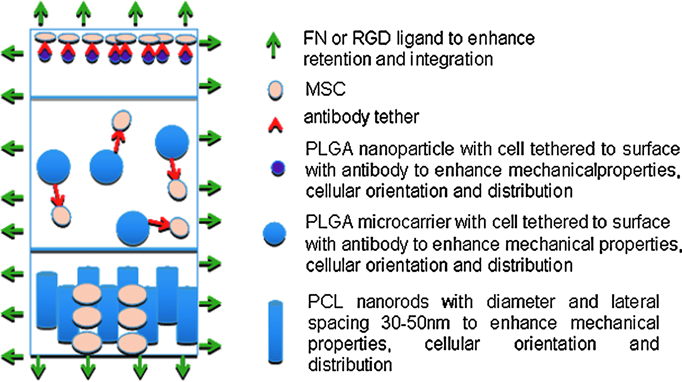
A combinatorial approach for a future cartilage repair strategy. Cartoon illustrating 3-layer zonally organized composite hydrogel, with biomechanical properties, cell orientation, and distribution tailored using nanoparticles in the superficial layer, microcarriers in the middle layer, and nanorods in the deep layer that mimics zonal cell orientation and distribution of native articular cartilage. Color images available online at www.liebertpub.com/teb
In summary, we have examined the use of hydrogels, proteins, peptides, antibodies, nanostructural cues, and microcarriers for cell localization, encapsulation, and support at the damaged cartilage tissue site. Moreover, we have also shown that none of these concepts alone possess the optimal combination of mechanical properties, biological functionality, or localizing potential. Therefore, by combining the technologies discussed, we propose a method that will advance the next generation of constructs for cartilage repair, with the ultimate aim of enhancing cell retention at the site of damage.
Footnotes
Acknowledgments
The authors would like to thank Drs. Jessica Hayes, Eric Farrell, and Sinéad D'Arcy, M.Sc., for the critical review of this manuscript. Funding Source: Science Foundation Ireland (09/SRC/B1794), European Framework 7 (HEALTH-F5-2010-241719-ADIPOA), Industrial Development Agency Ireland (RC255-126894-01/10/07) (143384-01/04/10).
Disclosure Statement
All authors have no conflicts of interest.