
Abstract
Microspherical particulates have been an attractive form of biomaterials that find usefulness in cell delivery and tissue engineering. A variety of compositions, including bioactive ceramics, degradable polymers, and their composites, have been developed into a microsphere form and have demonstrated the potential to fill defective bone and to populate tissue cells on curved matrices. To enhance the capacity of cell delivery, the conventional solid form of spheres is engineered to have either a porous structure to hold cells or a thin shell to in-situ encapsulate cells within the structure. Microcarriers can also be a potential reservoir system of bioactive molecules that have therapeutic effects in regulating cell behaviors. Due to their specific form, advanced technologies to culture cell-loaded microcarriers are required, such as simple agitation or shaking, spinner flask, and rotating chamber system. Here, we review systematically, from material design to culture technology, the microspherical carriers used for the delivery of cells and tissue engineering, particularly of bone.
Introduction
The function of cells that are populated on the microspheres is largely dependent on the sphere morphology and the material's physicochemical properties, as the microspheres are the supporting substrates and/or containers to hold and deliver cells, providing three-dimensional (3D) microenvironmental cues and consequently dictating their tissue development. To carry as many cells as possible, the microspheres are often designed to have macropores within their structure, or to encapsulate cells inside. The composition of biomaterials and the surface properties (chemistry and topography) can also significantly influence the fate of the supported cells. Because cells loaded in the spheres require bioactive factors and they also secrete necessary factors in the culture processes, the microsphere-carrying system is considered a sort of bioreactor and can be modulated to impart therapeutic effects on cell behavior.
To realize ex vivo engineered tissues that mimic native bone, the culture methodologies of the cell-containing microspheres are an important consideration. Due to the specific form of spheres, conventional two-dimensional (2D) static culture conditions are not considered satisfactory to attain uniformity in the cell population and functions. Unlike the general form of 3D porous scaffolds, the microspherical carriers are not fixed in place in devices, but rather are allowed to float in the culture medium. Upon culture in a designed apparatus, the cells experience mechanical fluid forces and collide with each other, which should affect the fate of cells that are delivered by the microcarriers.5–8 From the biomaterial designs to the culture methodologies, more studies on the microspherical cell culture system are needed to realize appropriate constructs for bone tissue engineering.
Here we review the spherical particulate forms of biomaterials developed for the purpose of delivering cells and tissue engineering, particularly of bone. Spherical designs to host tissue cells on the surface or within the carrier biomaterials are systematically reviewed. Their roles as bioreactor reservoirs in modulating cellular functions are described, and the culture methodologies to gain better cellular functions for tissue engineering are envisioned.
Designs of Microspheres
As the supporting and delivering matrices of cells, the compositions of microspheres should be chosen within the spectrum of biomedical materials that are not toxic, but compatible to cells and tissues, and even favor cell growth and differentiation into specific tissues. A variety of materials have been used for bone regeneration ranging from bioactive ceramics and degradable polymers to their composites.9–15 Because cells first recognize the surface of spheres, it is important to tailor the surface properties such as chemistry and micro-/nanotopology to allow cell anchorage and attachment. 16 Apart from the surface properties, morphological traits need special consideration in the design of microspheres to improve the cell delivery potential. Figure 1 depicts the possible designs of microspherical biomaterials for use in tissue regeneration. Compared to the conventional solid-filled microspheres (Fig. 1a), porous structured spheres are preferred to effectively host cells and carry them for tissue engineering (Fig. 1b). Moreover, in situ encapsulation of cells within the hollow inner space is one promising design of microspheres for cell carrier and reservoir.
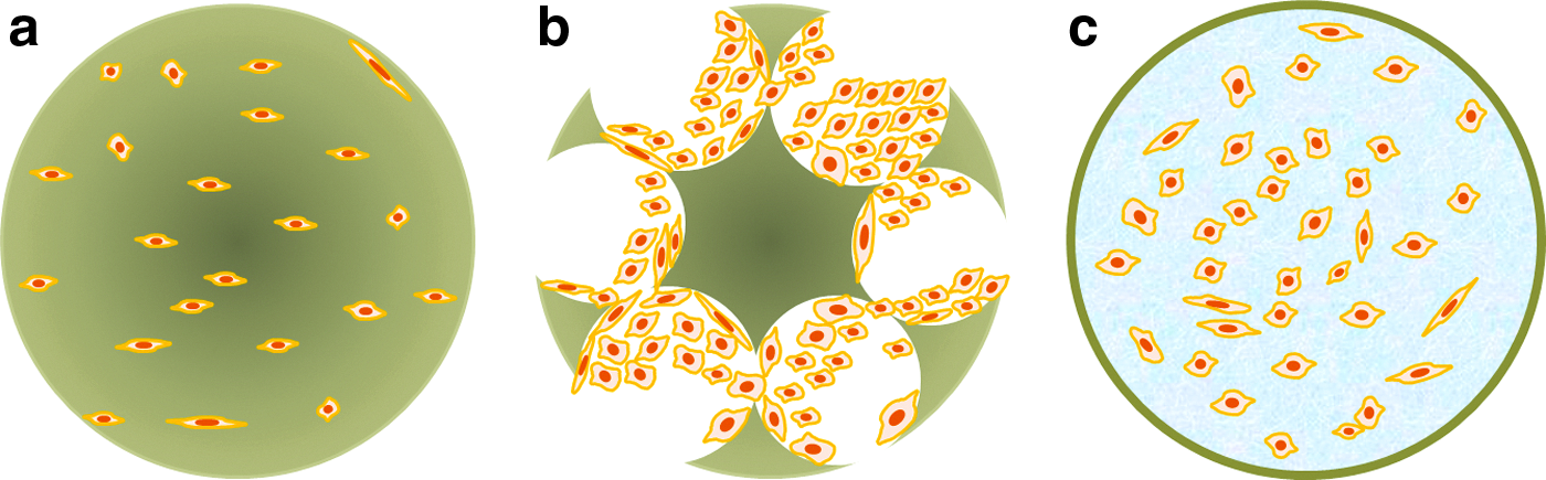
Microspherical biomaterial designs for use in tissue regeneration.
Solid-filled microspheres
Spherical formulation of materials used for biomedical purposes has been achieved using various compositions, including bioactive ceramics, degradable polymers, and their composites. Targeting bone tissue, bioactive ceramics such as hydroxyapatite (HA) and tricalcium phosphate (TCP), and glass/glass ceramics with bioactive compositions has become widely used in spherical particulates at tens to hundreds of micrometers in size.17–22 Bioceramic powders are generally mixed with binders such as poly(vinyl alcohol) or poly(vinyl butyral) to maintain structural stability and are then formulated into spheres by emulsification or spray-drying, followed by a high-temperature heat treatment to remove the binder and to consolidate (sinter) the ceramic powders.23–30 Because the resultant composition is similar to the bone mineral or the composition readily induces bone mineral phase in vivo, the bioceramic microspheres have been widely used to fill and augment the defective sites of bone. Some compositions of the bioceramics have proven in vivo compatibility, and their commercial clinical availability is being pursued. 31
While bioceramic phases generally require high temperature to consolidate their structure, biopolymers are more readily amenable to development into a microsphere form. Synthetic degradable polymers such as poly(lactic acid) (PLA), poly(glycolic acid), poly(caprolactone) (PCL), and their copolymers that are soluble in organic solvents can also be made into spheres by an oil-in-water emulsification with mechanical stirring.3,32–35 Surfactants such as poly(vinyl alcohol) mediate the sphere formation.36,37 The size of the spheres is largely dictated by conditions, including material content, surfactant type and amount, type of solvent, and stirring speed.37–40 The synthetic polymers, however, are hydrophobic and lack adhesive ligands for favorable cell adhesion. Therefore, the surface often has to be engineered to be hydrophilic or to be attractive to cells by modification with adhesive proteins, such as fibronectin or collagen.41–43 Furthermore, the synthetic polymers need to be processed in organic solvents, which markedly hamper the incorporation of therapeutic drugs and proteins. On the other hand, natural polymers, including collagen, gelatin, alginate, and chitosan, can be formulated in water-based solutions and, thus, are more hydrophilic than the synthetic polymers, allowing water uptake and cell migration to the surface of microspheres.44–46 However, the natural polymers often degrade easily and lose structural stability, which necessitates cross-linking of polymer chains, such as via glutaraldehyde treatment, which however raises the issue of cellular toxicity.
Of the available compositions, a particularly fascinating type is the composite of bioactive ceramics with degradable polymers. The approach exploits the excellent bone regenerative properties of the bioceramics and the sphere-forming ability of the degradable polymers. The use of HA powder within PLA microspheres has been demonstrated to markedly improve cell adhesion, cell growth, and osteoblastic differentiation. 47 In a similar way, poly(lactic-co-glycolic acid) (PLGA) was combined with HA to prepare microspheres, enabling better cell attachment and proliferation, and subsequent differentiation. 48 Poly(β-hydroxybutarate-co-β-hydroxyvalarate) was also combined with HA to support mesenchymal stem cell (MSC) culture. 49 A composite microsphere system has been successfully made of a degradable polymer PLA with bioactive glass granules. 50 The addition of 10%–30% bioactive glass has been shown to significantly improve the osteoblastic differentiation of marrow stromal cells as confirmed by alkaline phosphatase (ALP) activity. The incorporation of sol-gel-derived bioactive glass (40% weight) particles into poly(lactide-co-caprolactone) also demonstrated significant enhancement of osteogenic development of cells cultured on a spherical substrate. 51 Ions such as calcium, phosphate, and silicon can be released from the glass component and can influence cellular functions. 52 Therefore, the possible ionic control over osteogenic functions is considered one significant merit of using bioactive glasses in the composite microspheres with polymers. The addition of calcium phosphate (e.g., HA) powders within the collagen matrix is considered to better mimic the native bone structure and, thus, the potential to regulate osteogenic differentiation, therefore, being used in a variety of forms for tissue engineering.53–56 Mixtures of HA powder/collagen have been developed by an water-in-oil emulsion method with varying sizes.57–59 The prepared microspheres supported cell attachment, proliferation, and further differentiation into osteoblasts as demonstrated by matrix mineralization.57–59
Instead of directly adding the HA powder within collagen solution, the precursors of HA were coprecipitated with collagen, which was then formulated into collagen–HA nanocomposite microspheres. 59 In the study, the amino acid groups in collagen mediated the precipitation of HA nanocrystallites, with the ultrastructure mimicking the native bone. MSCs were able to grow actively on the nanocomposite microspheres and differentiate into an osteogenic lineage better than those on collagen microspheres, suggesting that nanocomposite microspheres mimicking the bone extracellular matrix (ECM) may be a good candidate material for bone tissue engineering.
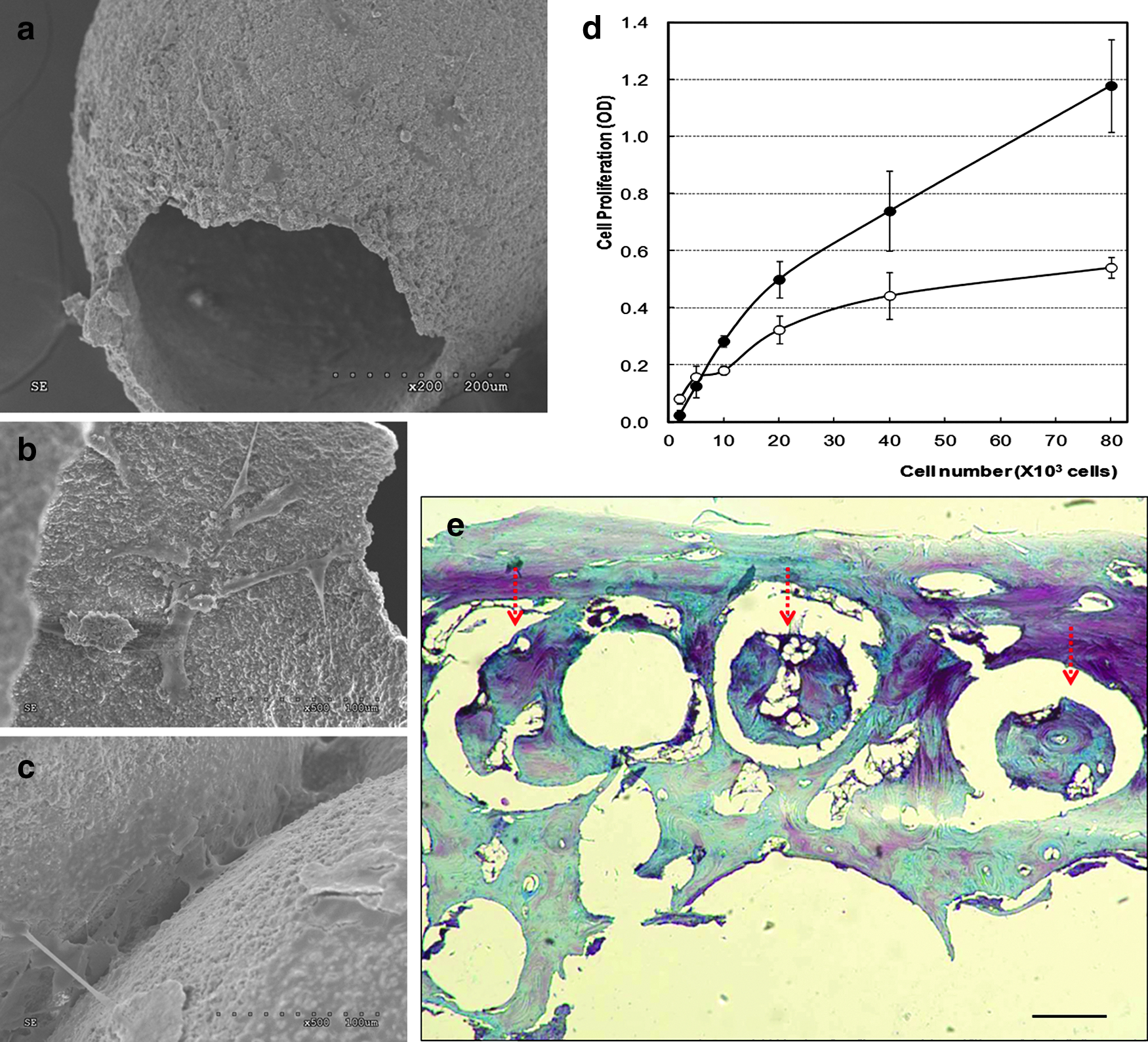
Calcium phosphate cements (CPCs) are an attractive bioceramic formulation for use as a microspherical carrier. CPCs self-harden in water-based solutions and transform into mineral phases mimicking bone, and so are regarded as potential bone repair materials. This self-setting property of CPCs has benefits over other bioceramic powders, allowing shape formation without a thermal treatment. This also facilitates the incorporation within the structure of bioactive molecules such as drugs and proteins, enabling the delivery of therapeutic molecules that will help the cells supported on the spheres engage in the bone-forming process. CPC-based microspheres using mainly an α-TCP precursor have recently been developed.16,60,61 A water-based mixture of the powder at relatively high liquid-to-powder ratios readily formed spheres in a water-in-oil emulsification condition. During reaction in water, the cement composition will harden, and phase transformation into calcium-deficient HA that mimics the native bone mineral can occur.16,61 Along with the general processing variables, such as stirring speed and oil viscosity, the initial particle size of the CPC also affects the size and properties of the microspheres. 16 CPCs obtained with different initial particle sizes result in different crystal sizes and microporosities, along with different cell proliferation and differentiation behaviors. 62 Natural polymers such as collagen can be incorporated in the CPC composition to improve the sphere formation associated with higher surface tension of the protein and to better mimic the natural bone composition.2,62 Collagen addition can improve sphere formation, while not altering the final mineral transformation (Fig. 2).2,62 The presence of collagen favors the proliferation of cells such as MSCs and osteoblast-like cells (SaOs-2) on microspherical substrates.2,62 In a similar way, gelatin can also be incorporated into the liquid phase of CPC, improving the initial cell adhesion, which was ascribed to the cell adhesive ligand present on the gelatin. 62 CPC-based microspheres were also proposed to deliver biological proteins for therapeutic efficacy. 2 The issue of biomolecule delivery systems will be detailed in the next section of this review.
Self-hardening microspheres made of calcium phosphate cement (CPC) containing collagen, used to carry bioactive molecules within the composition. The self-setting property of CPCs allows potential applications of microspheres for delivering proteins and the support of cell differentiation and maturation during osteogenesis. Mesenchymal stem cells (MSCs) derived from rat bone marrow favor the microspherical CPC surface and, as shown in the higher magnification view of a portion of the surface in the right panel, the formation of bone mineral-like crystallites was observed beneath the spreading cells.
In summary, when the solid form of microspheres of bioceramics, biopolymers, or their composites is developed, they can support tissue cells on the curved surface, guide cellular spreading and growth, and induce osteogenic differentiation under proper culture conditions. However, the surface area of the solid filled spheres is often limited in its capacity to hold a large quantity of cells that are more relevant to, and effective in, designing tissue-engineering constructs. Hence, the morphological exploitation of the spheres by endowing pores within where cells are contained is one strategy that has recently been pursued.
Porous microspheres
Compared to the dense microspheres where cells can only be supported on the outer surface, those containing pores can hold cells inside the pore structure. Moreover, the cells inside the pores will experience different microenvironmental conditions from those present on the outer surface in terms of fluid contact, nutrient gradients, and cell–cell contact. Therefore, the structure of pores is considered of special importance in determining the capacity for delivering cells and their functional development.
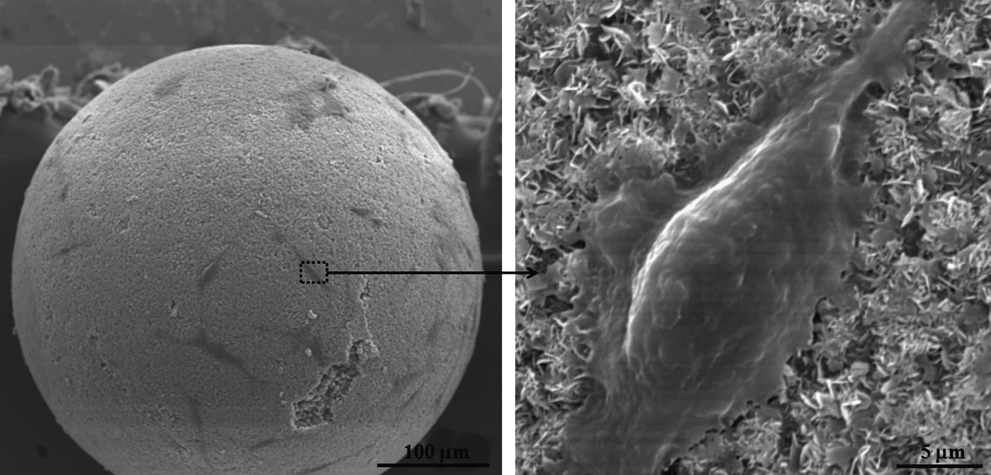
We recently developed peculiar-shaped microspheres composed of bioceramics such as HA and TCP.63,64 The inner part was totally evacuated to remain a thin shell with one end being open (Fig. 3a). This was easily generated by using an organic solvent (chloroform) in the mixture of bioceramic powders and PVB binder. During the sphere-forming process within a polyvinyl alcohol-containing water bath, the organic solvent easily evaporated to leave the inner part totally evacuated and one end open. The remaining shell part comprised just 34%, while the evacuated portion was as high as 66%. 64 Stem cells from human adipose or rat bone marrow cultured on the evacuated spheres proliferated actively, with levels significantly higher than those cultured two-dimensionally, demonstrating the role of 3D matrices and effective cell carriers (Fig. 3b–d). An in vivo experiment performed in rabbit calvarium demonstrated that the evacuated portion of the microspheres allowed osteoconduction, initiating a series of associated biological events, such as resident tissue cell anchorage, matrix synthesis, and bone formation (Fig. 3e). The evacuated microspheres should have much larger areas of newly formed bone per unit of material as compared to the filled microspheres because of the evacuated portion, suggesting that the evacuated microspheres have the potential for direct filling of bone defect with a relatively small quantity of material. The use of the evacuated spheres in combination with stem cells for tissue engineering thus shows promise.
Bioactive ceramic spherical microcarriers designed to be evacuated. Spherical thin-shelled microparticle with one end open is clearly seen
We have recently engineered a pore structure within the PCL biopolymer by introducing a new type of porogen, camphene.4,65 Camphene can easily sublime under ambient conditions and has an extremely high freezing point (around 40°C). Therefore, in our study, the PCL–camphene mixture easily solidified into microspheres during which the camphene and PCL formed a bicontinuous network. In the course of stirring, the camphene constituent of the microspheres freely sublimed to leave the PCL part a highly open-channeled structure. As PCL porous microspheres are hydrophobic, they float on the surface of the culture medium and do not easily allow cell ingrowth, so the surface of the spheres was subsequently mineralized with the HA phase (Fig. 4). A series of solution-mediated processes facilitated the surface modification with a bone-mimetic mineral phase and the tailored surface enabled tissue cells to adhere throughout the spheres, aiding their spreading and proliferation with time. The findings highlighted the role of 3D scaffolding for bone tissue engineering. In a successive study, composites made of degradable polymers with bioactive ceramics were also developed using camphene as the porogen. 4 Pore channels within the HA-PCL microspheres created using camphene became larger in size with increased camphene concentration, resulting in the generation of macropores exceeding 50 μm. Furthermore, confirming their potential for use as cell microcarriers, rat bone marrow MSCs cultured on the microspheres underwent active proliferation, and more interestingly, the MSCs were able to penetrate inside the pore channels. 4 Similarly, bioactive ceramic microspheres with macroporous structure were also produced, which enabled good anchorage of osteoblastic cells and their spread over the microsphere surface, demonstrating the ability to be used in tissue engineering. 66 A gas-foaming method using carbonate salt NH4HCO3 as a pore generator has been used to produce porous polymeric microspheres. Within the pores of PLGA microspheres, cells were able to grow, although the pore size was not sufficiently large (e.g., 20 μm). 67 Similarly, porous PCL microspheres were also produced, which were further mineralized to improve the bone cell responses. 68
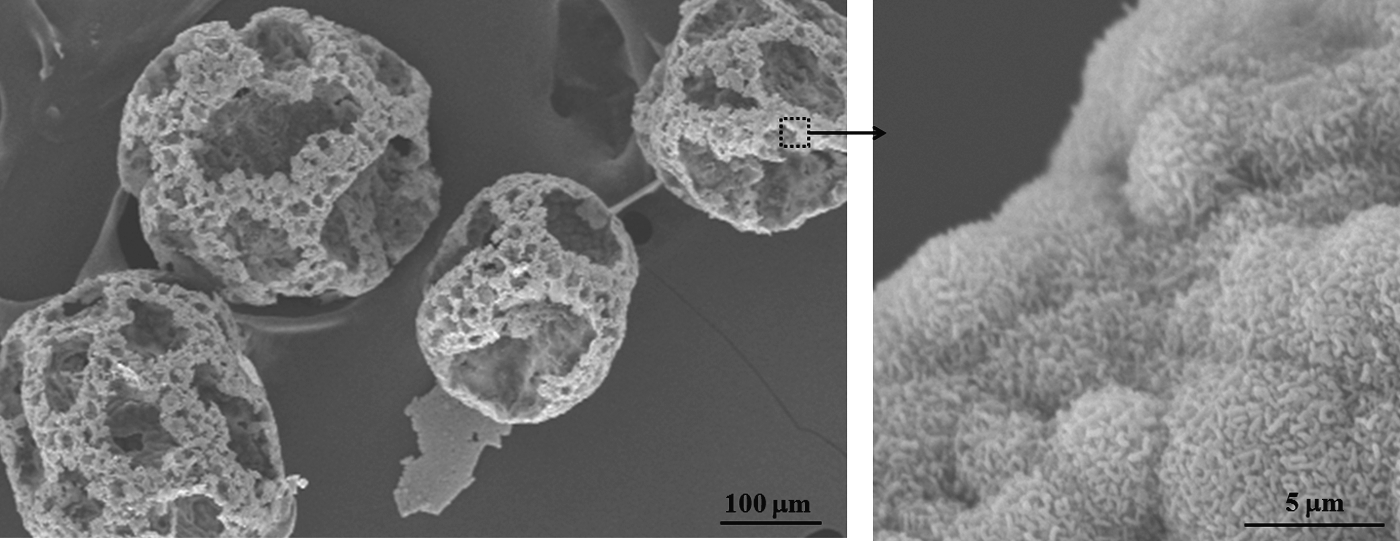
Microspherical particulates designed for bone tissue engineering. Poly(caprolactone) (PCL) polymer spheres were structured to have macroporous channels, and the surface was mineralized with the hydroxyapatite (HA) phase to improve osteogenesis of progenitor/stem cells. The higher magnification of the insert shows engineered HA mineral nanocrystallites.
Microcarriers potentiated by delivery of biomolecules
As the microspheres, either in solid or in porous form, should interact with cells as a scaffolding matrix, providing proper conditions during culture periods, the incorporation of bioactive molecules such as drugs and growth factors within the structure is believed to potentiate their biological properties. In this manner, some specific designs of the microspheres as drug delivery systems can be considered, and the ideas and methodologies can be shared in common with conventional porous scaffolds. In fact, polymeric microspheres have proven to be a popular system that has long been used for the loading and delivery of therapeutic molecules. The composition of polymers (to change degradation rate) and the morphology designs (microporous or hollow-structured) have been the most intensively studied with the aim of achieving controllable and sustainable release of the molecules. In such cases, the microspheres are directly implanted into bone-defective sites in vivo to treat the disease and repair tissue by the release of the incorporated bioactive molecules. For application as a cell-delivering carrier, the spheres should be large enough to carry cells, and the release kinetics of the incorporated drugs should be considered as related to the cells supported on the spheres and the ex vivo culture conditions.
Bioactive ceramic microspheres have widely been used as drug delivery systems for bone repair. However, due to processing barriers, incorporation of biomolecules has been limited. Instead, drugs are largely adsorbed onto the surface of the microspheres, but this is not suitable for sustainable long-term delivery. Therefore, designs of microporous or hollow bioceramic microspheres were proposed for high loading of drugs and their sustainable release. A solid microsphere of Li2O-CaO-B2O3 glass was immersed in a K2HPO4 solution for 2 days, which resulted in the formation of HA microspheres with hollow core.69,70 The release of bovine serum albumin (BSA) from the hollow microspheres was highly dependent on the temperature of post-thermal treatment; the quick release (1–2 days) was sustained up to 14 days when sintered at 600°C. 69 HA and β-TCP porous microspheres have shown prolonged (>8 week) release of bone morphogenetic protein (BMP), even though the initial burst effect was still prominent. 71 Similarly, diopside (CaMgSi2O6) microspheres were controlled in terms of micropore sizes (1–20 μm) by a carbon source; the release rate of dexamethasone could be changed over 2 weeks, even with an initial burst exceeding 80%. 72 Therefore, it is considered that even the microporous structure of bioactive ceramics has limits in the initial burst phenomenon of drugs. This is mainly due to the fact that incorporating drug molecules within the networked structure of bioactive ceramics is difficult; rather, the molecules were just adsorbed onto the surface microstructure. Therefore, the use of polymers together with bioactive ceramics has been shown to be effective for the sustainable release of drugs, as they are incorporated within the polymer networks.73,74
Nevertheless, some compositions of bioactive ceramics can incorporate drugs within the networked structure. Sol-gel-processed silica-based xerogels are one of the exemplar materials. Water-based sol-gel reactions involving hydrolysis and condensation allow easy and safe drug incorporation within the silica network, and microsphere formation was possible using a water-in-oil emulsification method (Fig. 5). 75 Vancomycin and bupivacaine, used as model drugs, were released over a 2-week period without an initial burst effect from silica-based sol-gel microspheres, 75 which were not readily observed in conventionally produced ground granules. As the silica xerogels contain a large quantity of mesopores of several nanometers in diameter, the control over this nanopore structure is considered to be important for controllable release of drugs. Moreover, bone-bioactive compositions by the addition of different types of ions, such as calcium and phosphate, may be a further area of research to follow when using the microsphere system for cell culture with drug delivery capacity.

Acid–base catalyzed sol-gel silica microspheres with potential use as drug delivery systems.
CPCs are considered a promising inorganic source to incorporate drugs inside the formulation, because of their self-hardening properties that allow direct incorporation of therapeutic molecules within the structure. Moreover, some valuable physicochemical properties, such as electrostatic charge, high surface area, and roughness, can improve the interaction with, and affinity to, biomolecules, allowing suitable matrices for drug delivery. Our recent study has highlighted the potential use of the CPC-based microspheres for this purpose. BSA used as a model protein was shown to be safely loaded within the CPC–collagen microspheres and released for a prolonged period (over a month). 2 Based on the findings performed on the CPC-based materials not in a microsphere form, where bioactive molecules (including gentamicin, vancomycin, indomethacin, transforming growth factor [TGF]-β1, or BMP2) could be effectively incorporated within the formulation during the setting reaction, with controllable and sustainable delivery possible over weeks to months, 76 more studies on the use of the CPC-based microspheres for drug delivery systems are required. This aspect could potentiate the cell delivery and tissue-engineering uses of CPC-based microspheres that target bone.
Compared to bioceramics, polymeric materials are more versatile for developing microsphere systems for drug delivery. For the synthetic polymers, strategies are more likely to incorporate biomolecules within the structure not to damage them, because the processing generally requires harsh conditions, such as using organic solvents. A hollow structured design is suitable and rational in allowing safe incorporation of biomolecules, and by controlling the outer shell boundary, release may be delayed. 77 Micro- or nanopores are often produced to control the release profiles. 78 Most of all, a compositional change in copolymers or blends has shown quite effective in controlling the release rate.12,79,80 Although the synthetic polymers have a broad spectrum for controllable and sustainable release of drugs, the poor cell affinity and lack of bone bioactivity frequently require the addition of bioactive ceramics, which is of particular merit in targeting bone tissue. The involvement of the bioactive ceramics (in powder or granular form) within the polymeric phase has been shown not to significantly alter the drug delivery performance of the polymers, while retaining potential of bone bioactivity related with the composition.81–85
Along with the synthetic polymers, natural polymers have also been developed into microspheres. In such cases, premature degradation has often been a problematic issue to achieve controllable and sustainable release of drugs. When the stability is secured, the incorporation of drugs is easier and safer than for synthetic polymers, as the process is generally water based. Approaches using affinity binding or induced ionic interaction have been developed to sustain the release rate,81–85 and functional groups to allow selective enzymatic degradation have also been introduced in the binding.82,85,86 The plethora of sources of polymeric materials for the microspheres and the development of methodologies to control the loading and release of therapeutic molecules have made polymeric microspheres an attractive choice for delivery of cells and bone-targeting drugs.
More recent interest in drug delivery systems has been concerned with designs that facilitate the multiple delivery of biomolecules. Another emerging issue is the stimulus-responsive actions of polymers. Utilizing these features of matrices in the system of cell-delivering microcarriers has great potential. Figure 6 illustrates those advanced strategies to deliver therapeutic molecules (drugs and growth factors) through the microspheres.
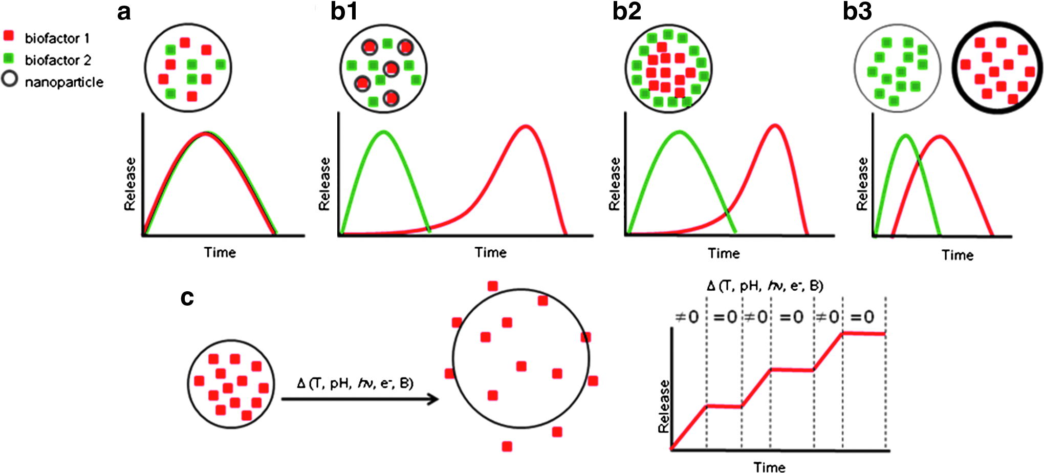
Advanced strategies to deliver therapeutic molecules (drugs and growth factors) through microspheres:
Delivery of multiple bioactive factors from porous scaffolds or hydrogels has recently been considered to better mimic the in vivo conditions and, thus, to improve the regenerative potential of tissues by the delivery systems. The synergistic action of multiple molecules has often shown better outcomes in bone repair.87–89 Microspheres that release multiple bioactive molecules in a timely fashion and at controlled doses should primarily aim to support appropriate cellular behaviors on the microcarriers, from stimulating initial attachment and proliferation to the later stage of differentiation and mineralization. For cartilage repair, delivery of the dual factors dexamethasone and TGF-β3 from PLGA microspheres improved the chondrogenic differentiation of MSCs. 90 For cardiac applications, gelatin microspheres incorporating both vascular endothelial growth factor (VEGF) and insulin-like growth factor-1 reduced the heart infarction of rats after 4-week treatment as compared to those delivering either growth factor alone. 91
Instead of adding the multiple biofactors in one compartment (Fig. 6a), making individual compartments of each different material or designed forms allows sequential delivery of the biofactors (Fig. 6b1–b3). As well as type of molecule, the doses that are precisely required at specific stages of biological reactions make it necessary to design drug-delivering scaffolds in a sequentially controlled way.92–95 Some elegant designs have been proposed in the porous scaffolds, which can also be possibly applied in a similar way to the microcarriers. Layering the microsphere structure from the inside to the outside with biofactors incorporated in different compartments can result in the release of one biofactor from the outer layer at earlier stage and another from the inner layer at a later period. Incorporation of small micro-/nanoparticles (<the size of microspheres) preloaded with one type of biofactor for later-stage delivery in microspheres delivering another biofactor can also be considered for this sequential delivery system. Utilizing different microspherical materials with different degradation rates can also provide a sequential release pattern of biofactors. While very few studies have reported on microsphere systems, some recent studies have utilized this sequential delivery system. The use of microspheres that have the sequential deliver potential of multiple factors in time-controllable manner for the culture of cells and tissue engineering will possibly provide the conditions favorable for progenitor/stem cells to be active in the initial proliferative stage and capable to later switching to an osteogenic differentiation, consequently recapitulating bone-engineered constructs.
The use of stimulus-responsive materials (mainly polymers) is also of great interest in designing next-generation platforms of microcarriers with drug delivery potential (Fig. 6c). Some natural and synthetic polymers are responsive to environment changes such as temperature, pH, enzyme, and magnetism, and the way of releasing molecules inside is primarily controllable and switchable by these cues. A well-known polymer for this purpose, poly(N-isopropylacrylamide) (pNIPAAm), is temperature-sensitive and undergoes a sol-to-gel transition at around 32°C, the lower critical solution temperature (LCST). 96 Below this temperature, it is water soluble, and so more apt to release drugs. pNIPAAm/PLA composite microspheres were shown to release BSA in a manner that was very dependent on temperature change, with a more rapid release below LCST switching to a very sustained release up to 3 weeks at 37°C. 97 A similar pattern was evident using copolymer microspheres made of pNIPAAm-co-acrylamide, which showed a switchable delivery of hydrophobic and hydrophilic drugs. 98 Microspheres that are sensitive to both pH and temperature were also prepared by combining poly(N,N-dimethylamino)ethyl methacrylate and polyethyl acrylamide, demonstrating the release of the model drug in a manner that was dependent on both temperature and pH. 99 Poly(β-amino ester) has also been shown to be thermo- and pH-responsive. In the delivery of Rhodamine, the microspheres showed lower initial burst at pH 7.4, but a much faster release at pH 5.1 and 6.5. 100 The incorporation of magnetic nanoparticles into pNIPAAm microspheres allowed them to respond in an on/off manner to an external magnetic field, which gives rise to a temperature increase and the responsiveness action of thermosensitive pNIPAAm. 101 Utilization of the stimulus responsiveness action has become an emerging field in drug delivery systems to achieve on-demand controllable release patterns. This smart action is considered potentially applicable for microspheres that are to deliver therapeutic molecules inside and consequently to dictate the behaviors of supported cells through the manipulated release action of the drugs. However, studies are still relatively few, warranting more in the near future.
Microcapsules as a Cell-Delivering Bioreactor
Microspheres for cell encapsulation have also been developed in an attempt to deliver tissue cells within a biomaterial container that is isolated, but is semipermeable to the outside environment. The encapsulating material should be permeable to nutrients and gas molecules, such as ions, growth factors, and oxygen, allowing cell interactions with the environment and removal of cell metabolites. The capsule is thus considered a sort of bioreservoir, providing cells with a microenvironment that supports proliferation and differentiation to specific tissues. Moreover, during culture, several biological molecules are secreted from the encapsulated cells, and the molecules can be accumulated and released from the microcapsule to the surroundings. Therefore, the cell-containing microcapsule is considered as a biofactor-eluting reactor that ultimately plays a crucial role in tissue regeneration when delivered in vivo. The roles of microcapsules in relation to delivered cells and the surrounding tissues are schematically shown in Figure 7.
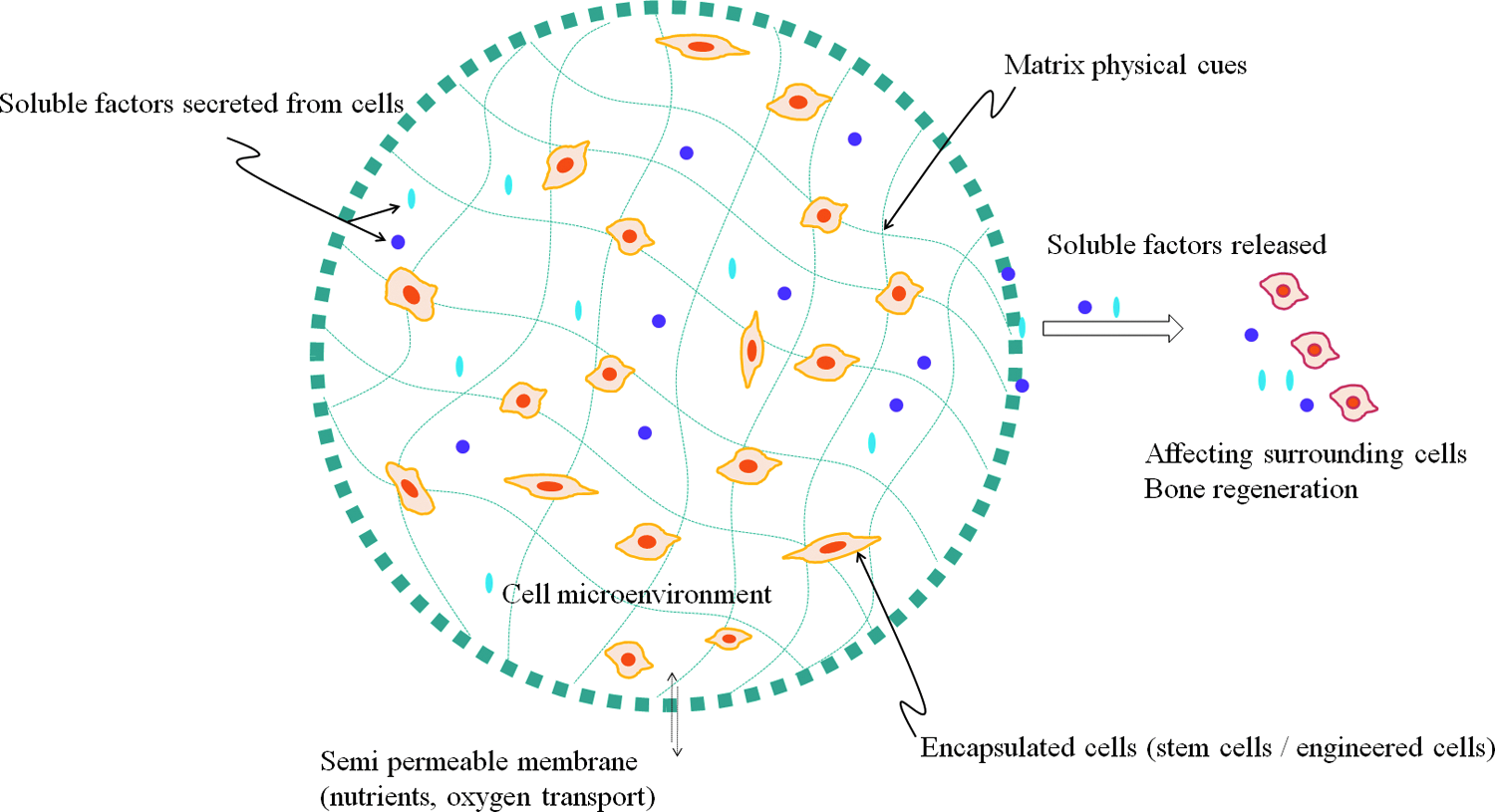
Encapsulating microspheres provide an internal environment that supports the cells being delivered. Soluble factors released from those cells, along with biofactors loaded into the scaffold, can have osteoinductive effects on surrounding tissue cells. Color images available online at www.liebertpub.com/teb
Biomaterials and strategies used in cell encapsulation
This cell encapsulation strategy has long been used for therapeutic treatments of diseases such as the production of deficient hormones including insulin in diabetes, factors VIII and IX in hemophilia, and erythropoietin in anemia.102–104 Primarily, this encapsulation strategy aims to safely deliver cells across the barrier of the body's immune system, which negates the use of immunosuppressive drugs, as these are generally known to have severe sideeffects. When cells are encapsulated, the host immune reaction is physically blunted and shielded, enabling successful cell transplantation. The increased knowledge of stem cells has also shifted the impetus toward tissue engineering. Stem cell sources or genetically engineered cells can also be delivered within the designed container to produce specific biological factors that are effective in tissue regeneration.
The most fascinating material choice for encapsulating microspheres has been hydrogel polymers such as alginate, collagen, gelatin, and agarose. This class of biopolymers is hydrophilic and cell friendly, permeable to nutrient diffusion, soft, and flexible, which makes the biopolymers easily amenable in fluid conditions without causing significant frictional force or mechanical irritation.15,105–109 Microcapsule formation is possible by the gelation or cross-linking of the polymer networks induced by the thermal or chemical change. Many different processes have been developed for the microencapsulation, including gelation, extrusion through a nozzle, and spray-drying.15,105–109
Among the hydrogels, alginate has been the most widely used biopolymer. Alginate, a type of natural polymer, is sourced from brown seaweeds and bacteria.
110
It is largely considered to be nontoxic and biocompatible and has gelling property easily absorbing water. Sodium alginate can cross-link in a reaction with divalent ions such as Ca2+, Sr2+, and Ba2+. Therefore, when an alginate–cell mixture solution is injected in a Ca2+-/Sr2+-/Ba2+-containing medium, it rapidly hardens to form a cell-encapsulating microsphere. During the process, the concentration of divalent ions should be appropriate to harden the alginate, but not be high to be toxic to the cells inside the capsule. Generally, Ba2+ forms more cross-links with alginate than does the more generally used Ca2+. The polyelectrolyte complexing of the alginate with poly(l-lysine) (PLL) has long been used to stabilize alginate in cell encapsulation.
105
PLL has generally been treated on the surface of alginate after the microencapsulation process. Charged PLL is known to be immunogenic. Thus, the final coating with alginate of the surface to form the alginate-PLL-alginate structure has been preferred.
106
While other cationic polymers such as chitosan, poly(
On the other hand, agarose is a thermally gelling polymer that has been used in microencapsulation of cells. Gelation is possible by reducing the temperature of agarose–cell solution. 113 Depending on the agarose concentration and cooling rate, the sol-gel transition temperature may vary in the range from 32°C to 47°C. 114 Taking this into account, the agarose fabrication is based on droplet breakup in a coflowing stream, where the agarose solution is extruded at 37°C or near into a liquid flow at low temperature (e.g., 2°C).108,109 The capsule diameter can be changed by the flow rate. 109 However, agarose also may not present adequate matrix cues for cells to grow and proliferate, so fibronectin and fibrinogen have been incorporated to improve the cell survival. 115 Similarly, osteopontin has also been added to the agarose microbeads. 116 One recent study highlighted the importance of control over the stiffness of the agarose capsule by varying the concentration, to provide different physical cues to the encapsulated cells. 108
Another important natural polymer for microcapsule is collagen. Collagen can gel easily upon pH change. 117 Because of its biocompatibility, collagen has been considered as a promising 3D scaffold for tissue engineering and cell delivery.53,118,119 The enzymatic degradation of collagen makes it appropriate as a degradable carrier, but collagen degradation is not easily controllable, which often remains as a challenge. Microencapsulation of MSCs within the collagen–agarose sphere was shown to maintain 75%–90% cell viability for up to 8 days. 120 Cell viability was increased with increasing collagen content, suggesting the potential use of collagen microcapsules in delivering MSCs and directing their differentiation. Construction of interpenetrating network polymers (IPNs) using two different characteristic polymers is another strategy to allow gelation. One recent study developed cell-encapsulating microspheres through IPN of collagen and hyaluronic acid. 121
Other types of biomaterials such as synthetic polymers, including poly(ethylene glycol) (PEG), polyacrylate, and pNIPAAm, have also been used as the encapsulating matrices. One example is the microencapsulation of cells within a photopolymerizable PEG diacrylate.122,123 pNIPAAm is also another type of thermoresponsive polymer, as it becomes insoluble on heating above a certain temperature (LCST) and undergoes gelation. The LCST value of pNIPAAm is around 32°C, being close to the body temperature, and can be controllable depending on the nature of substitute groups and their molar mass. 96 Cell encapsulation within pNIPAAm has been achieved by extrusion through a double-barreled needle. Cells were contained in the inner part, while the outer culture medium maintained at 37°C played a role in the gelling of the polymer extruded into a bath containing paraffin layer to form a cell-encapsulated microcarrier. 124
Microenvironment consideration in encapsulation system
The microencapsulation of tissue cells based on a variety of polymers has been possible by the gelation induced by the temperature or chemical change. Encapsulated cells survive within the capsules and interact with the outside environment through the membrane that is permeable to nutrients and gases. The inner matrix of the microcapsule is thus considered to provide a substrate for cells to adhere, migrate, and proliferate. Matrix properties, such as the chemical groups and physical stiffness, provide determinant cues to the anchorage-dependent cells in their survival and differentiation. In the course of propagation, cells encapsulated and supported on the matrices secrete biological molecules, which in turn affect the behavior of cells. Therefore, the microcapsule is considered a dynamic microenvironment of cells present inside and provides physical and chemical cues to control their fate. Moreover, as the membrane is permeable, allowing the transport of molecules and gas species, the soluble factors existing outside will freely pass to the cells inside, while the secreted factors from the inner cells can be delivered to the outer space, which possibly affects the behaviors of host cells in the tissue, allowing the therapeutic use of the solution. Figure 7 illustrates the relationship between the microcapsules and cells inside as well as the surrounding tissue cells.
Although the concept of the cell microencapsulation and the possible potential in stem cell therapy and tissue engineering has attracted increasing interest, control over the microenvironments and the use of microencapsulated cells for tissue regeneration purposes are in their infancy. It is conceivable that the behavior of encapsulated cells could be manipulated by adjusting the method of soluble factor addition to the culture medium. TGF-β1, among others, has been incorporated into a medium in which alginate beads containing MSCs were cultured in vitro; the MSCs formed the targeted cell phenotype (e.g., chondrocytes). 125 To observe the cardiogenic potential of embryonic stem cells (ESCs) encapsulated in PLL-coated alginate capsules, supplementation of the culture medium with BMP4 resulted in enhanced differentiation into cardiomyocytes, as characterized by the expression of cardiac markers by more cells. 126 Another study to promote survival of ESCs encapsulated in alginate microcapsules incorporated Y-27632, a Rho-associated protein kinase inhibitor. Results showed the promotion of cell survival, cluster formation, and proliferation, and their further differentiation into mesoderm, endoderm, and primitive gut tube, indicating the possible use of the microcapsule system for the ESC differentiation. 127
In addition to soluble molecules supplied from the surrounding medium, properties of the matrix upon which the encapsulated cells are growing provide crucial cues to determine the fate. One recent study explored the regulation of behaviors of microencapsulated stem cells by varying the elasticity of the matrix. 108 By using a high-throughput microfluidic droplet generator and different concentrations of agarose precursors, microcapsules with 35-fold variation in shear elastic modulus were developed, and the efficacy was demonstrated using mouse ESC lines (Fig. 8). Although in-depth characterization of cell behavior was not performed in the study, it is inspiring to note that modulation of the cell microenvironment within the confinement of encapsulating spheres may be possible by controlling physical parameters and not just chemical factors.
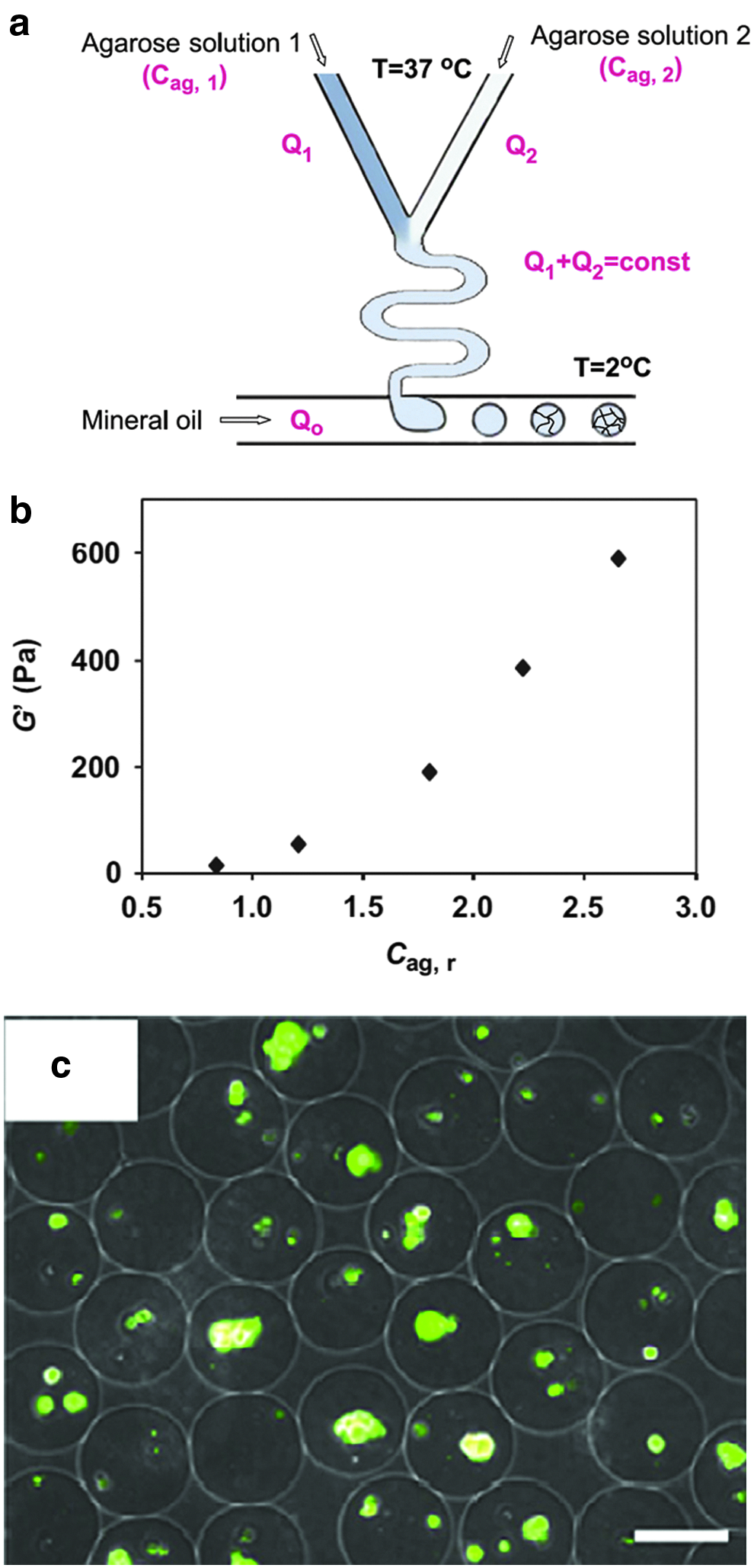
Agarose cell encapsulating microbeads with varying elasticity.
The secreted soluble factors from the encapsulated cells can be potent sources for therapeutic purposes. Moreover, when implanted in tissue defects, the eluted factors from the encapsulation device will directly influence the surrounding cells. Therefore, strategies have been established to genetically engineer the cells to encapsulate in an attempt to allow secretion of target factors. The factors produced by encapsulated cells provide trophic support to themselves and also to the surrounding exterior cells through the release of stimulating factors from the microcapsules. Therefore, controlling the pore size of the shell membrane and developing stimulus-responsive membranes to allow on-demand release of factors are possible strategies to achieve smart cell encapsulation systems, which have not yet been realized.
One study encapsulated PC12 cells that were genetically engineered to secrete dopamine within alginate beads, and demonstrated an effective dopamine-releasing system in rats with Parkinson's disease. 128 L929 murine fibroblast cells genetically engineered to produce VEGF were encapsulated in a hydroyethyl methacrylate–methyl methacrylate copolymer and then implanted for 1 and 3 weeks in vivo. Higher number of capsules and blood vessel formation in the case of the transfected cells were noted as compared to nontransfected cells. 129 A two-component system, consisting of cellulose sulfate and poly-dialyldimethyl ammonium chloride capsules, was designed to incorporate HEK-293T cells to express doxycycline- or erythromycin-inducible expression, 130 which provides precedent for creating microencapsulation systems capable of releasing multiple biofactors. 130
Examples targeting bone tissues
While the microencapsulation studies on such polymer-based in situ cross-linkable biomaterials have mainly focused on the therapeutic treatment of diseases such as diabetes and cell transplantation in soft tissues such as cartilage and nerves, those materials can also be used for the stem cell delivery for bone tissue. Abbah et al. evaluated the potential of alginate microcapsules in delivering adipose-tissue stromal cells and bone marrow stromal cells for use as an injectable bone substitute.131,132 When cultivated in the alginate microcapsule, the adipose tissue stromal cells showed significantly higher levels of osteogenic differentiation than those cultured in a monolayer. Alginate-microencapsulated system considerably increased the cell number (2-fold) after 7 days and maintained the cell viability over 21 days of culture. 131 Further osteogenic differentiation, including ALP activity and osteocalcin mRNA expression, produced a 6.9-fold increase with respect to monolayer culture, and the osteocalcin protein was detectable earlier in the encapsulated systems than in the monolayer culture. 131 Alginate microbeads coated with PLL also proved to be an effective substrate for supporting cell survival and osteogenic differentiation. The PLL coating significantly enhanced osteogenic differentiation of rabbit-derived bone marrow cells. 132 Genetically engineered adult MSCs that express BMP2 were encapsulated in alginate beads and then transplanted subcutaneously into bone defects. The MSCs were able to survive and secrete BMP2, suggesting that the secreted BMP2 should directly affect not only the MSCs inside but also the cells present in the tissue outside the microcapsules. While the MSCs inside the microcapsules differentiated into chondrocytes by an autocrine effect, newly formed bone was developed around the alginate microbeads, possibly resulting from a paracrine effect. 133
While the polymeric compositions are effective for development into microencapsulation system, the use of bioactive ceramics in concert with the polymers (a composite approach) is preferred in targeting bone tissue. The composite matrices that hold cells within the capsules better mimic the native bone ECM in terms of chemistry and physical properties, possibly stimulating osteogenic functions. Moreover, the ions that are released from the bioactive ceramics should significantly influence cellular behaviors within and outside the microcapsules, from the cell proliferation to differentiation and mineralization. MSCs treated with calcium ions will preferentially develop into an osteoblast lineage. 134 The ionic extracts from bioactive glasses can also significantly alter the differentiation behaviors of ESCs and MSCs.135–137 Trace elements such as zinc that are incorporated in the bioactive ceramics also importantly affect the mitosis, differentiation, and calcification processes of osteoprogenitor and bone cells. 52 These studies have indicated that ions that are eluted from the bioactive ceramics can be used for other key regulating cues, such as osteogenic biochemical factors.
In a particularly interesting study concerning the utilization of ion-releasing bioactive ceramics in microencapsulation, 138 45S5 bioactive glass, which induces the production of VEGF in fibroblasts, was added to alginate beads at varying concentrations (0.01, 0.1, and 1 wt%). Significantly enhanced production of VEGF was observed in cells encapsulated within alginate beads containing appropriate concentrations (0.01 and 0.1 wt%) of the bioactive glass, as assessed on the extract media (Fig. 9). 138 Moreover, endothelial cells cultured with a conditioned medium showed significantly increased proliferation. 138 Even though the study did not perform any bone-targeting assays, the materials used and the idea proposed may also be potentially applicable to the bone regeneration area. Instead of genetic modification of cells, addition of inductive materials such as ion-eluting bioactive ceramics within the microcapsules may also trigger the encapsulated cells to secrete specific biofactors. Therefore, strategies to trigger cells encapsulated within the microcapsules to secrete bone-stimulating biofactors can vary from the genetic modification of cells to the modulation of the chemical and physical cues of the capsule materials. In this sense, when targeting bone tissue, special care is needed in selection of capsules with proper properties and the type of cells to be encapsulated.
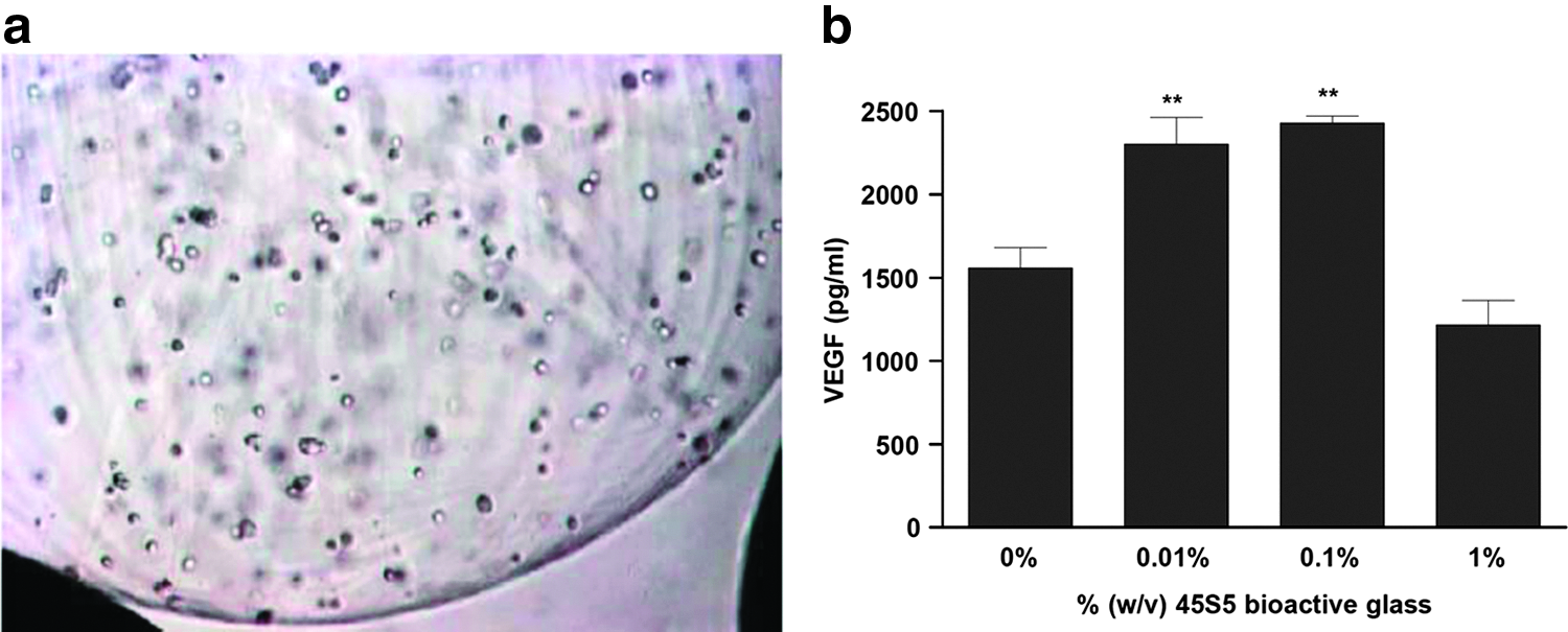
Vascular endothelial growth factor (VEGF)-releasing microcapsules of alginate added with bioactive glass (BG).
Culture Methodologies for Cell Expansion and Tissue Engineering
Cell culture for the manufacture of biopharmaceuticals such as recombinant human growth factors often utilizes fermentation, and so simply increasing the volume of the culture can increase the scale of production. The approach is feasible as cells used (e.g., Escherichia coli) are planktonic and therefore grow in suspension. Unfortunately, in the case of cell culture using anchorage-dependent mammalian cells such as those employed for human cell therapies, readily scalable fermentation approaches are not feasible, as the cells have to be grown on solid substrates. This limits the scalability of cell culture using T-flasks due to the surface area requirements for growth and medium volumes required.
For this reason, microcarriers have been extensively investigated for use as a scalable solution to cell expansion for tissue-engineering purposes. In fact, a variety of different culture methods have been explored for the purposes of cell expansion and bone tissue engineering on microcarriers, which include dynamic cultures under shake/agitation, rotation in spinner flasks, or in rotating wall vessels (Fig. 10). The most common type of culture for expanding cells on microcarriers uses stirred culture in the form of spinner flasks. While the shaking or agitation of culture plate possibly provides opportunities for cells in the medium to adhere to the microcarrier surface and subsequently colonize and proliferate, this methodology is considered to be limited primarily due to the inhomogeneous flow conditions and turbulence of the culture medium. 139 On the other hand, dynamic culture in a spinner flask or rotating wall vessel generates medium flow that can be precisely controlled, which allows prediction of flow dynamics.
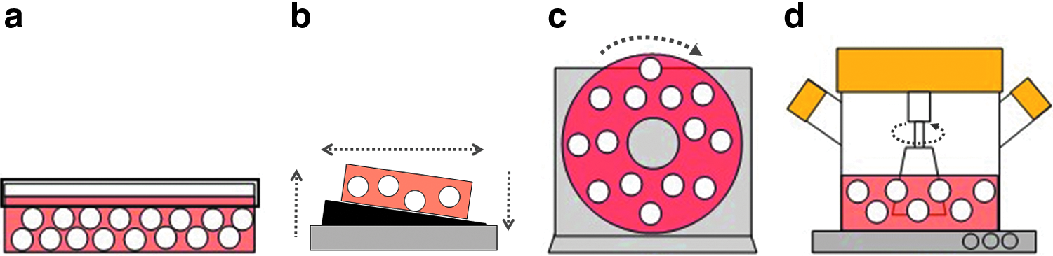
Culture methods utilizing microcarriers for cell expansion and tissue engineering.
The use of rotating wall vessels has been examined for 3D culture of stem cells and osteoblastic cells for tissue engineering purposes.140–143 Rotating wall vessels are essentially tube-shaped culture vessels that are positioned horizontally with the entire vessel rotating on a central axis, providing a suspension culture condition when cells are attached to microcarriers. They were initially seen as very promising for 3D expansion of cells because of the low shear stress and low turbulence they produced, while providing a good level of mixing to achieve homogenous dispersion of cells and nutrients throughout the culture.144,145 Epithelial cells have successfully been expanded on microcarriers using this method and form on ordered hierarchy and functional gap junctions. 143 From a skeletal tissue-engineering perspective, early studies exploring the rotating wall vessel successfully expanded chondrocytes on microcarriers.146,147 Osteoblastic cells cultured on buoyant polymer microcarriers in the rotating wall vessel expressed higher levels of ALP and more intense Alizarin Red S staining than in static culture, even though cell proliferation was slower. 141 Other research characterizing rat bone marrow stromal cells on microcarriers demonstrated that rotating wall vessels could be successfully employed to produce differentiated osteoblastic cells that bridge between microcarriers to form strings of tissue consisting of multiple cell-populated microcarriers. 142 After 14 days of culture on the microcarriers, cells expressed ALP and produced both type I collagen and osteopontin. The amount of shear stress experienced by cells at the surface of the microcarrier in suspension culture is directly dependent on the density of the microcarrier at a given rotation speed and fluid viscosity. 142
Compared to a rotating wall vessel, the spinner flask culture technique has been used more profoundly in the research fields for cell expansion and tissue engineering. The proof-of-principle demonstration of feasibility for cell expansion in spinner flasks has been made in numerous cell types, including human myoblasts, 148 mouse ESC-derived neural stem cells, 149 pancreatic stem cells, 150 primary astrocytes, 151 human intervertebral disc cells, 152 and articular chondrocytes, 153 in addition to pluripotent stem cells from human154–157 and mouse.158–162 Perhaps, most promising from the viewpoint of bone tissue engineering is the expansion and directed differentiation of MSCs into the osteogenic lineage. Stirred culture, predominantly using spinner flasks, can support expansion of MSCs.163–166
Many parameters are considered important in the spinner flask culture system, including flask and impeller geometry and size, medium quantity, microcarrier type and its size and quantity, cell concentration, flow rate, and culture methods.8,166–171 Flask and impeller size and geometry are well defined and can be customized. Modifying the size of the spinner flask also influences the cell behavior. The attachment kinetics of Vero cells on polymer microcarriers were much slower in the 15 mL than in the 100-mL flask.166,167 Depending on the microcarrier physical properties, such as elasticity and surface roughness, the flow shear force on the microcarrier surface will be significantly influenced,166,168 which consequently alters the initial response of cells. Initial cell-seeding density (or the relative cell quantity with respect to microcarriers) is also important. A study reported that seeding five MSCs per microcarrier generally yielded a higher cell number at the end of a fixed expansion period compared to seeding 10 MSCs per microcarrier. 166 This trend was consistent for two different impeller geometries and two different microcarrier concentrations in the starter culture. Nevertheless, when Chinese hamster ovary (CHO) cells were used, the optimum cell-to-bead ratio was 25 to 30 cells per bead. 169 A similar result also confirmed the need of a high cell/bead ratio for optimal cell culture. 171 Another important consideration is the performance of cell-loaded microcarriers in vivo, where microcarriers are required to aggregate to form a tissue structure. In fact, higher cell/bead ratios (up to 125 cells/bead) showed improved cell survival and aggregate formation as compared to the lower ratios (around 16 cell/bead). 8
The effect of impeller speed has also been researched thoroughly. The impeller itself influences cell behavior 166 due to the fluid flow shear stress created. Cell damage arises not only from the shear stress in laminar flow but also from collisions experienced during turbulence. 168 Therefore, reducing the turbulence and frequency of collisions can increase the cell viability. In this sense, studies have found relatively low speed (less than ∼100 rpm) is preferred for cell adhesion and survival. When CHO cells were cultured on microcarriers, the optimum speeds were between 40 and 60 rpm with favorable cell growth, which was considerably reduced at increased speeds of 150 rpm. 169 Similarly, Vero cells also behaved optimally when the speeds were low (e.g., 40 rpm). 171 Studies on human fibroblasts revealed that the optimum speed for cell proliferation was 50 rpm (vs. 30 or 80 rpm). The optimal speed is largely dependent on the type of microcarriers used, that is, the material properties, such as bulk properties like density (specific weight), and surface properties such as roughness, topography, and hydrophilicity. Furthermore, the hydrodynamic effect of the rotation speed was also highly dependent on the type of cells cultured. 170 In an experiment using different cell types (CHO, BHK-21, and Vero cells) cultured on the same microcarriers and at rotation speeds of 70, 110, and 200 rpm, the cell behaviors were completely different. 170 While the Vero cells and CHO cells were highly sensitive to the speed, showing an optimum proliferation at 70 rpm and a reduced proliferation at 200 rpm, BHK-21 cells were able to grow well at all speeds. 170
MSCs seeded onto microcarriers were found to be capable of proliferation and also of colonizing fresh microcarriers added to spinner flasks midway through a 28-day expansion protocol. 172 Furthermore, the newly colonized cells were capable of both osteogenic and chondrogenic differentiation over the same time period, as characterized by von Kossa stain and Alcian blue stain, respectively. 172 Therefore, the 3D expansion of MSCs on microcarriers was suggested as a beneficial alternative to conventional monolayer cultivation. This observation was supported by subsequent work on the MSC proliferation on degradable biopolymer microcarriers. 165 Compared to tissue culture plastic, spinner flask cultivation over 14 days yielded a superior cell growth rate while retaining cell differentiation capacity. 173 Particularly, the addition of fresh microspheres during spinner flask culture was shown to effectively allow bead-to-bead contact cell transfer and expansion, suggesting a promising tool to expand cells effectively by the spinner flask system. However, this technique was not always supportive for the expansion of MSCs, which largely depends on the type of microcarrier, indicating the importance of microcarrier compositions in the cell colonization via bead-to-bead contact. 174 Nevertheless, the differentiation potential was retained, and expanded cells were capable of multilineage differentiation. The same research group subsequently used biopolymer gelatin microcarriers in spinner flasks to successfully stimulate bone regeneration in a rat periodontal bone-defect model 175 and a rat critical-size calvarial-defect model. 176 However, the bead-to-bead contact expansion of cells was sometimes observed to be not so effective in osteogenic differentiation. Human bone marrow cells cultured on microcarriers in spinner flasks retained greater osteogenic and adipogenic differentiation potential in culture with a refreshing-only medium semicontinuously when compared to those where fresh microcarriers were added midway through the culture to provide additional expansion capacity. 177 Other researchers also demonstrated the effectiveness of the microsphere-based spinner flask cultures in the expansion of MSCs by refreshing a portion of the consumed medium, where they also used a xenofree culture medium that would be appropriate for clinical translation. 164
Culture regimes using spinner flask tend to use intermittent stirring during the initial culture period, with rest periods between to improve cell attachment to microcarriers. Yang et al. 178 used intermittent stirring for 3 h (40 rpm for 5 min followed by a 30-min rest) followed by continuous stirring at 40 rpm for 5 days in 50-mL spinner flasks. The study also used a thermosensitive-polymer-modified surface to enable the liberation of bone marrow stromal cells from the microcarriers without using proteolytic enzymes that damage cell surface proteins. Western blot analysis confirmed that cells liberated from the microcarriers after 5 days of continuous stirring had abundant intact fibronectin and laminin compared to cells trypsinized from conventional microcarriers. 178 More recently, MSCs were initially seeded onto microcarriers in 250-mL spinner flasks using intermittent stirring for 4 h (70 rpm for 3 min with a 30-min rest) followed by continuous stirring for 3 days. 179 In one study, stirred bioreactors with a working volume of 80 mL were used, and initial cell seeding was conducted using intermittent stirring for 24 h at 25 rpm for 15 min followed by 2 h static incubation, before being increased to 40 rpm for continuous culture. 164 On the whole, it seems that researchers favor an initial seeding stage that uses intermittent stirring at 20–35 rpm for periods of a few hours, followed by continuous culture for the remaining experiment duration of between 15 and 60 rpm.45,164,172,173,177 However, factors such as bioreactor diameter, stirrer geometry, and microcarrier concentration all have to be considered in addition to stirrer speed, as these all determine the resultant flow dynamics and the consequent movement of cells and microspheres and the shear forces at the microsphere/medium interface that affects the adherence of cells.
Concluding Remarks
Microcarriers have great potential to be used for scalable expansion of human stem/progenitor cells for tissue-engineering purposes. Particularly attractive is the fact that they can be processed using bioreactor methods that are, from a bioprocessing perspective, more akin to highly scalable fermentation methods than the somewhat-constrained monolayer culture methods typically adopted for human cell expansion. Many different forms of microcarrier are permissive for cell expansion, from solid-filled microcarriers that support cell growth on the outer surface to highly porous or even hollow evacuated microspheres that can be loaded with cells and bioactive molecules. Moreover, varying material composition and utilizing different fabrication methodologies can produce a multitude of different structures that have endless potential as dual-function cell expansion substrates/tissue-engineering scaffolds. The use of stimulus-responsive smart materials is effective for controlled release of drugs and bioactive molecules, and the compositional variation with ECM molecules confers further support either to early cell responses such as attachment, migration, and proliferation, or later responses such as directed differentiation. Furthermore, encapsulating hydrogel microspheres provides 3D internal environment that supports the cells being delivered, where soluble factors released from the encapsulated cells can have inductive effects on surrounding tissue cells, finding potential usefulness as therapeutic cell delivery systems.
In conclusion, microcarrier technology holds a great promise for cell culture and tissue engineering. To achieve success, two key areas need to be considered for successful tissue engineering using microcarriers in the future: (1) precise control of the microcarrier structural properties to best support cells and integrate in host tissue and (2) appropriate dynamic culture conditions for expansion and differentiation of cells for therapy.
Footnotes
Acknowledgments
This study was supported by grants from the Priority Research Centers Program (2009-0093829) and WCU program (R31-2008-000-100069-0), National Research Foundation, South Korea.
Disclosure Statement
No competing financial interests exist.