
Abstract
Hydroxyapatite is a biocompatible material that is extensively used in the replacement and regeneration of bone material. In nature, nanostructured hydroxyapatite is the main component present in hard body tissues. Hence, the state of the art in nanotechnology can be exploited to synthesize nanophase hydroxyapatite that has similar properties with natural hydroxyapatite. Sustainable methods to mass-produce synthetic hydroxyapatite nanoparticles are being developed to meet the increasing demand for these materials and to further develop the progress made in hard tissue regeneration, especially for orthopedic and dental applications. This article reviews the current developments in nanophase hydroxyapatite through various manufacturing techniques and modifications.
Introduction
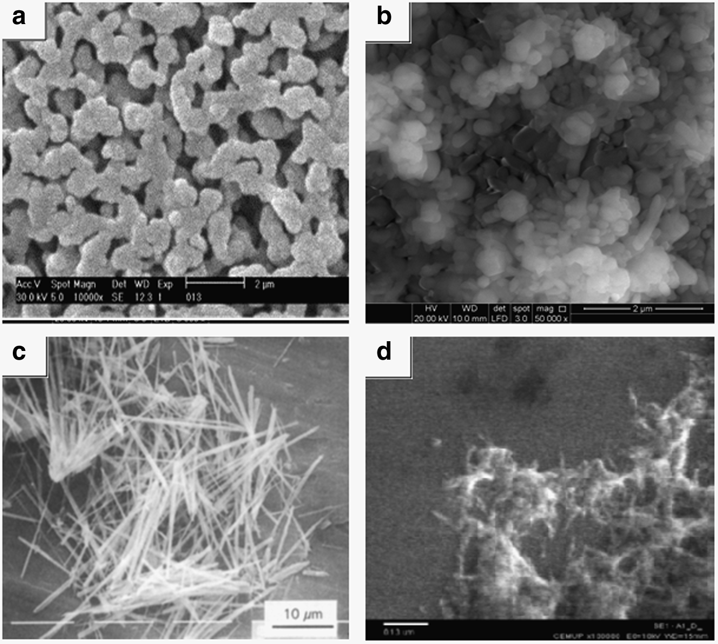
Hydroxyapatite (HA) is a synthetic biomaterial that has garnered widespread consideration in advanced hard tissue engineering. Its chemical composition is comparable to the mineral component of natural bone, and the bioactivity of HA supports the necessary bone regeneration. 7 Essentially, 70% of natural bone consists of nanocrystalline HA that is 20–80 nm long and 2–5 nm wide. 8 Synthetic HA that has been commercially produced in powders, bulk bodies, and fibers exhibits size greater than the size of natural HA. Nanophase HA is preferred over microphase HA due to several advantages that nanophase HA has over the latter. Figure 1 shows the typical transmission electron microscope (TEM) image of HA nanoparticles. 9 Such macro-sized HAs exhibit poor bioresorbability and brittle characteristics,10,11 which has made the development of nanophase HA increasingly imperative.12–14 Nanophase HA means HA either in the form of particles, fibers, or others that have a size of <100 nm in diameter of fiber/particle, width or length. Figure 2a–d show samples of scanning electron microscope (SEM) images of natural HA, 15 synthetic HA nanoparticles produced by electrospinning, 16 HA nanowhiskers by hydrothermal, 17 and HA nanoparticles by wet chemical precipitation, 18 respectively.
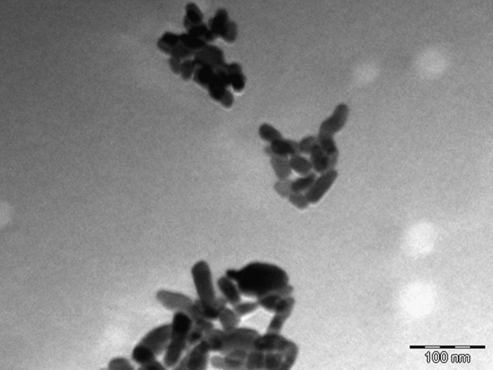
Transmission electron microscope image of hydroxyapatite (HA) nanoparticles 9 .
This article reviews recent progresses in HA development as a desirable biomaterial for hard tissue regeneration. The review begins with a biomaterial section that highlights the general properties of the biomaterials together with their classification. In the next section, the development of nanophase HA into a ceramic biomaterial is discussed, along with its general properties, synthesis, and surface modifications. The significance of HA nanoparticles in cell proliferation and the development of HA nanofibers is also included, and it is followed by demands on nanophase HA, especially for orthopedic and dental applications. The article concludes with a future outlook for this field.
Bioceramics in Hard Tissue Engineering
Bioceramics is one of the classes of biomaterials that is designed to substitute fragments of a living system and to harmoniously function with living tissues. The extent of the incorporation of biomaterials into body tissue depends on aspects such as biocompatibility of the material used, design, and material properties.
The widespread use of bioceramics such as calcium phosphate, 19 metallic oxides, 20 and glass ceramics 21 has been reported. Bioceramics can evoke three different biological responses. First, they can be bioinert, which means that they are non-adherent to the surrounding tissues. Second, they can be bioactive, which means that they can chemically react to form a strong bond with bone or soft tissue of the host. Third, they can be bioresorbable, which means that they actively contribute to the metabolic processes of an organism to reconstitute the tissue in part or in its entirety.
Typical examples of bioinert ceramics are carbon, alumina (Al2O3), zirconia (ZrO2), and single oxide ceramic. These materials do not form a bond with bone but they permit the development of thin fibrous capsules in the interface. Therefore, they are typically functional as bone plates, artificial heart valves, bone screws, femoral-head component, and artificial joints. 22
Generally, bioactive ceramics used in orthopedic operation are ceravital, bioglass, apatite-wollastonite (A-W) ceramic, and dense HA. Bioactive ceramic undergoes surface chemical reactions. 22 Ceramics with surface-reactive properties form solid links with bone (and soft) tissue after implantation. For some materials, the response of ion exchange between the bioactive implant and the body fluids results in the development of a biologically active carbonated apatite layer on the implant. 23 In general, bioactive ceramics have poor properties compared with bioinert ceramics except for A-W glass ceramic that has a better mechanical strength than cortical bone. Therefore, they are most commonly used as bone defect fillers provided in the structures of block, porous material and granules. 24
Examples of bioresorbable ceramics are calcium carbonate, calcium sulfate, carbonate HA, and β-tricalcium phosphate. Bioresorbable ceramic implants are designed to degrade constantly and be substituted with natural tissues. Instead of their replacement, it brings tissue renewal. 22 The advantage of applying bioresorbable ceramic is that the implant will be substituted by normal and functional bone, which then removes long-term biocompatibility difficulties. Commonly, this kind of ceramic is used for bone repair in the case of disease, bone defect, filler, and also as drug delivery devices. 22
For bone implantation purposes, calcium phosphate and bioglass are favored because they have the capability to produce a connection with bone tissue, while metallic oxides are nearly bioinert in the biological environment. 20 In general, biocompatibility and bioactivity are the key properties that synthetic biomaterials must have to be acceptable for use as bone replacement materials. 1 HA is known to possess these properties and can therefore serve as an exceptional bone replacement material. 25 Further, HA is one of the main minerals in human hard tissues. 26 Therefore, development of synthetic HA as hard tissue replacement materials has been extensively pursued.
Development of Nanophase HA as Ceramic Biomaterial
HA properties
HA, whose chemical formula is Ca10(PO4)6(OH)2, has been used in the medical field for implant resolutions. As introduced earlier, HA is the main mineral element of human hard tissues 26 and can be found in teeth and bone. Application of HA can lead to fast bone formation and solid biological fixation to bony tissue due to its encouraging osteoconductive and bioactive possessions. 17 Table 1 shows the comparison of the properties of HA and other calcium phosphates.27,28 Under normal temperatures, HA is the top stable calcium phosphate at pH values between 4 and 12. 29 In addition, HA does not show any cytotoxic properties. HA shows exceptional biocompatibility with hard tissues, skin, and muscle tissues. 30 Because of its good property of biocompatibility, it has often been the primary choice for the construction of bone fillers or as a coating on prosthetic implants to enhance implant incorporation with the host bone. 31 HA is also widely used in industries that manufacture fertilizers and pharmaceutical products, in protein chromatography applications, in catalysis, and in water treatment processes.32–35
HA, hydroxyapatite.
Synthesis of nanophase HA
Accuracy is important for most of the ceramic processing methods used to produce HA. Careful management of parameters, such as pH, reaction time, temperature, and concentration of the reactants, together with the proper selection of the precursor materials are extremely important during HA synthesis because these parameters and materials can affect the composition and properties of the final HA product. 36 Several procedures of preparing HA have been identified, involving hydrothermal, sol-gel, wet chemical, and biomimetic deposition methods as stated in Table 2.37–41
HA, hydroxyapatite.
Hydrothermal method
In the hydrothermal method, the reaction is conducted in the presence of water at a relatively high temperature and pressure. The reactants are heated in a sealed system in which the reactant mixture is autogenously pressurized as it is continuously heated. 37 The pressure varies from a small autogeneous pressure up to 100 MPa, and the temperature varies from 80°C–400°C. 42 Well-crystallized HA powder can be produced by the hydrothermal method. 43 This calcium-deficient HA can be converted into other forms such as tricalciumphosphate (TCP), which is unfavorable because it weakens the dense structure of HA. 43 In another study, 37 HA whiskers were synthesized by the hydrothermal method using beta-tricalcium phosphate and nitric acid. The hydrothermal experiment was conducted at 2 MPa and 200°C for 1 h. The HA whiskers were 20–30 μm long and 0.1–1 μm wide. The HA that was produced was slightly calcium deficient having 1.63 Ca/P molar ratio, and it included some carbonate (CO32−) in its structure.
Sol-gel method
The sol-gel process is favorable because it involves the molecular-level mixing of calcium and phosphorus. Molecular-level mixing improves the chemical homogeneity of the final HA product. 38 The sol-gel procedure to produce HA from a laboratory setting has been done by taking calcium nitrate tetrahydrate, ammonia, and phosphoric acid as the precursor materials as shown in Figure 3. 44 This method allows for the formation of apatite crystals under low temperature and pressure conditions due to high reactivity of the reactants. The high reactivity of sol-gel powders decreases the temperature required for calcining and sintering. 44 The main disadvantage of the sol-gel method is the time it takes to gel/precipitate HA, which can be excessively long. To reduce this time, the reaction is performed at either high pressure or high temperature or both. In a previous work, sol-gel-derived HA powder was obtained at 65°C–600°C by using triethyl phosphite and calcium nitrate as the precursors for phosphorus and calcium. 45

HA synthesis procedure by the sol-gel method 44 .
Phosphite was first hydrolyzed with water for 24 h, and then an aqueous nitrate solution was added. Between 300°C and 400°C, the gel (amorphous phase) was changed into a well-crystallized apatite. HA with diameter of 20–50 nm was produced after the calcination of the gels. The apatite exhibited low crystallinity with a carbonated apatite structure approximating that of human bone apatite. 46
Wet chemical method
The wet chemical process involves the mixing of an aqueous precursor solution or the hydrolysis of calcium phosphate. 47 The difference between the wet chemical and sol-gel methods is that the latter method involves gelation of the whole reactant mixture, whereas the former method involves precipitation of HA solids with the product remaining in the aqueous phase. The size, shape, and precise surface area of the HA attained using this approach are very sensitive to the reaction temperature and reaction rate. The wet chemical method involves a simple setup, low operating temperature, and high production of pure product.39,40 Typically, the HA that results from this method is poorly crystallized, irregularly formed, and inhomogeneous in composition. Although HA powder from this method may produce high surface area and fine particle size, it is still calcium deficient and low in crystallinity. To improve the properties of the material obtained, the hydrolysis is performed under reflux. Formation of HA (rather than tricalcium phosphate) with surface area of 89.58 m2/g, density of 4.05 g/cm3, and uniform grain size was successfully achieved under reflux using optimized conditions. 47
Nanoscale calcium phosphate with needle-like crystalized structure has been prepared using calcium nitrate and ammonium hydroxide as the precursors.48,49 These two starting materials were placed in an autoclave at 140°C and 0.3 MPa for 2 h. Crystals were obtained from this process with a Ca/P ratio of 1.5 to 1.67. After sintering at 1100°C, the crystals were found to exhibit a biphasic rod-like structure.
Biomimetic deposition method
The biomimetic deposition technique is one of the additional methods for the synthesis of HA. The growth and nucleation of carbonated and bone-like HA can be promoted by metastable simulated body fluid (SBF), which compositionally resemble human blood plasma. Biomimetic HA can be obtained via a chemical precipitation method by dissolving calcium nitrate tetrahydrate with diammonium hydrogen phosphate in SBF at 37°C. 41 The SBF retains the ability to facilitate the growth and generation of bone-like calcium apatite on immersed materials such as bioglass, silica gel, and titanium samples at a physiological pH and temperature. A carbonated apatite layer produced from the biomimetic process improved the cell differentiation in chondrocyte cell 50 and induced the subsequent bone matrix deposition that facilitates strong bonding to bone. 51 Nanosized HA and other Ca/P compounds can also be biomimetically coated onto porous implants by immersion in SBF during bone mineralization.52–54 An active agent such as an antibiotic may be enclosed to the supersaturated solution and precipitated with the calcium phosphate crystals to form a layer on the implant.55,56 This procedure results in the uniform incorporation of an antibiotic together with the biomimetic coating and avoids any local postsurgical infection. 57
Surface modification of HA
Surface modification of HA is an important part of the processing of biomaterials to obtain enhanced mechanical properties, remodeling interactions, and bone healing. Surface modification of HA contributes to enhanced HA bioactivity, improvements in colloid stability, and good interfacial bond formation. 58 In addition, surface modification can also affect the physical properties of HA by regulating the length scale, surface roughness, topography, and crystalline order of HA.59,60 Many chemicals have been manipulated to alter the surface of HA including acids, alcohols, bases, polymers, proteins, silane coupling agents, natural polysaccharides, and peptide sequences.61,62 Table 3 shows the compilation of some surface modification processes used for HA.63–69
Laser radiation
Laser radiation is a material surface treatment that is used to create a rough columnar surface topography that brings an increase in surface area.64–67 Laser treatment also preserves the mechanical and chemical integrity of the material. 70 In the work of Queiroz et al., 71 HA surface modification was performed using an excimer laser with a 248 nm radiation wavelength and a 30 ns pulse duration. The process was carried out in air by wide ranges of laser pulses and influence of radiation. The excimer laser successfully induced a high surface area on dense HA by altering the surface topography of the HA. Observation by SEM and laser profilometry presented that the surface roughness decreased as the number of laser pulses increased. HA treated with the process by 1000 pulses and a 1J/cm2 radiation fluence resulted in a twofold increase in the surface area. 71
Silane coupling
HA particles have a tendency to agglomerate when they are applied in a matrix. The surface modification of HA by silane coupling is an acceptable procedure for preventing agglomeration and for improving the interfacial bonding properties45,68 of the reconstituted materials (matrix). In addition, this procedure increases the mechanical strength of HA and reduces the surface energy of HA such that the HA dispersion in the matrix is expanded. The silane coupling agent serves as a bridge-linkage that connects the HA to the matrix materials, resulting in an enhanced interfacial bonding strength of the composite.72,73
It has been reported 68 that nanosized HA was treated with γ-amino-propyl-triethoxy-silane (KH-550) as a coupling agent to alter the HA surface. During the hydrolysis of KH-550, a multistage dissociation of molecules occurred that formed more Si-OH groups. During the drying stage, some hydrogen molecules bonded with the Si–OH group; subsequently, during the dehydration stage, these hydrogen molecules formed covalent bonds with the hydroxyl group. 74 Fourier-transform infrared (FTIR) spectroscopy and X-ray diffraction (XRD) analyses suggest that surface modification has no adverse consequence on the morphology and structure of the HA particle surface. The level of bioactivity of HA was preserved after the particles underwent the coupling procedure.
Water coupling polyelectrolytes
During the preparation of the HA composite, the formation of an interfacial bond with polymeric materials can be challenging.75,76 Weak interfacial bonding between the organic and inorganic phases can have an unfavorable consequence on the mechanical properties of the composites. Generally, excellent mechanical properties are obtained when there is a solid interfacial bonding between the two phases. It was reported 69 that the surface modification of HA with water-soluble polyelectrolytes such as poly(ethylene-co-maleic acid) (EMa) and polyacrylic acid (PAA) improved the interfacial bonding between HA and the copolymer polyactive™ 70/30. This surface modification shows a high absorbability onto the surface of HA.
In a previous article, 69 PAA and EMa solutions were made and HA particles were mixed in the solutions, stirred for 20 h and separated by centrifugation. The HA particles were cleaned with distilled water, heated at 100°C overnight, and dried in a vacuum oven at 80°C for 72 h. The HA modified with PAA and EMa exhibited almost no difference in particle size and surface morphology, as analyzed by SEM and a Coulter Particle Counter. The only obvious change after the surface modification was the sedimentation rate of the HA in distilled water. Surface-modified HA was capable of being suspended extremely well in water. The sedimentation rate was much slower than that of the unmodified HA particles. The introduction of the modifying agents onto the HA surface was found to significantly improve the interfacial bonding, strength, elongation, elastic modulus, and load transfer. 69
HA nanoparticles
Synthetic HA particles in nanometer size range have been continuously researched to develop new methods of HA nanoparticle fabrication for application in hard tissue regeneration. 77 This is due to the nano-structured HA that constitutes the main mineral in human hard tissues such as bone and teeth.
HA particles with sizes on the nanometer scale (<100 nm) are desirable for use as implant donor substances because they simulate the nanostructure of the inorganic phase in natural bones. 78 HA nanoparticles have found many uses in various medical applications such as the delivery of antibiotics, 79 growth factors, 80 anticancer drugs, 81 enzymes, 82 and antigens for slow-release vaccinations. HA nanoparticles are known to have good bioactivity, high biocompatibility, and a flexible structure,83–85 which are necessary qualities in biomedicine. The effect of nanosized HA particles on the host tissue response has been encouraging.
In various studies, HA nanoparticles were found to provide a high surface area per HA volume, resulting in increased cellular adherence, proliferation,86,87 and osteoblast adhesion.78,88 The ceramic materials with grain or fiber sizes of less than 100 nm enhanced the osteoblast proliferation in vitro78,89 and improved osteoblast and osteoclast behavior 88 in vivo. Such behavioral changes were not observed when conventional ceramics with large particle sizes were used. 90
In 2009, Shi et al. 78 reportedly synthesized and characterized HA nanoparticles with diameters 20 and 80 nm. The effects of these nanoparticles on human osteoblast-like cells in vitro were studied. It was found that both cell proliferation and cell apoptosis were closely correlated to the size of the HA particles. Specifically, it was observed that nanoparticles with a 20 nm diameter were better able to promote the growth of cells and inhibition of cell apoptosis and that cell proliferation was inversely related to the HA particle size. This favorable behavior of nanosized HA is most likely due to the better interfacial adhesion of HA nanoparticles with cells and the enhanced penetration ability of the smaller HA particles.
Despite the advantages of HA nanoparticles, their production can be complex, challenging, and expensive. Currently, commercially available HA particles are in the range of 400–1100 nm. Thus, methods to produce HA nanoparticles of less than 400 nm (HA nanoparticles) are being continuously researched. Apart from HA nanoparticles, HA nanofibers have also been extensively researched for use in biomedicine.
HA nanofibers
Nanofibers have been used in various applications due to their special qualities such as favorably high surface area and ability to be engineered into various forms. 3 They are of use in applications such as filtration, membrane technology, composite reinforcement, tissue engineering scaffolds, and vascular grafts. 26 Synthetic HA, especially in the form of nanofibers, is widely used in biomedical application specifically for bone regeneration. 3 The osteoconductivity, bioactivity characteristics, and composition of synthetic HA nanofibers are identical to those of hard tissue materials, making HA nanofibers useful for hard tissue engineering. 26
Fabrication of hydroxyapatite nanofibers via electrospinning
Electrospinning is one of the most attractive methods to produce nanofibers. The popularity stems from the unique characteristics and morphological appearance obtained. These fibers with nanoscale diameters have high surface areas, small pore sizes, and different types of useful surfaces. 26 In recent decades, electrospinning has gained popularity in research due to its ability to suit itself to specific applications and demands. 3 Nanofibers produced via electrospinning exhibit a high surface-to-weight ratio compared with conventional nonwoven fibers. 91 In addition, the high surface area, small pore size, and high surface-to-volume ratio of these nanofibers are advantageous compared with those of nanofibers produced using conventional methods. 91 The pore size can affect the fiber's mechanical properties such as the Young's modulus and tensile strength. 92 The assembly of nanoscale structures within nanofibers can increase the material strength, hardness, and packing density so that the nanofibers can be applied to biomedicine applications. 3
Nanofibrous HA can be used as an implantable material. HA nanofibers synthesized via the electrospinning technique for implantable materials can be prepared using polyvinyl butyral and ethanol as the precursors. 2 The obtained HA contains the polymeric precursors, which subsequently can be removed by calcination at 700°C to obtain pure HA nanofibers having an average diameter of 240 nm to 1.55 μm.
The electrospinning of a fibrous HA structure can be performed using polyvinyl alcohol with a based sol of calcium phosphate. Highly porous fibers with an average diameter between 200 and 800 nm were successfully fabricated after calcination at 600°C using the sol. The porosity level was reportedly controlled by manipulating the molecular weight of the polymer and the volume fraction of the sol. 93
In another report, 94 electrospun HA fibers were prepared using polyvinylpyrrolidone and cellulose acetate, and the resulting HA was intended for the construction of a structurally stable casing for prosthetic devices. The HA produced from the study was initially amorphous but later became crystalline after calcination at 439°C. 94
In addition to calcinated HA nanofibers, HA nanoparticles were mixed with a polymer solution to be directly electrospun into a nanofibrous composite and used as tissue engineering scaffolds or guided tissue regeneration membranes.9,95 The rationale behind this approach is that the combination of HA nanoparticles reinforces the polymer matrix and improves its bone-bonding ability and bioactivity.
Electrospun HA fibers can be prepared using polylactic acid (PLA) 96 to produce a scaffold for bone regeneration applications. HA was suspended in hydroxystearic acid to obtain a uniform dispersal of HA in the PLA matrix. The electrospun HA/PLA composite nanofiber scaffold was observed to give a more favorable effect than the pure PLA fibrous mats scaffold. The former scaffold exhibited a great cell attachment and improved the expression of alkaline phosphatase. 96
Collagen/HA composite nanofibers were also reportedly synthesized through electrospinning. 97 In this study, a newly modified procedure was proposed that slightly differs from the conventional method. Conventionally, dry inorganic powders were first obtained, followed by mixing with polymer solutions. On the other hand, in the modified procedure, the polymer solutions were mixed with the prepared HA precipitates followed by vigorous stirring for 24 h. Fibers with an average diameter of 60 nm were successfully constructed with a continuous and uniform morphology.
In one of our recent studies, 98 a composite electrospun scaffold was prepared from HA nanoparticles and poly-(lactide-co-glycolide) (PLGA)/polycaprolactone. It was shown that the integration of HA could slow down the degradation rate of PLGA-based materials in an HA-dependent manner. Further, incorporation of HA considerably enhanced the tissue response during 4 weeks of subcutaneous implantation in rats by resulting in less filtration of inflammatory cells and a decrease in the foreign body cell formation surrounding the scaffolds. The possible mechanism at play is that the weak alkaline HA particles neutralize the acidic degradation products of PLGA, which normally have an adverse effect on the host tissue response.
Nanophase HA in Hard Tissue Engineering Applications
Injuries or diseases related to bone fractures often require surgical operations. Biomaterials have been applied to improve the surgical procedures and to restore any loss of body functions, especially from orthopedic surgery.99,100 In the treatment of a bone defect, HA can be introduced as the so-called bone cement, which acts as a filler that hardens inside the defect. The bone filler promotes fast filling of the void by naturally developing new hard tissues. The new tissues eventually become part of the bone structure. This reduces the healing time compared to the situation where no bone filler is used. 101 Full restoration of the defects by artificial HA filler is possible 100 because HA is highly biocompatible, bioactive, and osteoconductive.102–104 HA filler also has the flexibility and fluidity to completely fill the voids created by the bone defects. 17 In addition, coatings of HA by some techniques such as biomimetic coating and electrophoretic deposition91,105,106 are regularly applied to metallic implants to modify the surface properties so that they are easily accepted by the host. 107 HA coatings can also increase the roughness of the implant surface, which further facilitates bone bonding. 82
In terms of nanophase HA, the osteoconductive properties of nanophase HA materials have been evaluated by Zhu et al., 108 who investigated the basis of the properties together with the potential for repairing bone defects. The animal model used was based on the bilateral radius of 39 New Zealand white rabbits. The rabbits were separated into three different groups including the experimental group in which bone defects were fixed with nanophase HA artificial bone, a control group in which bone defects were repaired with conventional microphase HA artificial bone, and a blank group with unrepaired defects. The results of the study show that the experimental group stimulated a callus that was bonier than those of the control and blank groups. These variations in bone conduction are statistically significant (p<0.05), and nanophase HA artificial bone has been shown to potentially be of use in bone defect treatment.
Shi et al. 109 described the biological response of osteosarcoma cells to two types of nanophase HA, nano-HA-S (average diameter of 20 nm) and nano-HA-L (average diameter of ∼80 nm), which have identical crystallinity and morphology but vary in size. Osteosarcoma is a primary malignant bone tumor that is mostly dominant in children and adolescents and that is extremely aggressive and ultimately lethal. The study shows that the suppression and apoptosis of osteosarcoma cells were proportionally related to the size of the nanoparticles. Within the collagen matrix, tens to hundreds of nanoblocks combined into self-assembled biomaterials. The materials showed significant physical and chemical features, such as insensitivity to growth, sole mechanical strength, and flexible structures.110,111 Smaller HA nanoparticles more strictly resemble the characteristics of natural HA during biomineralization than the characteristics of the larger HA particles. Additionally, nanophase HA may encourage osteoblast adhesion, proliferation, and eventually rapid repair of hard tissue injuries.112,113
A nanophase HA solution was also applied to the redevelopment of a micro-scratched tooth enamel layer. 114 It was reported that the enamel layers of damaged teeth were regenerated by nanophase HA powders with a mean particle size of 200 nm. An artificially scratched tooth was dipped in a nanophase HA powder suspension in deionized water at 37°C. Three months later, the scratched surface was finally inlaid with HA, and the roughness increased from 2.80 to 5.51. The precipitation of calcium and phosphate ions released from the HA powder dissolved in deionized water on the tooth produced cement pastes on the enamel surface, resulting in a solid and a new regenerated HA layer on the enamel. Moreover, this nanophase HA powder solution can also be used to cure decayed teeth and as tooth-whitening material.
Conclusions and Future Outlook
In conclusion, biomaterials are the preferred alternatives for the replacement of living tissues. Biomaterials are being extensively researched and developed to ensure that their biocompatibility is restored while other properties can be changed to cater to the specific functions of implant devices in orthopedics and dentistry. Nanosized HA particles are highly sought after in the development of synthetic HA because they are similar to the nanocrystalline HA found in natural bone. The extent of cell proliferation can be affected by the size of HA particles. Smaller HA particles (<100 nm) are desirable because they contribute to better interfacial adhesion, cell growth, and cellular adherence with the host cells.
The current production of synthetic HA follows methods such as hydrothermal, sol-gel, wet chemical, and biomimetic deposition. Factors such as reaction times, pH, temperature, and starting materials can significantly affect the composition and properties of the final HA products. On the other hand, the bioactivity, colloid stability, and interfacial bond formation of HA with natural hard tissues can be improved by surface modification.
HA nanofibers are also promising materials for use in biomedicine. Electrospinning is one of the techniques used to produce HA nanofibers. Due to the increasing demand for synthetic HA particles in the orthopedic and dental fields, either as the bone filler or coating, research and development of HA continue to progress to improve the HA properties and to discover more sustainable approaches to produce HA nanoparticles.
Footnotes
Acknowledgments
The authors gratefully acknowledge support from the Short Term Research University Grants provided by the Universiti Sains Malaysia.
Disclosure Statement
No competing financial interests exist.