
Abstract
Various cell types have been assessed for experimental periodontal tissue regeneration in a variety of animal models. Nonetheless, the efficacy of cell-based approaches for periodontal regeneration is still controversial. Therefore, the purpose of this study was to systematically review cell-based approaches for periodontal regeneration in animal studies including a meta-analysis to obtain more clarity on their efficacy. The results of this systematic review and meta-analysis revealed that cell-based approaches have a favorable effect on periodontal tissue regeneration, as displayed by the positive effect of cell-based approaches on new bone, cementum, and periodontal ligament (PDL) formation in periodontal defects. Moreover, subgroup analysis showed a favorable effect on PDL formation by PDL-derived cells, but not by bone marrow mesenchymal stem cells (BMSCs). However, meta-analysis did not show any statistically significant differences in effect between PDL-derived cells and BMSCs. These results provide important information for the implementation of cell-based approaches in clinical practice as a routine treatment for periodontal regeneration in the future.
Introduction
P
The ultimate goal of periodontal treatment is to prevent periodontal breakdown and to regenerate the destroyed periodontal tissues, including gingival connective tissue, PDL, alveolar bone, and cementum. 3 Conventional treatment for periodontitis consists mainly of oral hygiene instructions, scaling, and root planing. This approach is usually successful in preventing further disease progression, but the regeneration of already lost tissues remains a clinical challenge. 4 New therapeutic approaches include the introduction of bone grafts, alloplastic materials, guided tissue regeneration, and various growth factor-based therapies. 4 Still, these strategies fail to reliably regenerate the complete periodontium damaged by severe periodontitis.4–6
It has been postulated that stem/progenitor cells with the ability to self-renew and differentiate are the key factors in regenerative medicine.7,8 In view of this, recent research has been focused on the development of cell-based approaches for periodontal regeneration to overcome the limitations of existing treatments. Cells can either be injected directly into the defect as a suspension or delivered by biomaterial scaffolds or cell carriers.9–11 Various cell types, including but not limited to PDL-derived cells, bone marrow mesenchymal stromal cells (BMSCs), alveolar periosteal cells, dental follicle cells, and dental pulp cells (DPCs),12–14 have been assessed for experimental periodontal tissue regeneration in a variety of animal models. Despite numerous publications in animal models, the efficacy of cell-based approaches for periodontal regeneration remains controversial. Some studies demonstrated that cell-based approaches had a favorable effect on periodontal tissue regeneration compared to their controls (e.g., cell-carrier alone group when cell carrier was applied),9,15–17 while others reported that there were no significant differences between experimental groups with or without cells.12,18,19 These conflicting results have raised doubt about the validity and efficacy of cell-based approaches for the enhancement of periodontal regeneration.
To increase the value of animal experimental work as proof of concept preparative evidence for clinical trials, systematic reviews have been proposed as the standard method for analyzing preclinical studies. 20 By means of a systematic review (particularly if it includes a meta-analysis), information relevant for evaluating the efficacy of treatments that cannot be directly obtained from individual studies may be obtained.21,22 In the case of cell-based approaches for periodontal regeneration, a systematic review and meta-analyses of preclinical animal studies may provide valuable information for the implementation of cell-based approaches in clinical practice. Consequently, the purpose of this study was to perform a systematical review and a meta-analysis regarding cell-based approaches for periodontal regeneration in animal studies to obtain more clarity on their efficacy.
Materials and Methods
Search strategy and selection of articles
Original articles concerning the effects of cell-based approaches in periodontal regeneration were searched using two databases, PubMed and Embase (via OvidSP). Three components were included in the search strategy: cell, periodontal regeneration, and animal (see Table 1 for complete search strategy). Search filters to detect all animal studies were utilized in both PubMed and Embase.23,24 The search was performed on March 19, 2014. Furthermore, the reference lists of the selected relevant articles were screened by hand for potentially relevant new articles. No language restriction was used and articles in languages other than English were translated by native speakers of that particular language within Radboudumc.
For the selection of studies, two investigators (X.Y. and F.Y.) independently screened the titles and abstracts of the publications based on the inclusion criteria. Full texts of articles that were considered eligible for inclusion were obtained for further independent evaluation. Articles were included if they studied the effects of cells on periodontal regeneration and their outcome measures included the quantification of new bone, cementum, or PDL formation. The inclusion criteria made based on the study purpose and clinical experience were as follows: (1) The study was performed in animals in vivo; (2) Cell-based approaches should be used; (3) The study was an original article (e.g., not a letter or review etc.); (4) The study assessed periodontal tissue regeneration; (5) A periodontal defect model (e.g., not subcutaneous implantation etc.) was used; (6) Not a duplicate article; only one publication was included in case an article was found more than once in one of the databases; (7) Data should be acquired using an appropriate control group (e.g., data from cell-carrier alone group when cell carrier was applied); (8) Histomorphometrical data should be presented for new bone, cementum, or PDL formation; (9) Cells should be locally applied (e.g., not systemically applied cells). Criteria (1) to (5) were used only in initial screening phase based on titles and abstracts and criteria (1) to (9) were used in full-text selection phase. The inclusion criteria were specified in advance and documented in a protocol (www.umcn.nl/SYRCLE). Early Review Organizing Software version 2.0 (EROS, www.eros-systematic-review.org) was used for the selection process. Any disagreements between the two reviewers regarding inclusion of a certain publication were resolved by discussion.
Study characteristics and data extraction
The following study characteristics were extracted: animal species, strain, sex, age, and/or body weight of animals, defect type and size, description of experimental groups, number of defects per group, cell types, cell passage number, cell number per mL, cell number per defect, cell carrier/scaffold types, duration of treatment, outcome measures, number of defects excluded from statistical analysis, and reason for excluding samples (Table 2). Bibliographic details such as author, year of publication, and language were also registered.
advBMP-2, adenovirus-mediated BMP-2 gene-infected MSC+PF127 group; advbgal, adenovirus-mediated bgal gene-infected MSC+PF127 group; MSC, mesenchymal stem cell group; PF127, Pluronic F127 control group; PDLC, periodontal ligament cell; ABC, alveolar bone cell; GMC, gingival margin-derived cell; PDLSC, periodontal ligament stem cell; HA-TCP, hydroxyapatite-tricalcium phosphate; DBCB, deproteinized bovine cancellous bone; SRP, scaling and root planning; HAL, histological attachment level; JE, junctional epithelium length; CTA, connective tissue adhesion; SPDs, allogeneic stem cells isolated from miniature pig deciduous teeth; e-PTFE, e-polytetrafluoroethylene; DPSC, dental pulp stem cell; nHAC, nano-Hap-collagen; ALP, alkaline phosphatase; GF, gingival fibroblast; CDC, cementum-derived cell; ABBM, anorganic bovine bone mineral; DPSC, dental pulp stem cell; PAFSCs, periapical follicular stem cell; PRP, platelet-rich plasma; ACB, autogenous cortical bone; ECT, epithelium/connective tissue extension; ASC, adipose tissue-derived stem cell; APC, alveolar periosteal cells; AP-C-PLA, astragalus polysaccharides-chitosan/polylactic acid; PLGA, Poly(DL-lactic-co-glycolic acid; OPG, osteoprotegerin; BMSC, bone marrow mesenchymal stem cell; GTR, guided tissue regeneration.
The outcome measures used for the meta-analysis were the quantification of newly formed bone, cementum, and PDL. For all studies, outcome data for experimental and control groups were extracted if mean, standard deviation (SD) or standard error (SE), and number of defects per group (n) were reported, or could be recalculated. If SE was reported, this SE was converted to SD for meta-analysis. If data were only presented graphically, data were remeasured based on the distances of figures using universal on-screen digitizer software (Universal Desktop Ruler v3.6.3481; AVPSoft.com) when possible.
Assessment of risk of bias
Two investigators (X.Y. and F.Y.) independently assessed the risk of bias of the included studies using an adapted version of the risk of bias (RoB) tool for animal studies developed by Hooijmans et al. 25 Items 9 and 10 of the tool were not scored; two reporting questions were added: (a) Was it stated that the experiment was randomized at any level? (b) Was it stated that the experiment was blinded at any level? The risk of bias for question 4 was always considered to be low in cases where all experimental interventions were present in one animal. Given the nature of periodontal regeneration, the risk of bias for question 6 was always scored as low in cases where the outcome of intervention and control groups was assessed at the same time points.
Data synthesis and statistical analysis
Data were analyzed using Review Manager Version 5.2 (Copenhagen, The Nordic Cochrane Centre, The Cochrane Collaboration, 2012). Meta-analysis was performed for the outcome measures new bone, cementum, and PDL formation, by computing the standardized mean difference (SMD). If a study contained two or more cell types, or outcomes were measured at several time points, these groups were analyzed as if they were separate experiments. However, if the same control group served more than one experimental group, the number of defects in the control group was divided by the number of experimental groups served. 26 Forest plots were used to display individual and weighted overall effect sizes. Data were presented as SMD and 95% confidence intervals (CIs). To account for anticipated heterogeneity, a random effects model was used. 27 I 2 was used as the measure of heterogeneity.
To explore possible causes of heterogeneity and to assess the influence of several variables on cell-based strategy efficacy, subgroup analyses were performed. Only subgroups that contained more than three experiments were included in the subgroup analyses. The variables studied were animal species (dog, rat, or mini pig), sex (male or female), and cell type (PDL-derived cells or BMSC). Sub-subgroup analyses were performed if the SD between the SMDs of the subgroups was larger than 1, to determine whether the trend observed was also present within all sub-subgroups. Only sub-subgroups that contained more than three experiments were included in the sub-subgroup analyses.
Publication bias was assessed by visually evaluating the possible asymmetry in funnel plots. Finally, to assess the robustness of the findings, a sensitivity analysis was performed for pooling outcome data using different units of measurement (percentage, length, or volume of newly formed bone).
Results
Description of the included studies
The search strategy described in Table 1 retrieved 3742 articles (2011 articles in PubMed and 1731 articles in Embase). After initial screening based on titles and abstracts, 116 articles were included for full-text evaluation. After studying these full-text articles, 77 reports were excluded for reasons mentioned in Figure 1 and 39 studies were included (Fig. 1).9–12,15–19,28–57 Three of the included 39 studies were translated, as these were published in Chinese.42,44,53
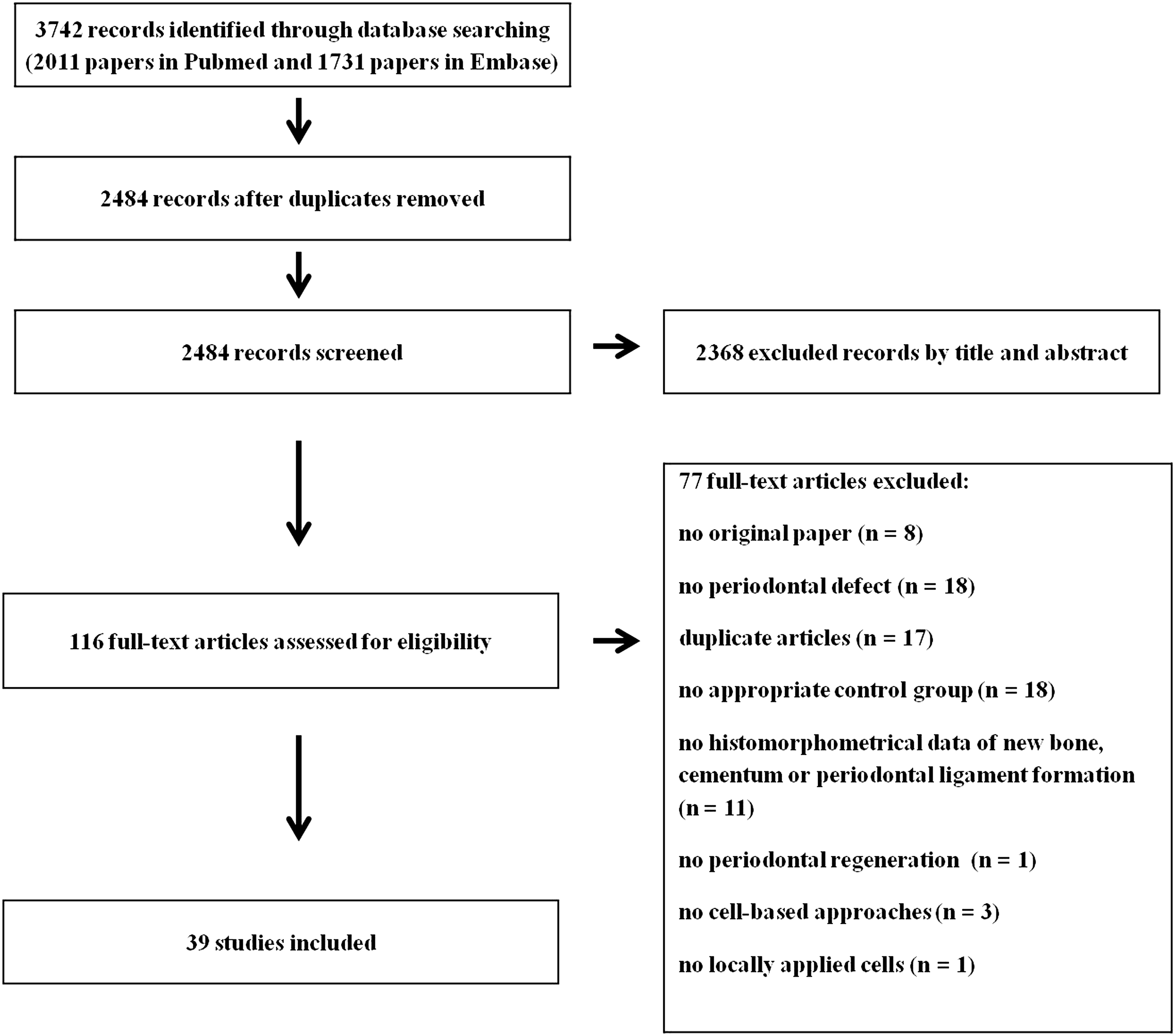
Flow chart of study selection. The number of studies in each phase is indicated between brackets.
The characteristics of included studies as shown in Table 2 varied substantially. Six different animal species were used: 21 studies were performed with dogs, 10 with rats, 5 with mini pigs, and 1 with rabbits, mice, or merino ewes, respectively. Fourteen studies used only male animals; 8 studies used only female animals; 1 study used both male and female animals; and 16 articles did not mention the sex of animals. A variety of defects were induced, including surgical dehiscence (2 studies), fenestration (11 studies), intrabony (7 studies), furcation (13 studies), and defects induced by chronic inflammation (6 studies). Twelve different cell types were evaluated in periodontal regeneration, including PDL-derived cells (19 studies), BMSCs (13 studies), gingival margin-derived cells (GMCs, 2 studies), alveolar bone cells (2 studies), stem cells isolated from deciduous teeth (1 study), induced pluripotent stem (iPS) cells (2 studies), periosteal cells (2 studies), DPCs (2 studies), cementum-derived cells (2 studies), periapical follicular stem cells (1 study), adipose-tissue derived stem cells (ASCs, 1 study), and follical cells (1 study). Also the passage number, cell concentrations, and number of applied cells per defect greatly varied between the studies, ranging from passage 1 to passage 7, from 4×10⁵ to 1.25×10⁸ cells/mL, and from 9×10⁴ to 2×10⁸ cells per defect, respectively. Moreover, multiple types of cell carriers or biomaterial scaffolds were used, including natural (e.g., collagen, platelet-rich plasma, etc.) and synthetic biomaterials. The included synthetic biomaterials were ceramics (e.g., hydroxyapatite-tricalcium phosphate), polymers (e.g., polyglycolic acid), and composites (e.g., apatite-coated silk).
Risk of bias and quality of reporting
Figure 2 shows the overall results of the risk of bias assessment of the 39 studies included in this systematic review. Regarding selection bias, 64.1% of the included studies reported randomization of the experimental units across treatment groups. However, only 23.1% of the studies mentioned the method of randomization used, so that the adequacy of the method could be judged. 76.9% of the articles described that intervention and control groups were similar at the start of the experiment. With regard to the selection bias item “allocation concealment,” only 7.7% described whether or not the allocation to the different groups during the randomization process was concealed. In addition, 64.1% and 97.4% of the included studies were scored as unclear risk of bias with regard to performance bias items “random housing” and “blinding,” respectively. For the detection bias item “random outcome assessment,” 100% of the included studies were scored as low risk of bias, because the outcome of intervention and control groups were assessed at the same time points. 28.2% of the studies reported that the outcome assessment was blinded based on the item 7 of the RoB tool. For attrition bias, 51.3% of the studies were scored as low risk of bias, as they adequately addressed incomplete outcome data.
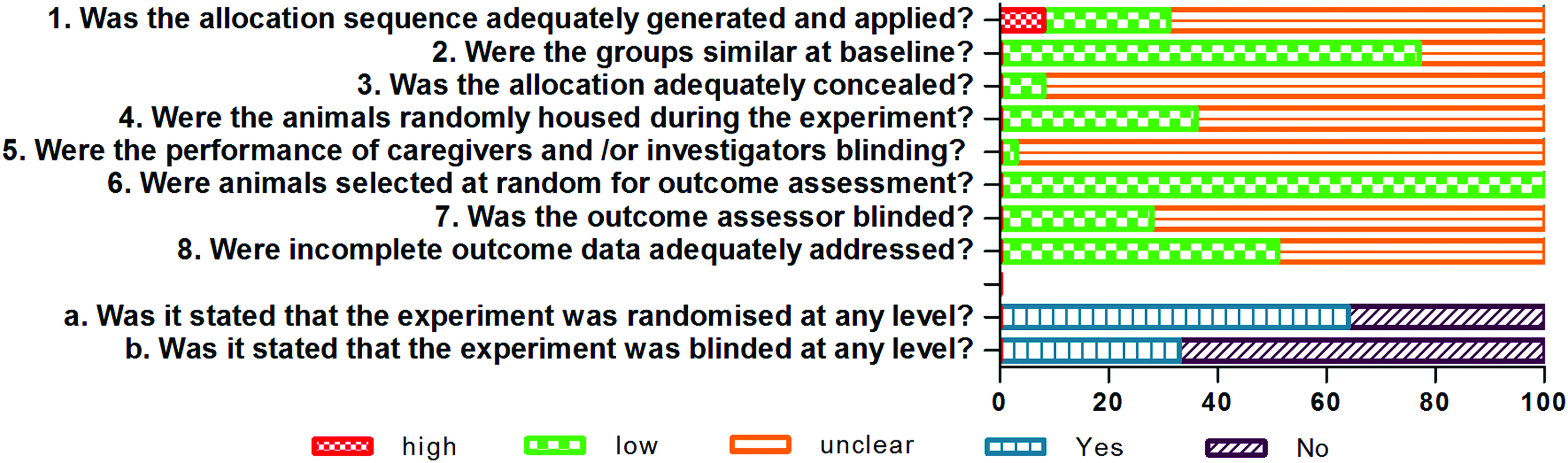
Risk of bias, averaged per item. Color images available online at www.liebertpub.com/teb
Meta-analysis of outcome measures
New bone formation
Thirty-eight studies of the included 39 studies evaluated the effect of cell-based strategies on new bone formation in periodontal regeneration, 37 of which could be included in the meta-analysis, as in one study the number of defects per group was not reported and could not be retrieved by contacting the authors. 38 The analysis contained 60 experiments or experimental groups, including data from 647 defects. In 30 experiments, the SMD and 95% CIs indicated that implantation of cells significantly increased new bone formation in the defect area (Fig. 3). None of the studies found a statistically significant negative effect of cell implantation on new bone formation. In the remaining 30 experiments, no statistically significant results were found. Overall analysis showed that cell-based strategies enhance new bone formation in periodontal regeneration, as displayed by the overall SMD and its 95% CIs (1.58 [1.19, 1.97]). However, overall study heterogeneity was moderate to high (I 2 =66%).
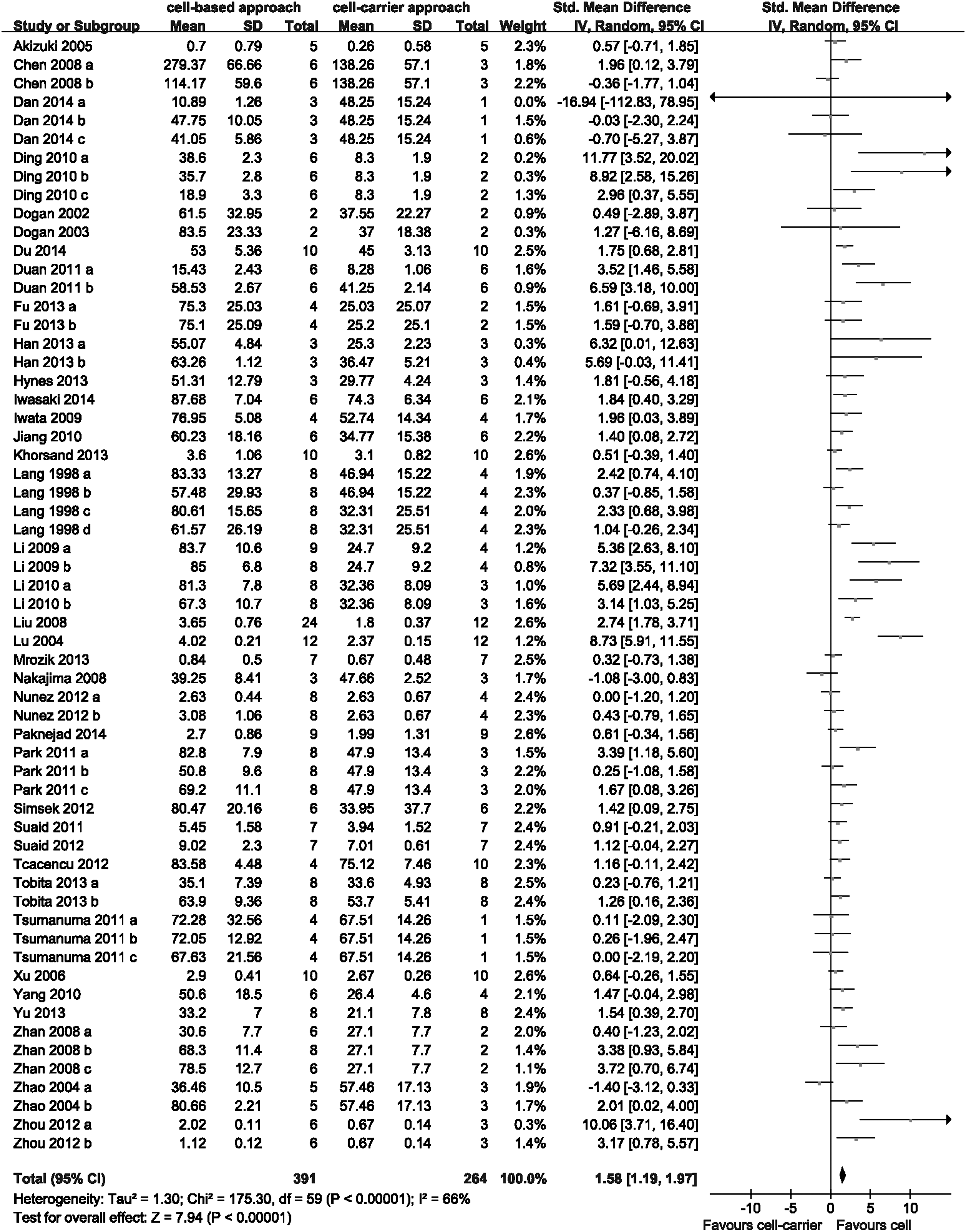
Forest plot of the included studies for outcome new bone formation. The forest plot displays relative weight of the individual experiments, the standardized mean difference (SMD), and 95% confidence intervals (CIs). The diamond indicates the global estimate and its 95% CI.
Subgroup analysis showed a beneficial effect of cells for all subgroups. No statistically significant differences between the subgroups were found for any of the examined variables (species, sex, cell type; Table 3). Two subgroups showed differences between SMDs larger than 1 SD (mini pig>rat, female>male). Only the results of the sub-subgroup analysis for sex was consistent with the trend found in the subgroup analysis. However, because of the small number of experiments in the subgroups and even more so in the sub-subgroups, these results must be interpreted with caution.
The effect estimate was displayed by the standardized mean difference (SMD) and 95% confidence intervals (CIs).
PDL, periodontal ligament.
New cementum formation
Twenty-three of the included studies evaluated the effect of cell-based strategies on new cementum formation, 22 of which could be included in the meta-analysis, as in one study the number of defects per group was not clear. 38 The analysis contained 33 experiments, including data from 344 defects. In 16 experiments, the SMD and 95% CIs indicated that implantation of cells significantly increased new cementum formation in the defect area (Fig. 4). In the remaining 17 experiments, no statistically significant results were found. None of the included studies showed a statistically significant negative effect of cell implantation on new cementum formation. Overall analysis showed that cell-based strategies enhance new cementum formation in periodontal regeneration, as displayed by the overall SMD and its 95% CIs (1.74 [1.13, 2.35]). However, overall study heterogeneity was moderate to high (I 2 =69%).
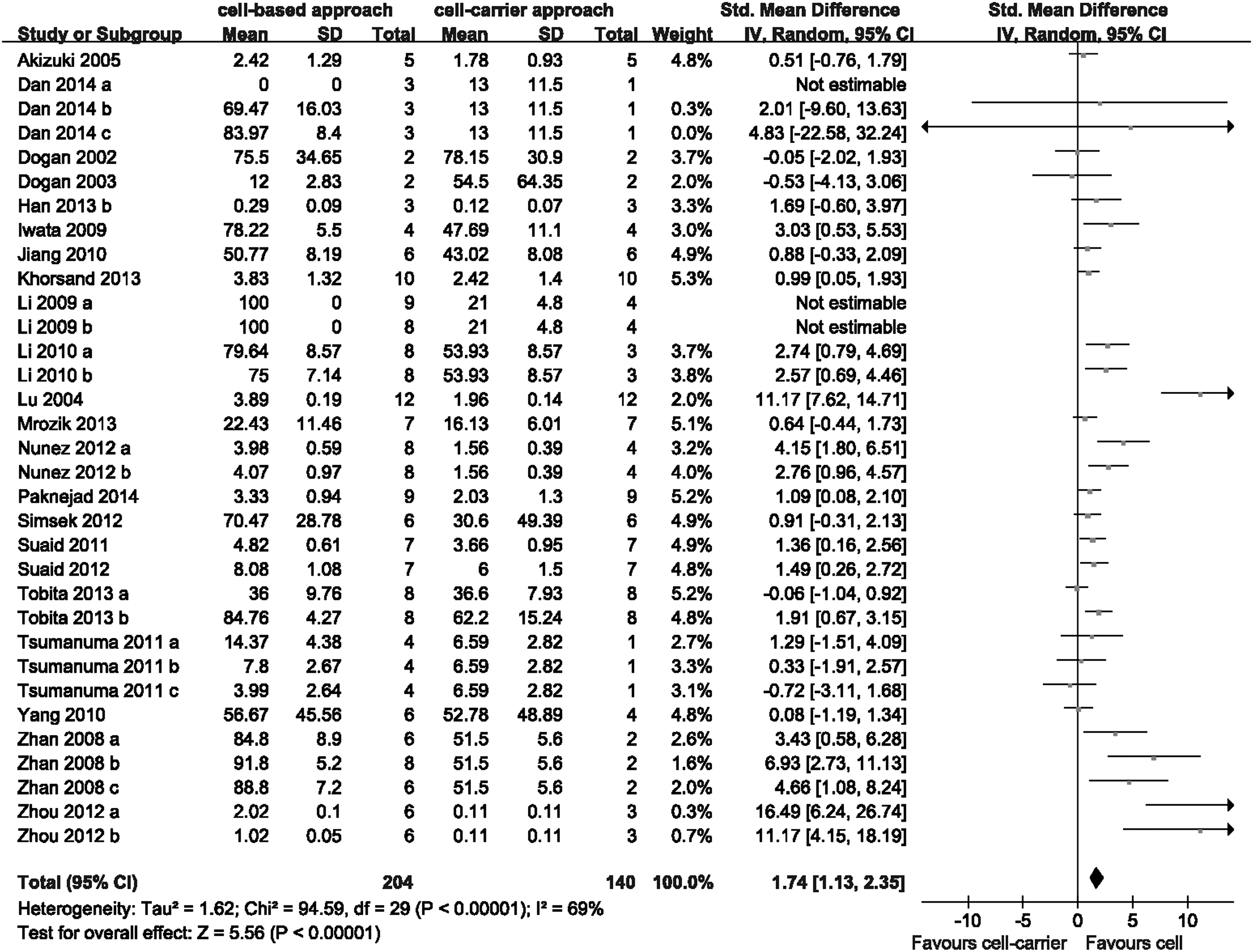
Forest plot of the included studies for outcome new cementum formation. The forest plot displays relative weight of the individual experiments, SMD, and 95% CIs. The diamond indicates the global estimate and its 95% CI.
Subgroup analysis showed a beneficial effect of cells for all subgroups, except for rat and female. No statistically significant differences between the subgroups were found for any of the examined variables (species, sex, cell type; Table 4). Two subgroups showed differences between SMDs larger than 1 SD (dog>rat, male>female). However, no sub-subgroup analysis was performed because of the small number of experiments in the sub-subgroups.
The effect estimate was displayed by the SMD and 95% CIs.
New PDL formation
Nineteen of the included studies evaluated the effect of cell-based strategies on new PDL formation in periodontal regeneration. The analysis contained 29 experiments or experimental groups, including data from 318 defects. The SMD and 95% CIs from nine experiments indicated that implantation of cells significantly increased new PDL formation in the defect area (Fig. 5). One experiment found a statistically significant negative effect of cell implantation on new PDL formation. 57 In the remaining 19 experiments, no statistically significant results were found. Overall analysis showed that cell-based strategies enhance new PDL formation in periodontal regeneration, as displayed by the global estimate SMD and its 95% CIs (1.25 [0.64, 1.86]). However, overall study heterogeneity was moderate to high (I 2 =71%).
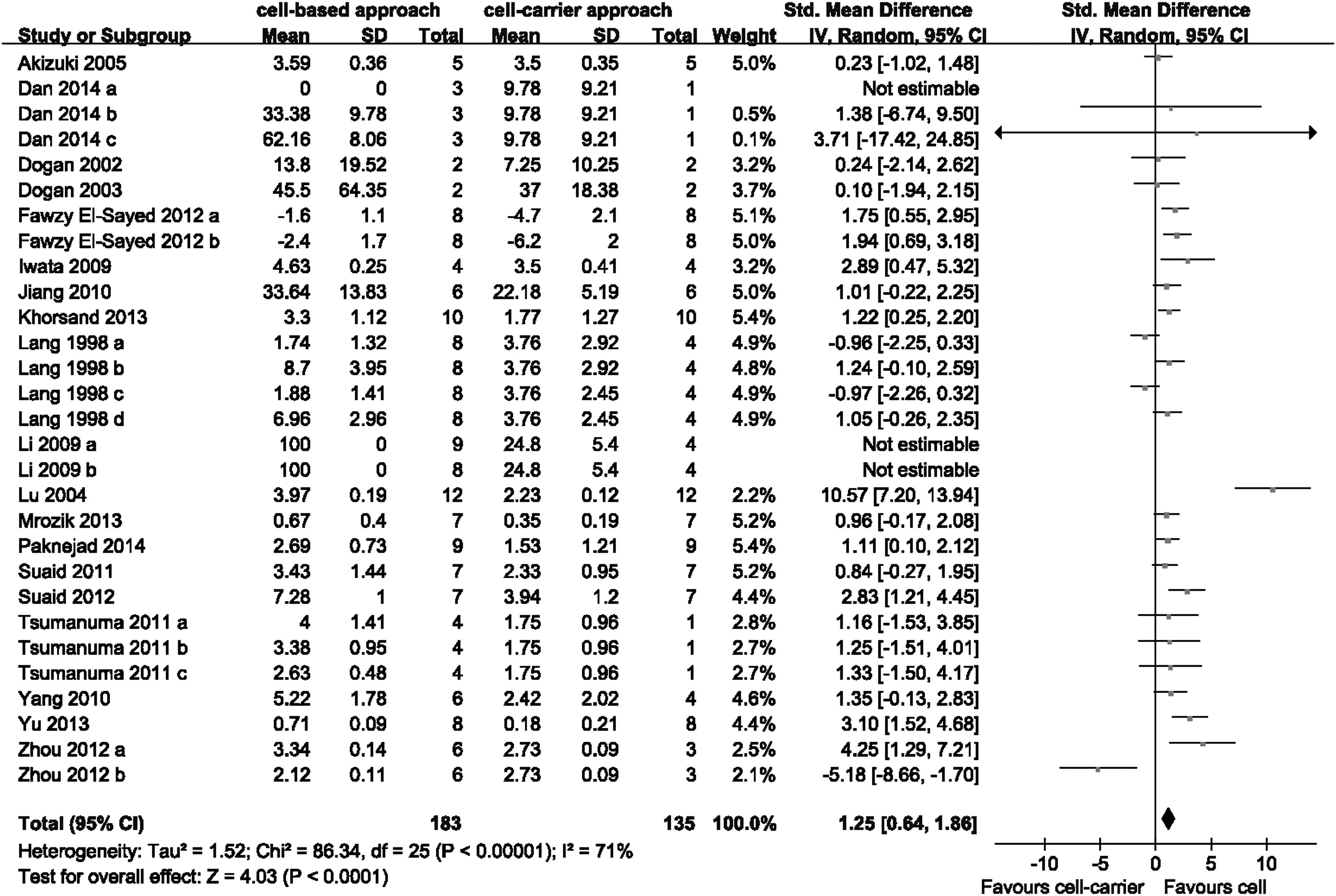
Forest plot of the included studies for outcome new periodontal ligament formation. The forest plot displays relative weight of the individual experiments, SMD, and 95% CIs. The diamond indicates the global estimate and its 95% CI.
Subgroup analysis showed a beneficial effect of cells for all subgroups, except for mini pig, female and BMSC. No statistically significant differences between the subgroups were found for any of the examined variables (species, sex, cell type; Table 5). One subgroup showed a difference between SMDs larger than 1 SD (rat>mini pig). However, no sub-subgroup analysis was performed because of the small number of experiments in the sub-subgroups.
The effect estimate was displayed by the SMD and 95% CIs.
Publication bias and sensitivity analysis
The presence of publication bias was assessed for all outcome measures by visual analysis of funnel plots. As shown in Figure 6, the largest studies are plotted near the average (the dashed line) for all outcome measures. Funnel plot for new bone (Fig. 6A) indicates that small, negative studies appeared to be underrepresented (within the red box), while funnel plots for new cementum (Fig. 6B) and new PDL (Fig. 6C) were quite symmetrical.
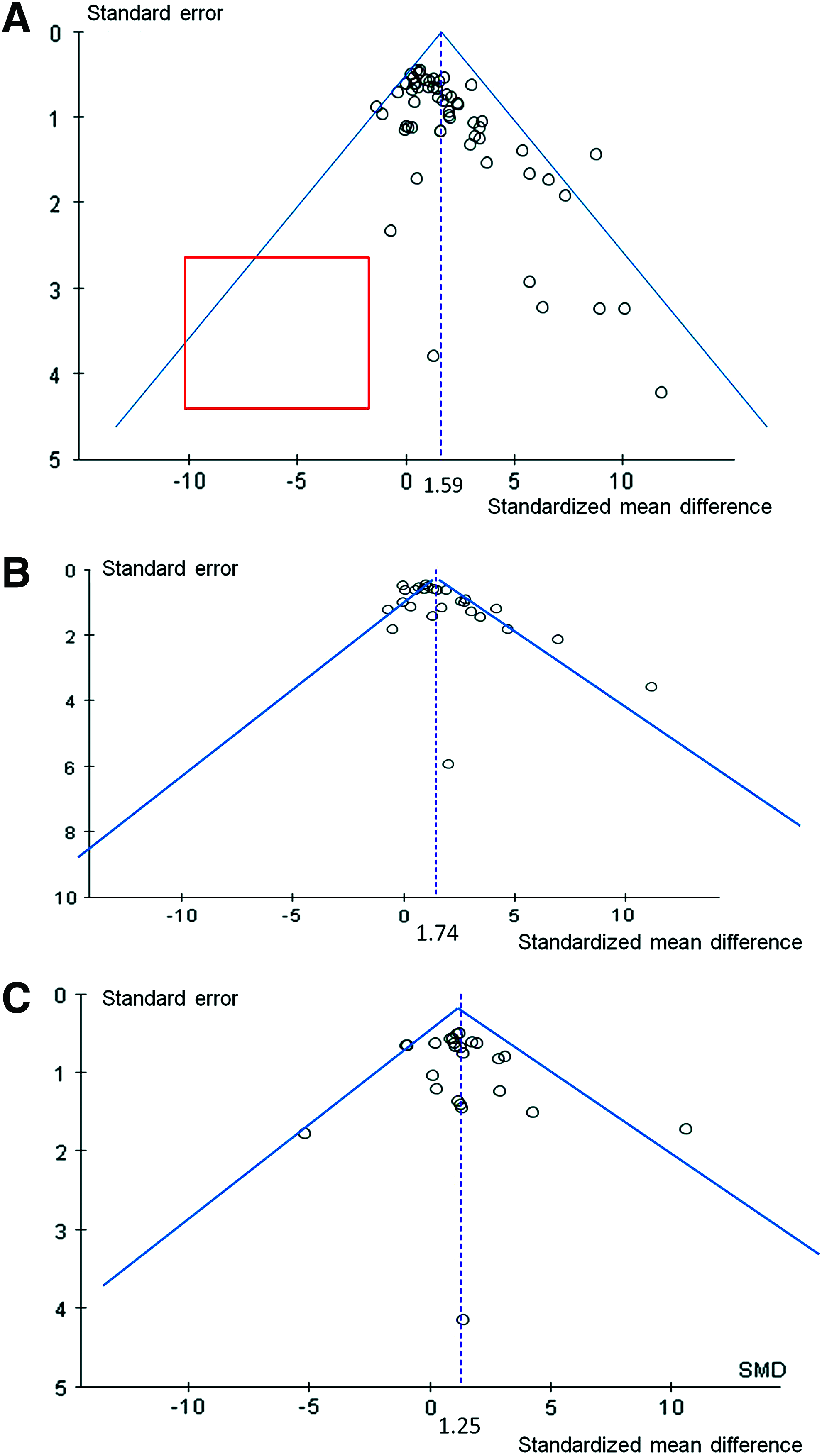
Funnel plot of the included studies for outcome new bone formation
To assess the robustness of our findings, sensitivity analysis was carried out by separately assessing the different units of measurement for expressing new bone formation (the percentage, length, or volume of newly formed bone). The effect sizes did not significantly differ between these groups (data not shown).
Discussion
The aim of this study was to systematically evaluate the current evidence for the efficacy of cell-based approaches for periodontal regeneration in animal studies. The results of this systematic review and meta-analysis revealed that cell-based approaches have a favorable effect on periodontal tissue formation, as displayed by the positive effect of cell-based approaches on new bone, cementum, and PDL formation. Moreover, subgroup analysis showed a favorable effect on PDL formation of PDL-derived cells, but not of BMSC. However, meta-analysis did not show a statistically significant difference in effect between PDL-derived cells and BMSC.
The currently used approach of systematic review allowed the inclusion of a large number of studies, which enabled us to perform the meta-analysis and explore the effect of several subgroup variables. However, there are some potential limitations related to this approach. First, preferably, all experiments should be performed in a similar manner when their results are being combined in a meta-analysis. However, the publications display experimental variability for the utilized animal species and sex, the periodontal defect type and size, the used cell types and passage number, the number of cells per defect, the biomaterials applied as cell carrier, and the healing time after cell transplantation. Not surprisingly, substantial statistical heterogeneity was found, which might be explained in part by experimental variability.58,59 To account for the expected heterogeneity, a random effects model was used. Moreover, subgroup analyses (animal species, sex, and cell types) were performed in an attempt to explain this heterogeneity, but these subgroup analyses did not notably reduce the heterogeneity. Second, the results of the meta-analysis may be subject to publication bias, as visual analysis of funnel plots revealed that small negative studies appeared to be underrepresented. The true effect of cell-based approaches may hence be smaller than the effect found in our meta-analysis. Importantly, funnel plot asymmetry can either result from non-publication of negative results, or be caused by other factors, such as true study heterogeneity, or differences in study quality. 60 Third, the study quality assessment (assessment of risk of bias) revealed that poor reporting of animal studies in scientific publications is of serious concern. Key measures to avoid bias, such as randomization and blinding, were infrequently reported. This seriously hampers drawing reliable conclusions from animal studies. For example, only 23.1% of the studies provided sufficient details to judge the adequacy of the method of randomization and only 28.2% of the studies reported that the outcome assessment was blinded. If the methods of randomization used in the other studies were inadequate and if the other studies did not actually blind the outcome assessment, there is a substantial risk that the effects of cell-based approaches in periodontal regeneration have been overestimated. To generate reliable and unbiased data, it is suggested that the standards of animal experiment reporting should be more like the standards routinely applied in human randomized controlled trials. 61 Despite these limitations, the combined analysis of the included studies still generated extra and valuable information that could not be derived from individual studies.
To assess the influence of several variables on cell-based strategy efficacy, subgroup analyses (animal species, sex, and cell types) were performed. No statistically significant differences between the subgroups were found for any of the examined variables. As shown in this systematic review, dogs, rats, and mini pigs are the mostly used animal species for preclinical trials and hence subgroup analyses of these species were performed. Some subgroups did show differences between SMDs larger than 1 SD, for example, mini pig>rat in new bone formation, dog>rat in new cementum formation, and rat>mini pig in new PDL formation. However, the sub-subgroup analysis for animal species was either inconsistent with the trend found in the subgroup analysis or could not be performed because of the small number of experiments in the sub-subgroups. For the subgroup analyses of sex, we also observed that some subgroups showed differences between SMDs larger than 1 SD (female>male in new bone formation and male>female in new cementum formation). And the results of the sub-subgroup analysis for sex in new bone formation were consistent with the trend found in the subgroup analysis. However, because of the small number of experiments in the subgroups and even more so in the sub-subgroups, these results must be interpreted with caution and cannot be used for drawing any final conclusions. Still, it might be interesting to study the effect of sex on periodontal regeneration in future experiments.
One of the most important issues for clinical application of cell-based approaches is the type of cell used. As shown in this systematic review, PDL-derived cells and BMSCs are the mostly used cell types for preclinical trials and hence subgroup analyses of these two cell types were performed. Previous studies compared the regenerative potential between PDL-derived cells and BMSCs, and the results suggested that PDL-derived cells are the more suitable cell population for periodontal regeneration, as significantly more well-oriented periodontal PDL fibers, newly formed bone, and cementum were observed upon transplantation of PDL-derived cells.12,13 A recent systematic review on PDL-derived cells for periodontal regeneration reported that 12 out of the 17 included studies demonstrated a statistically significant improvement in periodontal regeneration. 62 However, no meta-analysis was performed and no information of BMSC in periodontal regeneration was provided in this systematic review. 62 In our systematic review, 12 out of 23 included studies reported a statistically significant positive effect of PDL-derived cells on new bone formation. For BMSCs, 10 out of 18 included studies demonstrated a statistically significant improvement in new bone formation. The subgroup meta-analysis revealed that PDL-derived cells and BMSCs had a similar effect on the enhancement of bone and cementum formation. However, only PDL-derived cells but not BMSCs showed a favorable effect on new PDL formation, although there is no significant difference between these two subgroups. The enhancement of PDL-derived cells in PDL regeneration is probably caused by the fact that PDL-derived cells contain several subpopulations of cells, including osteoblasts, fibroblasts, and cementoblasts, and the combined subpopulations of cells can simultaneously synthesize both hard and soft periodontal tissues.30,63,64 Our results indicate that PDL-derived cells may provide a promising treatment to regenerate periodontal tissues including bone, cementum, and PDL. However, for the implementation of cell-based approaches in clinical practice, either PDL-derived cells or BMSCs can be applied based on the specific condition of each individual patient (e.g., accessibility of cells and health condition of donor tissues).
The meta-analysis provided evidence for the enhancement of periodontal regeneration by implantation of either PDL-derived cells or BMSCs. It has to be emphasized that treatment of periodontal defects requires a large number of cells (9×10⁴ to 2×10⁸ cells are needed for one defect), which sometimes would be difficult to obtain from a single human subject. Although the in vitro expansion of stem/progenitor cells is necessary, those cells typically reduce their ability to self-renew and proliferate during passaging. 7 Therefore, other sources of stem/progenitor cell types need to be sought. Among the evaluated 12 different cell types, GMCs, iPS cells, and ASCs are potential alternative sources. GMCs, which can be readily obtained during oral surgery, have mesenchymal stem cell properties and can differentiate into osteogenic phenotype under certain inductive conditions.32,65 In addition, iPS cells, obtained after transfection of certain stem cell-related genes into adult somatic cells, have the ability to differentiate into a variety of cell types. 7 Moreover, ASCs can be recovered easily in large numbers by liposuction under local anesthesia and show large similarity with BMSCs regarding gene expression and osteogenic capacity. 66 Regarding these three cell types, however, only few studies were included in this systematic review (two, two, and one for GMCs, iPS cells, and ASCs, respectively). Both iPS cells17,31 and ASCs 52 have given promising results in periodontal regeneration compared to cell-carrier only groups, while GMCs18,19 did not promote any significant periodontal regeneration. Nevertheless, to further ensure the efficacy of the aforementioned cell types in periodontal regeneration, additional studies are required in future experimental work.
Cell passage number is another important factor for clinical application of cell-based approaches. For instance, primary PDL-derived cells used at early passages have the advantage of maintaining the rich phenotypic and functional heterogeneity of fibroblasts characteristic of the original tissue. 67 Nevertheless, characteristic changes of primary cells have been observed during passage. Alkaline phosphatase activity of PDL-derived cells gradually decreased and the expression of tendo/ligamentogenesis-related genes was downregulated as the passage number increased. 68 However, in the current review, only less than half of the included studies (18 out of 39) provided information on exact cell passage number, which made it difficult to perform subgroup analysis based on cell passage numbers. Therefore, it is highly recommended that the passage number of implanted cells is clearly reported in future studies.
Animal studies included in this systematic review indicate that cell-based approaches are effective for periodontal regeneration, providing some support for the implementation of cell therapy in clinical practice as a routine treatment in the future. Nevertheless, the longest evaluation time within the included studies was 12 weeks. Therefore, long-term in vivo studies are required to ensure preclinical safety. Moreover, various animal models were utilized in the included studies. An optimal preclinical animal model plays a crucial role in the evaluation of cell-based strategies for periodontal regeneration. Convenience and biologic similarity of the periodontium to that of humans are important considerations for the selection of an appropriate animal model. For animal species, six different animal species were utilized in the included studies and the most frequently used species were dogs (21 studies) and rats (10 studies). In general, defect size characteristics and biologic similarities favor the selection of large animals in periodontal regeneration before moving to clinical trials. In a study that compared bone composition, density, and quality in bone samples derived from seven vertebrates (human, dog, pig, cow, sheep, chicken, and rat), rat bone was most different whereas canine bone best resembled human bone. 69 The more commonly used animal models in periodontology have been canine and periodontal disease in rats has not been found to be as closely related to the human varieties. 70 However, due to ethical issues, the use of large animals should be reserved for the last phase of validation of a new treatment. Rodents are inexpensive and easy to house, and are often used as a starting point for preliminary screenings, followed by verification in large animals. 69
To create periodontal defects, either chronic 9 or acute 11 periodontal defect models have been utilized. In chronic defect models, formation of natural periodontitis lesions can be induced by placing wires, ligatures, or impression material around the teeth. Although the clinical, histopathological, and microbiological features of chronic models were similar to those in human periodontitis, the onset of the lesions is difficult to predict and the defect area is difficult to be standardized. 71 In contrast, the acute defect models can be surgically created including surgical dehiscence, fenestration, intrabony, and furcation defects, to mimic the defect morphology in humans and meet different experimental purposes. In this systematic review, periodontal defects were mostly surgically generated (33 studies) and only few studies used chronic inflammation (6 studies). To combine the advantages of chronic and acute models, periodontal defects have been created in an acute-chronic way. For instance, Yan et al. created an acute-chronic Class III furcation defect in beagle dogs by placing a cotton ball saturated with anaerobic bacteria in surgical defect to cause inflammation. 72 Still, acute-chronic animal models cannot mimic some fundamental features including genetic background and risk factors (e.g., aggressive bacterial flora, tobacco, systemic diseases of host, etc.). 6 Consequently, these features can only be evaluated in future clinical trials.
Conclusion
The current systematic review and meta-analysis indicates that, based on animal experimental work involving periodontal defect models, cell-based therapies have a favorable effect on periodontal regeneration compared to cell-carrier only therapies. Moreover, subgroup analysis showed a favorable effect on PDL formation of PDL-derived cells, but not of BMSC. However, meta-analysis did not show a statistically significant difference in effect between PDL-derived cells and BMSCs. These results provide important information for the implementation of cell-based approaches in clinical practice as a routine treatment in the future.
Footnotes
Acknowledgments
The authors thank the Royal Netherlands Academy of Arts and Sciences (KNAW; project number PSA 08-PSA-M-02) and China Scholarship Council (No. 2010622061).
Disclosure Statement
The authors report no conflicts of interest related to this study.