
Abstract
In the field of tissue engineering, there is a need for methods that allow assessing the performance of tissue-engineered constructs noninvasively in vitro and in vivo. To date, histological analysis is the golden standard to retrieve information on tissue growth, cellular distribution, and cell fate on tissue-engineered constructs after in vitro cell culture or on explanted specimens after in vivo applications. Yet, many advances have been made to optimize imaging techniques for monitoring tissue-engineered constructs with a sub-mm or μm resolution. Many imaging modalities have first been developed for clinical applications, in which a high penetration depth has been often more important than lateral resolution. In this study, we have reviewed the current state of the art in several imaging approaches that have shown to be promising in monitoring cell fate and tissue growth upon in vitro culture. Depending on the aimed tissue type and scaffold properties, some imaging methods are more applicable than others. Optical methods are mostly suited for transparent materials such as hydrogels, whereas magnetic resonance-based methods are mostly applied to obtain contrast between hard and soft tissues regardless of their transparency. Overall, this review shows that the field of imaging in scaffold-based tissue engineering is developing at a fast pace and has the potential to overcome the limitations of destructive endpoint analysis.
Introduction
T
Data obtained by real-time nondestructive methods often lack spatiotemporal information since these methods treat a tissue-engineered construct as a bulk material. 2 Therefore, there is a growing interest in novel methods to be able to nondestructively, and ideally noninvasively, assess the performance of tissue-engineered constructs with a sufficient spatiotemporal resolution upon in vitro culture before implantation.3–5 By gaining more insights in cell fate and tissue growth during culture, higher efficiencies and faster procedures could be realized.
Tissue engineering approaches can be generally divided into two main classes, which consist of (1) cell-based approaches without the presence of any supporting material and (2) combinatorial approaches in which cells are introduced into biological or synthetic materials supporting proliferation or differentiation. In this review, we will outline current methodologies and challenges with respect to the characterization of this second class of combinatorial scaffold-based tissue engineering approaches. We will specifically focus on the applicability of imaging modalities to assess cell fate when cultured on scaffolds fabricated of solid materials for musculoskeletal tissue engineering approaches.
Recent reviews have been published on studies that have addressed the importance of nondestructive assessment of tissue constructs' quality by the application of imaging methods.6–10 However, the majority of these studies focus on scaffolds' geometry characterization before cell seeding in vitro or cell labeling and tracking after implantation in vivo. 11 These in vitro and in vivo studies unfortunately do not all address other challenges faced when monitoring the scaffolds combined with cells in vitro.
The limited penetration depth for most microscopy techniques forms one of these major constraints. 12 Conventional imaging techniques such as laser confocal microscopy or transillumination microscopy require the use of thin samples or suffer from spatial resolution, respectively, to study the cell–scaffold interaction, cellular distribution, and proliferation during scaffold population. 12 So, the applicability of imaging modalities highly depends on the type of scaffold used and the biological components to be detected.
First, we will highlight some of the most used scaffold materials and fabrication methods and their limitations with respect to monitoring tissue growth, cell fate, and/or cell metabolism. Second, we will summarize conventional methods for tissue growth, cell fate, and cell metabolism assessment. Subsequently, imaging modalities, which are potential candidates to monitor the previously mentioned parameters, will be introduced.
Scaffold-Based Tissue Engineering
In scaffold-based tissue engineering, biological or synthetic materials are processed into three-dimensional (3D) constructs or particles that enable cell growth within the final structure. Depending on the nature of these materials, the constructs can be hydrogel based or comprise solids. Hydrogels mostly retain optical transparency, whereas many solid scaffolding materials can be autofluorescent, semitransparent, or opaque. These differences in optical properties obviously result in different challenges faced when monitoring cell fate, tissue growth, and cell metabolism by imaging modalities. In this study, we will shortly outline two classes of conventional scaffold materials, followed by scaffold fabrication methods that have shown their relevance in musculoskeletal tissue engineering.
Scaffold materials
Natural materials
In many tissue engineering approaches, natural materials such as bioceramics, bioglasses, and biopolymers have gained interest because of their chemical properties, processability, and availability. 13 Biological materials can be divided in several classes, namely materials retrieved from inorganic materials or plants, from animal species (xenogeneic), or from allogeneic and autologous sources. Generally, when materials for scaffolds are retrieved from inorganic substances or a plant, a certain component is isolated and purified, after which, in most cases, the macrostructure will be lost.
Materials of interest vary from relatively brittle and stiff materials such as ceramics to relatively soft materials such as biopolymers, which can be processed into hydrogels. Some commonly used bioceramics such as hydroxyapatites find their potential in bone tissue engineering because of their known osteoconductive and/or osteoinductive properties.14–17 Biopolymers, which can be divided in polysaccharides, proteins, and polyesters, are also of interest in several tissue engineering approaches. 18 For example, in cartilage tissue engineering research, hydrogels comprising collagen, alginate, hyaluronic acid, or chitosan, among others, are widely applied as ECM-mimetic materials.19–26 Furthermore, silk, marine shells, and corals have been used as sources for biological materials in several tissue engineering approaches.27–32 Bioceramics are crystalline and therefore opaque materials, whereas bioglasses and biopolymers are semicrystalline or fully amorphous, permitting a certain degree of transillumination.
Decellularized ECM scaffolds from xenogeneic, allogeneic, or autologous sources are opted as promising nonimmunogenic materials for several tissue engineering approaches since their composition mimics the native tissues to a great extent.33–36 However, it is well known that the structure of the native ECM may be modified or even disrupted in the decellularization process.37,38 Therefore, in soft tissue engineering, these obtained ECM components are mostly processed as a hydrogel,39–42 which offers a matrix for cell encapsulation and permits cell fate monitoring by optical imaging modalities. In other works, some scaffold fabrication techniques such as electrospinning have been applied to regain cell-instructive structural elements of the native ECM after decellularization, which leads to the fabrication of nanofiber meshes mimicking the structure of native ECM.43–45
For musculoskeletal tissue engineering and especially bone tissue engineering, hard tissue replacements are favored, which will require sufficient mechanical stability and structural integrity to sustain the loading conditions in the implantation site. Therefore, combinatorial approaches of synthetic materials with decellularized ECM have been investigated to retain both the native tissue properties of the ECM components and the mechanical properties of the (synthetic) scaffold material.46–48 Several processes of decellularization and their tissue sources and applications are well reviewed elsewhere.49–52
Synthetic materials and composites
Synthetic materials represent a large class of widely used materials in tissue engineering approaches. Upon material synthesis, mechanical properties, surface chemistry, and degradability can be tailored. Furthermore, synthetic materials are generally easily processed into scaffolds by various methods, which will be outlined in the next section. Synthetic polymers have received considerable attention and are widely studied in cartilage and bone tissue engineering. 53 Natural polymers are often difficult to process and pathogenic risks may be involved with natural polymers retrieved from xenogeneic or allogeneic sources. Synthetic polymers exclude these drawbacks and exhibit tailorable, predictable, and reproducible physical, chemical, and degradation properties. 54 Depending on several parameters such as the degree of branching, hydrophilicity, and molecular weight of the polymer, synthetic polymers can be processed both as hydrogels and as solid materials. Some well-known polymers used for hydrogel fabrication are poly(acrylic acid), poly(ethylene oxide) (PEO), poly(vinyl alcohol), and polypeptides. 55 Examples of the most popular and most widely studied biodegradable synthetic polymers in bone tissue engineering are poly-L-(lactic acid) (PLA), poly(glycolic acid) (PGA), and their copolymers poly-(lactic-co-glycolic acid) (PLGA).53,56 Furthermore, other synthetic polymers have been investigated for cartilage and bone reconstruction such as poly-(ethylene glycol) (PEG)-based polymers,57,58 poly-ɛ-(caprolactone) (PCL), 59 polycarbonates, 60 polyfumarates, 61 polyanhydrides, 62 and poly-(ethylene oxide terephthalate)/poly-(butylene terephthalate) (PEOT/PBT) 63 copolymers or blends thereof.64,65
Even though many polymer materials have been synthesized and investigated, to date, no single polymer can meet all the requirements set for scaffolds in cartilage and bone tissue engineering. 64 All polymers have their own characteristics, benefits, and drawbacks. Composite materials often show improved characteristics compared with their individual components with a sufficient balance between strength and toughness. Taking into account the composition of native bone, in which both organic and inorganic components are found, makes composite materials a logical choice for bone tissue engineering scaffolds.53,66 Combinatorial approaches, in which synthetic materials are used together with biological materials, offer both a high degree of tailorability in mechanical properties and enhanced instructive properties due to their biological activity.67,68 These instructive properties can result in enhanced cell attachment, proliferation, and differentiation.18,48,69–73
Similar to decellularized matrices and biological materials, the optical properties of scaffolds made of synthetics are dependent on their crystallinity, autofluorescent properties, absorption coefficient, processed geometry or porosity, and scaffold dimensions.
Scaffold fabrication methods
In the past two decades, a library of scaffold fabrication methods for tissue engineering applications has been developed. For nearly all common scaffold-based applications, reviews on fabrication processes can be found.54,74 In a review from Weigel et al., several commonly used fabrication methods are outlined with examples of specific applications per method. 75 In the following section, we will shortly introduce not only some well-known methods but also some recently developed scaffold fabrication methods and their implications in imaging methods. A resulting example from each method is given in Figure 1.
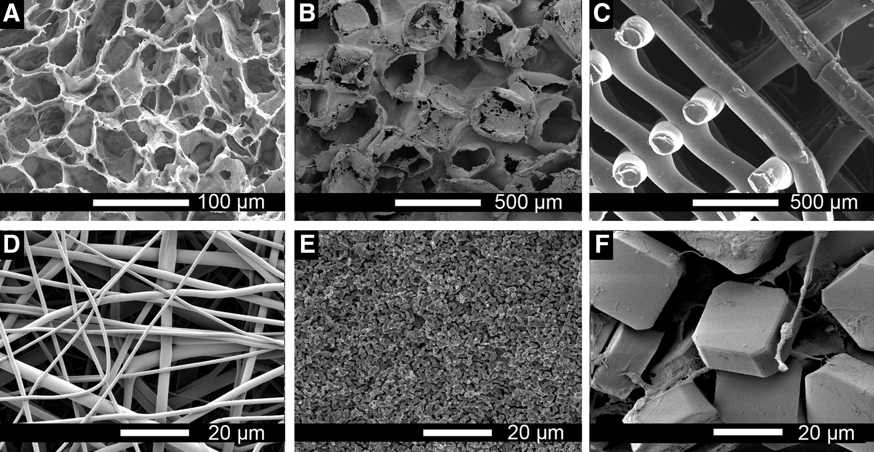
Resulting scaffold structures from various fabrication methods.
Phase separation methods
One of the earliest scaffold fabrication processes is based on phase separation principles (Fig. 1A). The methods and parameters in phase separation processes have been reviewed recently. 76 Highly porous scaffolds obtained by phase separation techniques find their use in a broad range of applications from soft scaffolds to study vascularization to sponge-like constructs for cartilage regeneration and stiff scaffolds for bone tissue engineering.77–80 The porosities obtained by these fabrication methods vary per polymer type and concentration, but mostly range from ∼60% to 97.5%. 76
The morphology of the obtained pores is often torturous, irregular, and the pores generally show a large pore size distribution up to 10-fold within one construct. Generally, the pore sizes can range from submicrometer to a few hundred microns and are, besides the degree of interconnectivity, largely dependent on the applied chemicals and the process parameters. 76 One specific benefit of phase separation methods is the possibility to create longitudinal pore shapes 81 or to introduce pore size gradients within the scaffold. 82
The transparency of the material used will be a major determinant in the applicability of optical imaging techniques. Yet, even with transparent materials, the torturous geometry of the pores introduces many interfaces in the optical path, which will inevitable result in enhanced scatter and diffraction. The high porosity, and therewith low bulk density of these constructs, is expected to complicate imaging by nonoptical methods. Specifically, methods in which contrast is based on the material's nature with a spatial resolution limited to several microns will suffer from the limited thickness of the material layers that are separating the pores.
Particle leaching
Particle leaching methods result in 3D porous matrices, in which the pore sizes are controlled by the size of the particles (typically in the order of tens to hundreds of microns) and the porosity and interconnectivity are determined by the concentration of particles per scaffold volume (Fig. 1B).83–86 Particle-leached scaffolds are fabricated by casting a solution of a ceramic or polymer, for example, with solid particles.83,87 After the casted solution turned solid by evaporation of the solvent or by cooling down under the melting temperature, fully solid constructs are retrieved. Subsequently, the particles are leached out by immersing the full construct in a solvent for the particles, but a nonsolvent for the chosen polymer. This results in scaffolds with porosities in the range of 30% to 98%. 88 The most commonly used particles are sugar or salt based because of their good solubility in water.
The types of scaffolds retrieved with this method are besides many other applications also widely applied in musculoskeletal tissue engineering, membrane-like scaffolds for urological purposes, and in in vitro vascularization research. 89 The limitations in imaging these types of scaffolds are similar to those of scaffolds fabricated by phase separation methods. This similarity is caused by the comparable nature of the scaffold's geometry, introducing many thin layers per volume in highly porous scaffolds.
Additive manufacturing
Additive manufacturing techniques allow researchers to generate scaffolds with outer geometries based on computer-aided design (CAD).90–92 The field of additive manufacturing includes fused deposition modeling of thermoplastic materials,93–95 3D printing of pastes or liquids, 96 selective laser sintering (SLS),97–100 and stereolithography applied to photosensitive materials.101–104 Additive manufacturing techniques gained interest because of the capability to create scaffold geometries with well-defined parameters and high reproducibility (Fig. 1C). 74 This results in scaffolds with porosities and pore size virtually ranging in the whole possible spectrum. Typically, manufactured scaffolds with porosities from 30% to 95% and with pore size ranging from tens of microns to millimeters have been reported. 74 Another degree of freedom is enabled by the stepwise or layer-by-layer processing in additive manufacturing methods, which allows combining multiple materials into one construct.
Due to the often relatively large pores and the geometries introduced by these methods, many scaffolds only block the applicability of imaging methods to a certain extent, even if the materials are opaque. One can imagine that the regular structure of the construct enables observations through longitudinal pores. When transparent materials are used, the number of interfaces that optical light will have to cross is much lower than for scaffolds produced with phase separation or particle leaching methods. However, high resolution at greater imaging depths will still be limited, mainly due to the numerical aperture of the objectives that will be utilized.
Electrospinning
Electrospinning (ES) is a commonly used scaffold fabrication process to obtain nano- to microfiber meshes with high porosities, mimicking the collagenous morphology of native ECM (Fig. 1D).105–107 The method is based upon charging a solution of a chosen material and subsequent extrusion through a capillary tip or needle. A jet is drawn from the needle toward a collector due to the presence of an electric field. Varying process parameters such as electric field strength distance between needle and collector, polymer concentration, and extrusion speed allow tuning of the fiber diameter and morphology. Fabricated scaffolds are typically meshes or membranes with porosities varying from 80% to 99%, but with small pore sizes typically in the range of tens of nanometers till tens of micrometers. 106 Since the ES process is mostly applied to fabricate sheets or discs with limited thicknesses (the majority between 200 and 500 μm), no major additional implications are expected to play a role in the applicability of imaging methods compared with other fabrication methods.
Sintering
Sintering is mainly applied for hard tissue engineering and is based on the heat treatment of a powder to make the nano- or microparticles partially fuse with each other.108–110 Sintered scaffolds cover a wide range of porosities from 5% to 80% with pore size ranging from tens of microns to submillimeter. 88 Sintering is traditionally applied on ceramics, but it has also shown its potential for other materials such as metals, glasses, and certain polymers and composites (Fig. 1E).
Sintering can be applied locally with the use of lasers (SLS), which allows the fabrication of scaffolds with CAD geometries, as previously described in the section on rapid prototyping technologies. 97 When sintering is applied to obtain a homogeneously dense construct of ceramic materials, imaging possibilities will be restricted to nonoptical material penetrating methods, such as microcomputed tomography (CT) or magnetic resonance imaging (MRI). When amorphous materials are utilized, optical methods could be applied. However, the multiple interfaces of material with culture medium upon in vitro culture will limit the achievable resolution and imaging depths due to scatter and diffraction.
Bottom-up approaches
Bottom-up approaches have been recently introduced to overcome hurdles faced with mm-sized 3D scaffolds caused by limited access of the scaffold for surface or bulk modification and functionalization, as well as for nutrient availability. These hurdles include inhomogeneous cell distribution, 111 necrotic cores, 112 a limited remodeling capacity, and a limited control of cell fate. In bottom-up approaches, materials and cells are first combined at the cellular level and allowed to form so-called building blocks before assembling into larger clinical relevant-sized constructs. The formation and secondary assembly of these building blocks can be both cell and material guided. Bottom-up designs have shown the potential to construct tissues with defined properties, including spatial and temporal control at a cellular level.113,114 The porosities and pore sizes are determined by the physical properties of the individual units and by the assembling properties and can in theory be ranging from ∼10% to 90% and from nanometers to hundreds of micrometers, respectively. With the freedom in modulating the sizes and shapes of the building blocks, the most optimal porosity and pore size per application can be easily fabricated. 88 By creating micrometer-scaled building blocks from cells combined with biomaterials eventually comprising instructive capacities, complex tissues can be created and mechanisms of tissue development can be studied (Fig. 1F). 115 When using monodispersed spheres, compaction will be limited, introducing a high interconnectivity. 116
Bottom-up approaches based on gel-like materials allow the cells to reside in an ECM-mimicking matrix, retaining their rounded morphology.114,117 The use of hydrogels and engineered micro-objects has shown a high potential in injectable systems to reduce the invasiveness of surgical procedures in vivo and allow for controlled assembly to achieve complex tissues in vitro. The applicability of imaging methods to monitor cell fate and tissue growth in bottom-up engineered scaffolds is dependent on the optical properties of the materials used, the ratio of material versus tissue, and the density of the obtained 3D constructs.
Imaging Modalities
Imaging methods have gained growing interest over the past decades in the field of tissue engineering. The importance of noninvasive monitoring of cell and tissue behavior during culture, to be able to more rationally predict the success of the construct upon implantation, has been widely acknowledged. 118
Conventional destructive quality assessment procedures such as histological analysis are still the golden standard, mostly because of their high spatial resolution and specificity. Yet, for these analyses, the engineered construct is sacrificed, which constrains the ability to instantly and accurately adapt culture conditions. Nondestructive technologies can overcome this inefficient and costly approach. Imaging-based nondestructive technologies enable real-time determination of parameters such as the cell number and distribution and the tissue type, content, and distribution. With these insights and understanding of culture conditions, immediate optimization of culture parameters could be carried out, which ultimately results in faster and more efficient procedures.
Generally, imaging modalities require changes in interactions of electromagnetic or mechanical energy among various substances to be able to retrieve contrast between these substances (Fig. 2). These changes in interactions include changes in energy due to absorption, refraction, or scattering.119,120 Parameters such as imaging depth, contrast, and spatial resolution achieved by a given imaging modality are largely based on the type and frequency of energy employed. The benefits and drawbacks of several imaging methods in the context of monitoring in vitro tissue-engineered constructs are described in the following sections and summarized in Table 1. Additionally, to obtain an impression of the capabilities of imaging methods, an overview with examples of obtained imaging results is given in Figure 3. In this study, we will outline some commonly used and some more promising imaging modalities for assessing in vitro tissue engineering constructs.
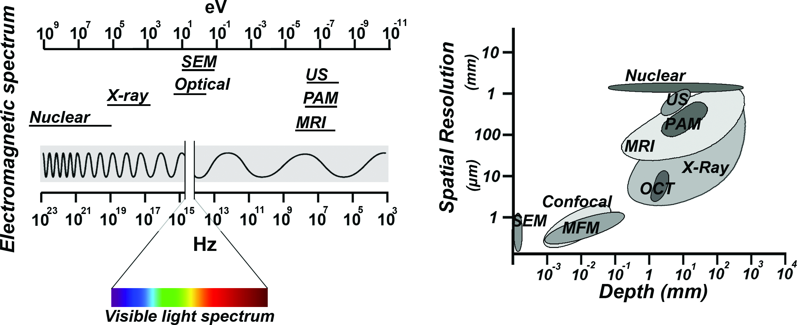
The electromagnetic properties of various imaging modalities (left). Spatial resolutions and imaging depths obtained by conventional imaging methods applied on tissue samples (right). Color images available online at www.liebertpub.com/teb
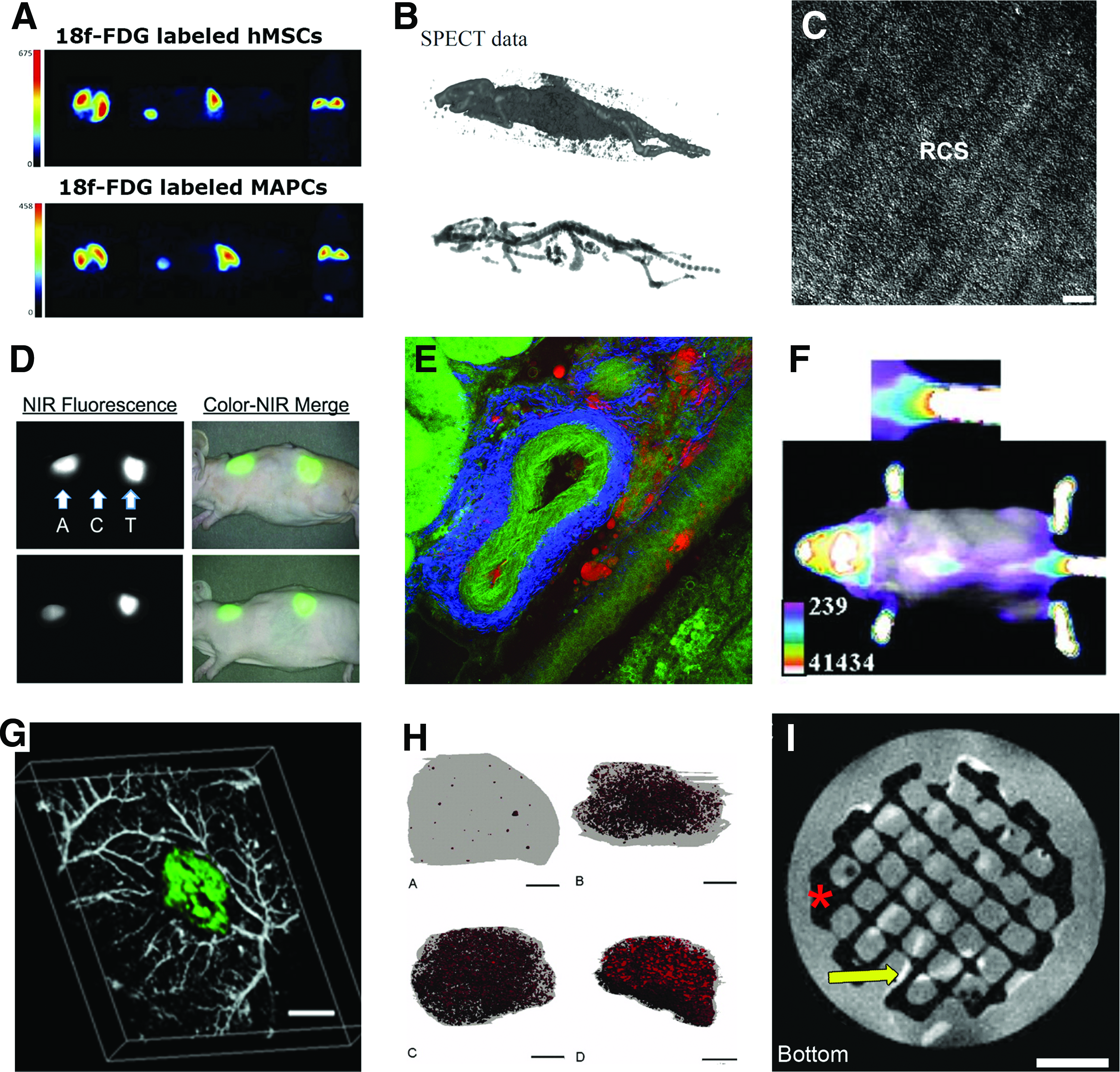
Examples of results of various imaging methods applied in vitro and in vivo for cell tracking or tissue or scaffold characterization.
The symbols indicate applicability and/or quality of the method in the given situation. These indications are based on the authors' interpretation of reported results.
—, not applicable/very low quality; –, limited applicable/low quality; ±, somewhat applicable/reasonable quality; +, applicable/good quality; ++, very applicable/very good quality. CT, computed tomography; DIC, differential interference contrast; MPM, multiphoton microscopy; MRI, magnetic resonance imaging; OCT, optical coherence tomography; PAM, photoacoustic microscopy; PAT, photoacoustic tomography; PET, positron emission tomography; SEM, scanning electron microscopy; SHG, second harmonic generation; SPECT, single-photon emission computed tomography; TEM, transmission electron microscopy; US, ultrasound.
Electron beam imaging
The two most widely used electron beam-based imaging methods in tissue engineering are scanning electron microscopy (SEM) and transmission electron microscopy (TEM). SEM can provide high-resolution images up to a few nanometers, but has limitations with respect to penetration depth. 121 In tissue engineering, SEM is mostly applied to assess scaffold quality after fabrication before cell culture or implantation and after culture or implantation. SEM is often referred to as destructive since conventional SEM requires dehydration and fixation of biological samples.9,122 However, there are SEM devices that have an environmental (ESEM) modality such as cryo-SEM, in which hydrated unfixed tissues can be studied.121,123–125 In a study of Doyle et al., cryo-SEM was applied to study the morphology of colonies of fibroblasts on PLA. 126 Yan et al. imaged cell growth of mouse osteoblast cells on plasma-treated PCL nanofibers in wet mode SEM. 127 This method can not only be applied on large mm-sized constructs but also ESEM will only reveal information from the exterior surface of the construct.
Despite the benefits ESEM can offer over conventional high-vacuum SEM, it is not extensively applied in the field of tissue engineering, whereas it is a commonly used imaging method in food industry and plant sciences. TEM requires sample fixation and processing into thin sections and is therefore considered a destructive endpoint imaging method. 128 Therefore, TEM has in our view no potential in noninvasive monitoring of tissue growth or cell fate in 3D constructs.
Nuclear imaging
Nuclear-based imaging techniques have shown potential in many in vivo applications such as visualization of lymphatic and vascular systems. 129 For in vitro applications, however, to date, nuclear-based methods are not yet commonly applied. The lateral resolution of nuclear imaging modalities is limited since most available devices are mainly optimized for human-scale use with a resolution of 1–2 mm. 130 To be able to achieve contrast in nuclear-based imaging methods such as single-photon emission computed tomography (SPECT) and positron emission tomography (PET), radioactive agents are required.
Although PET and SPECT cannot yet provide single-cell resolution, which would be desirable in monitoring cell fate in tissue-engineered constructs, both methods could still be valuable tools enabling cell cluster localization and cell function in vivo and in vitro. For example, by labeling cells with radioactive tracer molecules such as fluorine-18-radiolabeled fluorodeoxyglucose ( 18 F-FDG) or fluorine-18-radiolabeled 9-[4-fluoro-3-(hydroxymethyl)butyl]guanine ( 18 F-FHBG) for PET imaging, noninvasive cell tracking is aided (Fig. 3A).131,132 A drawback of 18 F-FDG is the high tracer elution rate from the cells, 133 which makes this tracer mainly applicable for short-time monitoring. 134 In addition, for SPECT, a library of tracers is developed, which can be targeted to specific cell surface receptors, metabolites, or genes.135–137 The addition of CT to PET or SPECT imaging has shown to allow precise anatomic coregistration of functional information with anatomy in small animals. 138 Recently, micro-SPECT was developed and compared with micro-CT to image a whole-mouse skeleton; however, anatomical atlas-based segmentation and image processing were required to be able to reconstruct 3D models of the detected skeleton (Fig. 3B). 139 Furthermore, applying tools such as pinhole collimators to improve SPECT resolution has the consequence that longer scanning times are required due to reduced photon sensitivity. 138 Overall, PET and SPECT have high potential in clinics to add functional information on to anatomical structures imaged by other imaging modalities. Yet, the applicability of nuclear-based imaging methods on in vitro tissue-engineered constructs with a sub-mm scale resolution is to date still limited.
Optical imaging
Optical imaging is based on systems that measure the interaction of light (∼10 up to 400 nm for ultraviolet, 400 nm up to 710 nm for visible light, and 710 nm to 1 mm for infrared light) with matter. These interactions can show differences in scatter, absorption, or luminescence, which depending on the imaging modality, can be optically visible or can be displayed after computing retrieved spectra into images.
Epi- and transillumination microscopy
In epi- and transillumination microscopy, visible light is projected onto a specimen and contrast or colors are dependent on absorption and reflection, scatter, and breaking of the light. Epi-illumination modalities such as stereomicroscopy are applied on opaque specimens, from which reflected light is captured revealing information from the surface of the specimens only. Commonly used transillumination microscopy-based imaging modalities are bright-field microscopy, phase-contrast microscopy, 140 and differential interference contrast (DIC) (Fig. 3C) 141 microscopy, for which the applicability is restricted to imaging two-dimensionally (2D) and in 3D to thin (<300 μm) and virtually transparent constructs. 9
Bright-field microscopy renders most detail of living cells invisible since there are no large differences in absorption by the structures inside cells. Phase-contrast microscopy is based on differences in refraction, which lead to a phase delay in some of the light causing contrast in the images. DIC imaging allows detailed microscopic examination of the matrix texture and cell orientation in the plane of the construct 141 and can be applied to study dynamics in living cells since this method is noninvasive and continuous. 142 For several types of rapid prototyped scaffolds with longitudinal pores cultured with cells in vitro, bright-field microscopy can still be applied to gain some insight in tissue growth in the early phase. However, when the formed tissue becomes denser, complete absorption of the light will inhibit further assessment with transillumination-based methods.
Fluorescence microscopy
Optical fluorescent imaging has developed into an important tool in biomedical research both in vitro and in vivo. Fluorescence microscopy allows for noninvasive cell and tissue monitoring, in which contrast is offered by differences in autofluorescent properties of tissue constructs due to endogenous fluorophores present in all tissues,143–145 endocytosis or transfection of fluorescent probes, 146 or targeted fluorescent exogenous probes.147,148 Depending on the fluorescence modality applied (e.g., epifluorescence, confocal fluorescence, multiphoton fluorescence) and the excitation wavelengths utilized, imaging depths between submicron up to whole-animal imaging in the cm scale can be achieved.149,150 The majority of studies on cell fate, in which fluorescence microscopy is applied in 3D, are focused on in vivo cancer metastasis, cell homing, and cell differentiation. 151
Martin et al. showed that combining bright-field microscopy with fluorescent microscopy on ex vivo slices of unstained bone scaffolds enables the quantification of formed bone matrix by autofluorescence detection. 144 Cowles et al. applied near-infrared (NIR) optical imaging with a bone-specific NIR-targeted probe to noninvasively study mineralization of tissue-engineered bone constructs over time in vivo. 152 By the application of NIR, imaging problems related to autofluorescence from the native tissue can be eliminated. 138 In another study, NIR fluorescent imaging was applied to monitor scaffold degradation in vivo. 153
For in vitro cell studies, de Mel et al. showed that biofunctionalized quantum dots have great potential for live monitoring of endothelial progenitor cells seeded in mm-sized polymeric scaffolds and cultured in a pulsatile flow bioreactor for vascular tissue engineering applications. 154 Although, whole-body imaging of mice was shown to be feasible, the spatial resolution with such imaging depths drastically decreased from submicron to mm scale, in which only clusters of cells were detectable.149,155 Recent studies have shown in vivo assessment of cell migration in living mice with a subcellular resolution.156,157 Although the applied methods are classified as noninvasive and label-free, the continuous expression of fluorescent proteins to monitor cellular migration over time with a subcellular resolution requires genetic modifications in the applied cell lines or animals. For more information on the current state of the art in live cell imaging in whole animals by the application of fluorescent proteins, one can refer to a review from Hoffman. 158 Most conventional fluorescent microscopes found in tissue engineering laboratories have limited penetration depths of both the excitation and emission wavelengths when applied on dense tissues such as cartilage and bone and on opaque materials.
Multiphoton microscopy and second harmonic generation
Fluorescence microscopy has long been used for visualizing cells and angiogenesis in scaffolds as described earlier. However, due to strong light scattering, especially in the presence of blood, conventional fluorescence microscopy shows poor tissue penetration of several hundred micrometers. Multiphoton microscopy (MPM) has shown to partly overcome this limitation and allows imaging two to three times deeper into specimens and enables optical sectioning into stacks for 3D reconstruction.159,160 For MPM, some specialized optical effects can be applied to enable label-free imaging of cells within scaffolds without the requirement of sample preparation. 12 Imaging with second harmonic generation (SHG) requires high photon densities and, to avoid tissue damage, short pulses of laser light, as provided by picosecond or femtosecond multiphoton lasers. 142
Conventionally available multiphoton lasers generated light with wavelengths ranging from 700 to nearly 1000 nm. The emission detection for SHG will be in the range of 350–500 nm when these lasers are applied. In biological specimens, SHG signals are obtained from proteins that contain a high amount of higher-order structures, for example, from alpha-helical structures, which are present in collagens and elastic fibers. 161 Because of its high resolution and penetration depth, yet with only moderate phototoxicity, this technique holds strong promise for future developments in live cell and matrix research exploiting nonlinear molecular characteristics of molecules and tissues.
Sun et al. have applied a combinatorial modality of MPM with SHG imaging to investigate the nonlinear optical properties of five commercially available scaffold materials: a collagen composite scaffold, a bone graft matrix strip (collagraft), an open-cell PLA scaffold, a PGA scaffold, and a nylon mesh. 162 They showed that MPM is effective in providing spectrally resolved morphological information of the materials and hypothesized (yet did not show) that this method can be used to study cell–matrix interactions when cells are cultured on these scaffolds. In other studies, MPM was applied on tissue-engineered constructs and was combined with SHG imaging to noninvasively retrieve insights on the interaction between scaffolds and tissues or between scaffolds and cells in vitro and in vivo.118,163,164 For example, in the study of Lee et al., ECM formation on PGA electrospun scaffolds was followed and showed to initially align along the scaffold fibers, while after inducing chondrogenesis for several weeks, scaffold reorganization was observed. 118
Pena et al. have also shown scaffold remodeling during culture, in this case, in collagen matrices seeded with fibroblasts. 164 Furthermore, combining SHG with MPM showed that among other structural parameters, the molecular orientation of the sample and the overall beta sheet content could be detected.145,165 Overall, combinatorial imaging modalities based on MPM and SHG provide information about materials present in tissue-engineered constructs, ECM components such as collagens, and cells simultaneously in separate channels.145,166 This allows for adequate (automated) image analysis of specific (labeled) components per channel. Ultimately, this enables novel approaches to directly modify culture parameters to induce or prevent certain cell and tissue fate-related processes.
Bioluminescence
Bioluminescence (BLI) uses light emission produced through enzymatic catalysis of, for example, luciferin by a luciferase enzyme. The principle has proven useful to monitor gene expression and cellular activities both in vitro and in small animal models in vivo.167–169 Several studies have shown the correlation between luciferase activity and the number of cells within tissues in vivo or scaffolds in vitro.170–173 Logeart-Avramoglou et al. have shown the quantification and characterization of luminescence from live cells and cell lysates after culture on polymeric translucent soft hydrogels and on opaque hard ceramics. 174 Olivo et al. reported a further correlation between luciferase activity, the number of cells, and bone formation in vivo. 175 In another study, osteogenesis was assessed by monitoring gene expression-dependent BLI by coupling of luciferase reporters to the promoter of osteocalcin.172,176–178 Because of its high sensitivity, BLI technology has recently proven its usefulness in tracking stem cells on material scaffolds transplanted in live animals for tissue engineering purposes. 178 One of the limitations of BLI is the limited resolution, which can range from tens of microns to millimeters. Furthermore, to the best of our knowledge, the current technology to image BLI does not allow for retrieving 3D imaging stacks of tissues and tissue constructs, thereby limiting the reconstruction modalities of 3D tissue development.
Optical coherence tomography
Optical coherence tomography (OCT) was first introduced by Huang et al. for the assessment of biological tissues. 179 OCT has been used for label-free imaging of tissue/scaffold constructs at a relatively high resolution in the submicron range. 9 Depending on the type of scaffold, it can be rather difficult to distinguish between the tissue and the scaffold when their refractive indices are similar.180,181 A light beam from a broad bandwidth light source is focused into a sample and the time that the light takes to return from the specimen to the detector is measured. This echo time-of-flight information enables the determination of the depth in the sample from where the light was scattered.120,179,182
OCT is applied in the field of tissue engineering to characterize the architecture of scaffolds, including porosity, pore distribution, and interconnectivity before cell culture. 183 In deep tissue imaging, OCT is one of the most used optical imaging modalities for its high spatial resolution of <15 μm in scattering matter at depths up to 2 mm.184,185 Wang et al. applied OCT to assess scaffold-assisted wound healing in mice and compared their results with conventional H&E staining. 186 Tissue development in vitro was monitored with OCT by detecting an increase in backscattered light over time. 187 In in vitro tissue engineering, OCT combined with Doppler velocimetry has been applied for characterization of flow in engineered tissues, such as artificial blood vessels, by increasing the obtained contrast compared with conventional OCT.188,189
In another study of Liang et al., three other combinatorial modalities have been introduced and evaluated for tissue engineering applications, namely OCT and multiphoton microscopy (OCM/MPM), optical coherence elastography (OCE), and spectroscopic OCT (SOCT). 181 OCM/MPM enables imaging at greater depths in highly scattering tissues by combining the spatial optical sectioning capabilities of confocal microscopy with coherence gating and rejection of multiply scattered photons within one modality, resulting in high sensitivity and high contrast. 181 This integrated system allows to obtain microstructural and functional properties of engineered tissues simultaneously and to display the data within one representation.
OCE reveals information on the biomechanical properties of tissues by applying mechanical stimulation to the material with simultaneous OCT detection. Dependent on the optical properties of the tissues, the main features of OCE can include millimeters of penetration depth and spatial resolution in the micron scale. SOCT is based on spectral analysis and intensity analysis of backscattered light from tissues. Similar to OCM, OCE, and conventional OCT, SOCT can obtain spatial resolutions in the micron scale. 190 In conventional OCT, no exogenous fluorophores are required since OCT relies on variations in indices of refraction and optical scattering for image contrast. 185 Therefore, OCT can be considered a noninvasive, label-free imaging modality, enabling cellular imaging within living specimens over time without loss of viability. 191
Raman microspectroscopy
Raman microspectroscopy is a label-free spectroscopic technique, which does not require special sample preparation and can be used for noninvasive characterization of cell and tissue biochemistry.192,193 Careful selection of suitable laser wavelengths and laser intensity can eliminate cell damage, allowing for the study of cells without inadvertently changing their phenotype or behavior caused by photo damage.194–196 Raman spectral studies have been performed for structural analysis of ECM components such as collagen 197 and proteoglycans.192,198 Raman peaks at distinct wavelengths exist, for example, for different types of carbon–carbon bonds, amide, carboxyl, sulfhydryl, and phenol groups. 120
In some relatively complex samples, Raman peaks could be characterized by entire molecules, such as carotenoids, glucose, and hydroxyapatite. 120 Spectral information retrieved by scanning specimens over a certain area, optionally confocal, can be computed and displayed as 3D cluster images. 199 Raman spectroscopy and Raman spectroscopy-based imaging have been successfully applied in several tissue engineering applications, for example, by monitoring chondrocyte behavior on bioactive scaffolds. 200 Boyd et al. have utilized a benchtop macro-Raman spectrometer with high-throughput screening capability to examine spectral differences between well-characterized cell lines (two types of osteosarcoma cells, human dermal fibroblasts and human embryonic lung epithelial cells) and the effects of cellular death. 201
Ultrasound
Ultrasound (US)-based imaging is in principal similar to OCT except that acoustic waves are used instead of NIR light. 182 Compared with OCT, clinical US has a lower resolution, but higher penetration depth. Although US imaging is widely applied in the clinics, not much research has been reported on applying US methods on in vitro cultured scaffolds. US-based monitoring methods have, similarly to X-ray-based micro-CT, OCT, and MRI, already been applied to nondestructively assess scaffold properties during and after fabrication.202,203
Mather et al. applied ultrasonic pulse-echo reflectometry to monitor changes in acoustic impedance of poly-(D,L-lactic acid)-based scaffolds during supercritical scaffold fabrication. 202 Kim et al. introduced US elasticity imaging on tissue-engineered constructs to monitor scaffold degradation in vitro and in vivo with an axial and lateral resolution of ∼250 and 500 μm, respectively. 203 Yet, both these methods did not provide sufficient spatiotemporal information on the scaffold structure to be able to draw conclusions on tissue development throughout a 3D construct.
Rice et al. opted to use US to monitor cartilaginous matrix development in chondrocyte-seeded PEG hydrogels in vitro. 204 Kreitz et al. have also evaluated the potential of US for quantitative in vitro monitoring of tissue development in a hydrogel-based 3D tissue-engineered construct. They showed a correlation between the gray-scale values of the obtained images with hydroxyproline content, which is a marker of collagen formation. 205
In a study of Fite et al., US backscatter microscopy (UBM) was combined with time-resolved fluorescence to follow the progression of tissue maturation along the chondrogenic lineage by monitoring collagen type-2 production and by detecting changes in mechanical properties of PLGA-based constructs upon in vitro culture. 206 UBM can provide structural information, respectively, with an axial and lateral resolution of ∼30 and 65 μm that can be correlated with tissue microstructure and construct stiffness, which can be a measure for the functionality of cartilage and bone-like tissue constructs. 206 Overall, the applicability of US-based imaging methods to monitor tissue growth in mm-sized tissue-engineered constructs highly depends on the aimed resolution and imaging depth, the targeted tissue type, and the sound transmission properties of these tissues and scaffold materials.
Photoacoustic tomography and photoacoustic microscopy
Photoacoustic tomography (PAT) is a relatively novel, but fast developing, clinically applied noninvasive and nonionizing method to monitor tissues in vivo and to detect, for example, blood oxygenation in situ.207–210 The method is based on the collection of ultrasonic waves resulting from heat expansion of tissues after absorption of laser irradiation within those tissues. 211 Large imaging depths up to ∼7 cm can be achieved since the light only has to travel in one direction and will result in a sound wave upon absorption. 212 In PAT and photoacoustic microscopy (PAM), the spatial resolution correlates with the imaging depth and depends on the application, yet a high depth-to-resolution ratio is maintained. 213
Recently, a lateral resolution of 5 μm with an imaging depth of 1 mm was obtained in highly scattering soft tissue. 214 By modulating the US detection frequency after laser irradiation, PAT enables high-resolution imaging of biological structures with strong optical absorption contrasts. 215 Although PAT and PAM are promising imaging methods for in vivo applications, to date, just a few researchers have investigated the potential of these methods for the field of tissue engineering. 130 Recently, PAM has been applied to retrieve structural information on tissue engineering constructs in vitro216,217 and in vivo.122,214
Cai et al. incorporated carbon nanotubes in PLGA scaffolds to be able to obtain detectable contrast between the polymeric scaffold and surrounding tissue. 216 Other approaches have introduced NIR fluorescent proteins in vivo to enable multicontrast next to already present endogenous contrast agents such as hemoglobin. 211 Such contrast agents could also be applied to cells in vitro in tissue-engineered constructs. To be able to extract more information from tissue-engineered constructs, multimodal imaging techniques can be applied. PAM could, for example, be combined with US-based imaging as was already done in vivo. 218
PAM has some limitations when imaging bony or air-filled tissues caused by limited transparency and the absence of a US wave transporting medium. 214 Despite these two major limitations, we still consider PAT and PAM to be promising in analyzing biomaterial–tissue interactions in soft tissue engineering applications in a completely noninvasive and nonionizing manner.
X-ray-based micro-CT
Previously, X-rays were clinically applied as a fast diagnostic tool to obtain direct 3D whole-body projections on 2D photosensitive films. Currently, X-ray imaging combined with CT can be applied in scanning mode at distinct focal planes and with varying projection directions, enabling 3D reconstruction of the information.119,219,220 Depending on the spot size of the generated X-ray, the beam properties, and mostly the detector properties, nanometer scale resolution has been achieved. 220
One of these CT modalities is micro-CT, which enables tissue engineers and material scientists to evaluate the morphological structures of their dry materials and fabricated scaffolds nondestructively with high resolution and accuracy.221–223 However, when polymeric scaffolds are immersed in physiological fluids in vitro, or embedded in vivo, wherein fluids perfuse through the scaffolds, the contrast of micro-CT images has shown to be poor. 216
Similar to MRI, micro-CT is also capable of distinguishing soft tissue material from harder mineralized tissues.216,224 However, instead of contrast resulting from changes in proton dynamics, X-ray contrast is the result from differences in absorption, refraction, and/or scattering properties of the materials. Bone, fibrous tissue, and ceramic scaffolds present different coefficients of X-ray absorption, therefore their 3D structures can be separated and corresponding quantitative data such as bone volume, thickness, growth, destruction, remodeling, and changes in bone density can be obtained.223,225 However, contrast between distinct types of soft tissue with similar X-ray attenuation is limited 226 and requires high cell densities. 224
One approach to improve contrast between various soft tissues is by the application of contrast agents. 11 Tissue engineering studies that do not assess mineralization often require those toxic contrast agents or sample processing before imaging. Unfortunately, most stained scaffolds are no longer usable for culture due to high toxicity of the applied contrast agents. Another approach to improve contrast is by combining micro-CT with, for example, X-ray phase contrast. When these contrast-related challenges can be overcome, monitoring of soft tissues and live cells during scaffold culture can be a major future application.6,227,228 Zhu et al. have shown the application of X-ray diffraction-enhanced imaging (DEI) on the characterization of rapid prototyped scaffold geometry, chitosan scaffold structure, and on muscle tissue morphology. 228 With the latter, they showed the outstanding capacity of DEI to better image low-contrast soft tissues compared with both radiography and in-line phase-contrast imaging.
Magnetic resonance imaging
Applications of MRI are often described when methods for 3D noninvasive imaging of nontransparent biological materials in the mm scale with relatively high resolution are reviewed.4,119,229 MRI can be applied either label-free or with targeted magnetic beads, for example, to be able to track or localize specific components or cells.230–232 Contrast in MRI images is based on proton densities and differences in the spin phase and relaxation time of these protons, among others, due to variations in tissue hydration or water/lipid ratios. 233
MRI has shown to be a promising imaging modality for the assessment of musculoskeletal tissue-engineered constructs.7,234–239 In a study of Washburn et al., for instance, an inverse relationship between MR relaxation times and mineral concentration was found after culturing osteoblasts on poly-(ethyl methacrylate) scaffolds. 240 Chesnick et al. showed that similar collagen mineralization results in cartilage tissue engineering. 241 Other studies have shown a correlation between collagen orientation and T2 relaxation times in articular cartilage242,243and in tendons. 244
Recently, advances have been made in the realization of MRI-compatible bioreactors to be able to assess cell fate and tissue growth longitudinally.245,246 Implementation of contrast agents in MRI enables cell tracking both in vitro and in vivo.232,247–250 Immobilization of contrast agents on nanoparticles is required for cell labeling through endocytosis.251,252 Upon endocytosis, accumulation of the agent leads to a darker or brighter signal, which will either reduce or increase the contrast between the labeled cells and the scaffolds, depending on the type of contrast agent used. 253
Although MRI seems to be very promising in noninvasive tissue construct quality assessment, this technology has not yet become part of standard laboratory equipment. 254 To increase the signal-to-noise ratio in a voxel, and consequently increase the spatial resolution, the use of high-field MRI and long scanning times are required. Devices capable of applying sufficient magnetic field strengths for micro-MRI are expensive and not yet developed to fit in standard tissue engineering laboratories.255,256
Discussion and Future Outlook
The need for novel approaches to monitor the cell and tissue-related parameters that play a major role in the success and quality of tissue-engineered constructs is well acknowledged. This monitoring is not only pivotal for the assessment of the engineered construct before implantation but can also aid in the optimization of the culture conditions. By gaining real-time insights in cell and tissue fate, direct interventions could be foreseen, which would increase the efficiency and decrease the costs of the chosen tissue engineering approach. These insights are expected to be of even greater value when spatiotemporal information is revealed.
In the past few decades, many scientists have put efforts in developing and optimizing noninvasive methods to assess tissue growth, cell fate, and scaffold integrity in vivo and in vitro. Despite all these advances reviewed here, the most applied method for the evaluation of the performance of a tissue-engineered construct still remains destructive histological analysis. One of the main reasons for this being that the golden standard has wide applicability on different types of tissues, materials, and cells. Moreover, histological analysis allows for multicolored labeling of tissue sections, providing functional information with high accuracy, specificity, and resolution. Yet, some imaging modalities have already shown to perform similar or even better in the characterization of cell functionality in specific samples. Depending on the application, location, and size of the tissue-engineered construct, a specific imaging modality could be chosen and optimized to obtain the desired information without the need to disrupt the sample.
Recent developments in multimodal imaging will help to overcome specific method-based limitations by combining the benefits of each individual imaging method. Furthermore, incorporation of sensor-based information can further move forward the comprehensiveness of retrieved information from a single sample during culture. One can think of cell localization and oxygen concentration by multimodal imaging within a 3D construct cultured in a bioreactor 257 and subsequent cell functionality determination by detecting protein secretion with protein-specific circuitry integrated sensors.258,259 Wireless nanoscale biosensors have been developed, which in theory could be incorporated within a scaffold, revealing information with a certain spatiotemporal resolution.260,261 Ultimately, these sensors could be combined with actuators, resulting in a responsive autonomous system capable of locally providing instructive signals.
Parameters that are currently already monitored by sensor–actuator-based systems are pH, oxygen saturation, and temperature. However, observing recent advances in sensor–actuator technology, we think that protein secretion, surface marker expression, or even gene expression will be included in the next generation of targeted parameters.
The combination of this information could allow a tissue engineer to adapt its culture conditions while following the outcomes in real time. Ultimately, monitoring to automate production successfully would require integrated control systems with built-in data analysis and programmed parameter adaptations.
Footnotes
Acknowledgment
The authors gratefully acknowledge the funding from the Netherlands Institute for Regenerative Medicine (NIRM) through the grant number FES0908.
Disclosure Statement
No competing financial interests exist.