
Abstract
In the past few decades, the field of tissue engineering combined with rapid prototyping (RP) techniques has been successful in creating biological substitutes that mimic tissues. Its applications in regenerative medicine have drawn efforts in research from various scientific fields, diagnostics, and clinical translation to therapies. While some areas of therapeutics are well developed, such as skin replacement, many others such as cartilage repair can still greatly benefit from tissue engineering and RP due to the low success and/or inefficiency of current existing, often surgical treatments. Through fabrication of complex scaffolds and development of advanced materials, RP provides a new avenue for cartilage repair. Computer-aided design and three-dimensional (3D) printing allow the fabrication of modeled cartilage scaffolds for repair and regeneration of damaged cartilage tissues. Specifically, the various processes of 3D printing will be discussed in details, both cellular and acellular techniques, covering the different materials, geometries, and operational printing conditions for the development of tissue-engineered articular cartilage. Finally, we conclude with some insights on future applications and challenges related to this technology, especially using 3D printing techniques to recapitulate the complexity of native structure for advanced cartilage regeneration.
Introduction
A
Current clinical therapies for articular cartilage defects involve nonsurgical interventions (e.g., physical treatment, pain control) and surgical techniques (e.g., microfractures, autografts, and allografts implantation). While such techniques improve the life quality of patients, they are not capable of regenerating functional cartilage akin to native tissue and are often highly case dependent and, therefore, are not effective cures for the cartilage damage. Tissue engineering has enabled the development of biological substitutes that restore, maintain, or improve tissue functions for therapeutic purposes in the past few decades. 16 For cartilage, in particular, the components do not require vascularization or multiple cell–cell interactions, making tissue engineering an attractive method for cartilage regeneration. Previous studies in cartilage tissue engineering using various materials are presented in Table 1.17–38 Nevertheless, the organized zonal structure adds difficulties in terms of recapitulating the mechanical properties of the native cartilage. A few studies have revealed attempts at creating multilayered structures that mimic the zonal zone with varying levels of collagen II, for example, lamination of multiple material layers with varying properties,17–19 application of gradient in mechanical environment,20,38 and chemical modification of materials. 22
Even with the advancement in tissue engineering, the intrinsic properties of the cartilage create barriers and challenges for a satisfactory solution to cartilage repair. For example, relatively isolated chondrocytes have low proliferation rate and experience dedifferentiation rapidly after expansion. 39 To maintain desired cell phenotypes and to guide the cells to form functional tissue structure, three-dimensional (3D) scaffolds have been frequently used in the field of tissue engineering.40,41 Numerous studies have demonstrated the importance and success of using 3D scaffolds in supporting chondrogenesis of mesenchymal stem cells (MSCs), indicating early formation of articular cartilage,42,43 thus justifying the need for a method to fabricate precise and accurate 3D structures for cartilage regeneration applications.
With the development of computer-aided design (CAD) techniques and the understanding of biomaterials, solid free-form fabrication or what we recognize today as rapid prototyping (RP) has become a popular and advantageous way to create 3D scaffolds with complex structures or simultaneously distributed cells.44,45 A more general term that evolved from RP concept called 3D printing has been commonly used for applications in tissue engineering, which will also be used in the content of this review. The technology allows the fabrication of 3D objects of any shape constructible in a CAD digital model, layer-by-layer, using two-dimensional slices of the computer model. Three-dimensional printing provides many advantages, including highly reproducible well-controlled architecture (size, shape, interconnectivity, branching, geometry, and orientation) and materials compositional variations. 46 The tunability of the mechanical property, biological effects, and degradation kinetics of the printed scaffolds45,47,48 is also an attractive quality for yielding desired biomimetic structures.
In tissue engineering, the ideal 3D-printed structures would provide structural, mechanical support, and sufficient nutrient supply, therefore enabling growth and migration of cells to form a functional tissue that can actively remodel once implanted. This is where the concept of 3D printing can be utilized to recapitulate the complex properties of native cartilage. Topographically correct structures in the different zones achieved by 3D printing can provide the right cues to guide cell growth and alignment, induce cell differentiation, consequently affecting the deposit of ECM, and thus ultimately forming a functional tissue. The development of materials for 3D printing also enables the fabrication of structures that ultimately exhibit relevant properties of articular cartilage and can recapitulate the complexity of the ECM that is present in vivo due to influence of biomechanical load and the microenvironment. 49 Overall, 3D printing provides a solution to engineer cartilage tissue while capturing both the biochemical and mechanical complexity that is otherwise difficult to achieve with conventional cartilage regeneration using zonal chondrocyte subpopulations. 50 Combined with imaging techniques, 3D printing serves as a powerful tool to produce customizable scaffolds for particular applications or for specific patients.51,52
Various 3D printing techniques have been developed, each associated with its own range of operating parameters and compatible selection of materials. The desired qualities of the 3D printing process typically include the high resolution of the printed scaffold, small processing time, and compatibility with cells. 51 Parameters to be considered in 3D printing process for tissue engineering include material degradation rate, mechanical strength of the printed structures, morphology and topography of the printed products, and the capability of vascularization of the formed tissue. One of the biggest challenges of 3D printing techniques is the limited types of materials that can be used, which have led to thorough research on the development of materials for 3D printing.
While extensive studies have been conducted on 3D printing for other tissue engineering applications,53–55 such applications for cartilage tissue engineering, while growing, are still relatively rare due to the difficulties associated with the complex nature of the tissue. For the first time, in the review, we discuss recent advances in the development of 3D printing techniques, specifically for applications in articular cartilage regeneration. We focus on the most widely used techniques for cartilage regeneration among the many in 3D printing technology, namely extrusion-based printing and light polymerization44,56,57 (Fig. 1). In extrusion-based printing, materials are loaded into cartridges that are typically made of plastic or stainless steel depending on the process condition, and then fed into an attached nozzle or needle. The loaded material is maintained at a certain temperature to allow formation of continuous jet onto a platform. Different solidification methods such as photocrosslinking, temperature change, and pH change can be applied during material deposition.58,59 Light-induced polymerization generates solid constructs from a reservoir, where liquid material that has photocrosslinkable groups is premixed with a photoinitiator and exposed to light on the platform. As the platform vertically moves, the 3D construct is built with increased height. We will discuss choices of materials for each technique that will address major obstacles in cartilage tissue engineering. The two approaches to create cell–scaffold structure aiming at repopulating the zonal cell distribution using printing technology will also be discussed. One is to seed cells directly on the printed scaffolds and the other one is to directly print materials with encapsulated cells for homogeneous cell distribution (Fig. 2). Finally, we conclude with some design consideration, future challenges and applications of this technology.

Commonly used printing techniques for cartilage regeneration applications.
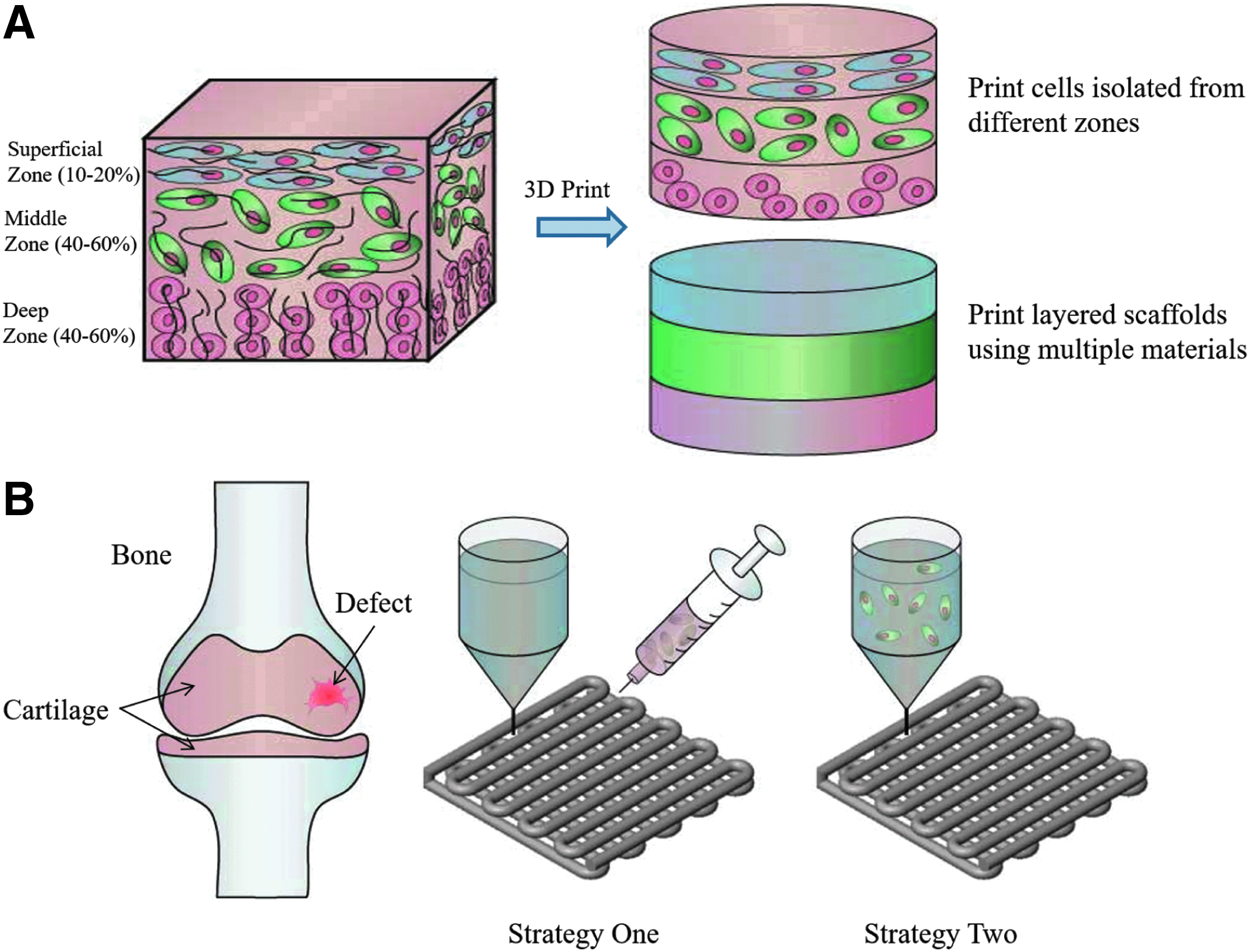
Zonal property of native cartilage and current 3D printing approaches for cartilage defect repair.
Acellular Cartilaginous Scaffolds: Microstructure to Create the Complexity
The cartilage regeneration field has benefited from the advancement of fabrication of acellular scaffolds. From the engineering aspect of 3D printing, the acellular scaffold allows more choices of materials and processing conditions. The unrestricted fabrication process supports the potential of generating scaffolds with complex structures, which will benefit the cartilage tissue structure recapitulation at a small scale. Since the printing of acellular cartilage scaffold does not require suitability for bioactive components during the fabrication, the choice of material has more flexibility and is often based on different application specifications and desired printing techniques. For example, common synthetic thermoplastic material such as poly(caprolactone) (PCL) can be chosen for an easy melt fabrication process. 60 Materials that can be modified with photocrosslinkable side chains such as gelatin methacrylamide are good candidates for UV-based gelation process.
The use of acellular scaffolds is attractive for clinical cartilage repair applications because it can avoid the potential immune response from transplanting allogeneic cartilage tissue and extra maintenance of autologous chondrocytes associated with the current treatments. One method to fabricate these scaffolds is extrusion-based printing, which is the most popular method to fabricate cartilage tissues. Using this method, complex 3D structures can first be virtually sectioned to layers using an associated software, and then created layer-by-layer, as materials are deposited on the printing platform. The fabrication time usually ranges from minutes to hours depending on the material extrusion efficiency and construct dimensions. There are different options to solidify the materials, such as photocrosslinking, temperature, or pH change. Compared to casted gels, extrusion-based printing offers another advantage for the cartilaginous scaffold, which is to provide additional interconnected pores at the scaffold level that supports local cell growth with the help of the CAD model.48,61
During the scaffold building process, various input parameters such as different patterns and curing methods can be tuned to give rise to a range of mechanical properties for the printed fibers, which can be applied to create zonal structures in the cartilage scaffolds, considering that native chondrocytes have a unique response to the mechanical signal to regulate their metabolic activities and phenotypes. 62 The fiber dimension and orientation can be controlled precisely, with a resolution limit (the minimum width of the fibers) of around 100 μm. These parameters greatly influence the mechanical properties of the fibers. 63 Notably, although the yield resolution is in theory mainly dependent on the extrusion needle size, a needle size below 100 μm often brings practical problems such as extreme printing environment where conditions such as extremely high temperature and pressure may be harmful to the material or beyond the capacity of the machine.
One category of the extrusion-based methods, fused deposition manufacturing (FDM), or melt extrusion has been used as a convenient fabrication method for thermoplastic material. The main components include a computer that controls the scaffold model and nozzle motion with condition parameters, a heater that can be set to the desired temperature to melt the loaded material, and a compressed gas supply to apply pressure for injection. The melted material can be deposited onto a platform with room or lower temperature to solidify and form 3D structures. PCL is a commonly used thermoplastic material in 3D printing scaffolds for cartilage and bone repair. Different architectures can be manufactured with clean fibers with different yield mechanical strength. Notably, as a reference to the future applications with controlled mechanical properties, it was demonstrated that the stress–strain behavior mostly depends on the porosity, rather than the lay-down pattern and channel size. 64 The stable polymer structure makes PCL very resistant to high temperature during melt extrusion, but this could be an obstacle for in vivo cartilage regeneration because of the low degradation rate in the order of magnitude of years, 65 which further creates a barrier for the integration in native tissues. To overcome this problem, biocompatible thermoplastic materials that have relatively faster degradation rates have been explored. Polylactic acid (PLA) has been commonly used in cartilage tissue engineering for a favored degradation property.66,67 However, although printing temperature is molecular weight dependent, PLA in general requires a higher processing temperature than PCL.
While synthetic polymer with 3D printing allows fabrication of cartilage scaffolds with desired architecture that better mimics the native tissue, the ultimate goal is to achieve desired cellular response. The first matter to consider would be the effective cell attachment. For instance, Hsu et al. indicated that the printed PLA scaffolds with less complex structure and small interval of fibers allow better cell adhesion and proliferation, which might be due to easier cell trapping and larger surface for cell attachment. 68
Although commonly used thermoplastic materials such as PCL or PLA yield a relatively high resolution when building a cartilage-like structure, the lack of elasticity remains a potential disadvantage in cartilage regeneration since the difference in mechanical modulus also plays a role on cell activity. Therefore, block copolymers such as polyethyleneoxide terephthalate and polybutylene terephthalate (PBT) have been investigated as a printing material due to their viscoelastic properties that mimic the native cartilage tissues. Different mechanical properties can be achieved by varying the internal structure of a scaffold. 63 The main advantage of poly(ethylene glycol)-terephthalate (PEGT)/PBT as a biomaterial is that it is amphiphilic by combining hydrophilic PEGT and hydrophobic PBT, allowing tailored swelling and mechanical strength. A similar study utilized and characterized 3D PEGT/PBT copolymer scaffolds fabricated using the fiber deposition technique. 21 The additional glycol in the compound prevents the material from crystallizing and becoming breakable. This class of material has been proven to promote chondrogenesis both in vitro and in vivo after being seeded with chondrocytes. As a follow-up study, scaffolds fabricated with pore-size gradients were further evaluated as a model to recapitulate the zonal structure of native cartilage. 69 Despite the improved mechanical strength of PEGT/PBT, the required high processing temperature (>200°C) does not make this type of material attractive for printing. Taking into account the practical pressure and temperature the printer can apply, printing with a small diameter needle that yields a more detailed cartilage structure can be even more challenging. Considering the high processing temperature required for the above materials, these printed scaffolds can only be coated with additional natural ECM components for better biological function such as chondrogenesis for cartilage tissue engineering.
Common thermoset synthetic materials usually require a heating temperature higher than 100°C for extrusion. Interests in low temperature printing have grown considering the potential to incorporate natural proteins or live cells in the printing process to add biological functions to the cartilage scaffold. Since chondrocytes have limited cell–cell interaction compared to most of the cell types, the sustained release of bioactive components becomes very important in chondrogenesis. To ensure the activity of growth factors after printing, a temperature below 70°C is recommended although different proteins may have various responses and resistance to temperature.
70
The addition of solvent is one of the most widely used methods to reduce the printing temperature. A low temperature 3D printing method using dioxane was developed to form scaffolds in a refrigerator.
71
While typical printing processes for commonly reported materials such as PCL, poly-
In addition to solvent-based printing, the water-based printing system using synthetic material has also been recently developed as a modification to regular synthesized polymer. For example, water dispersion of polyurethane nanoparticles can first be synthesized and then printed with the presence of polyethylene oxide as a viscosity enhancer. 74 The hydrophilic surface of the polymer provides good cell adhesion and a nontoxic environment for chondrocyte proliferation. The water-based system could also allow incorporation of bioactive molecules such as growth factors for future cartilage tissue fabrication applications. However, one of the drawbacks of this current water-based system is that the printing process requires more synthesis steps and thus increases the complexity of the fabrication process.
In the cartilage tissue engineering field, hydrogels have been considered as a promising scaffold material for cell encapsulation because it offers better diffusion of hydrophilic substrates such as growth factors and nutrients for living cells. However, the intrinsic diffusion is limited by distance so that a macroscale printed porous structure is usually required in a 3D scaffold. Traditional fabrication methods such as molding and casting paste to create hydrogel block are not capable of fabricating patterned internal porous structure to support successful chondrogenesis. RP-based thermoreversible hydrogel scaffold production using 3D dispensing techniques was first introduced in year 2000, using agar and gelatin. 58 This study, incorporating agar as the plotting material and gelatin as the plotting medium, demonstrated the feasibility of such printing technique, although some level of deformation was observed due to gravity for a 1 cm3 scaffold as a result of the low mechanical strength.
Chemical modification of the hydrogel is a common way to improve or expand its suitability for 3D printing cartilaginous tissue by enabling the materials to cross-link with other materials, therefore improving its mechanical strength or biological potential. Chemical modification to make photopolymerizable and thermosensitive poly(ethylene glycol) (PEG) gel was reported by synthesizing thermosensitive short copolymer chains and methacrylated groups onto the PEG polymer. 75 In addition, Skardal et al. developed a photocrosslinkable hyaluronan (HA)-gelatin hydrogel for bioprinting. 76 By adding the methacrylate group, the HA-gelatin formed an extrudable gel solution, and therefore, the fabrication can be completed in a two-step printing. Both cell free and cell laden printed scaffolds showed good biocompatibility and cell viability, demonstrating the potential to use this type of synthetic material for chondrogenesis in combination with different cell sources and other techniques. Although chemical modification expands the methods to solidify the hydrogels and makes various hydrogels suitable for printing, it usually has no or very limited impact on improving the weak mechanical strength of hydrogel scaffolds. Future studies aiming to solve this intrinsic problem of hydrogel may consider new material synthesis that provides much stronger mechanical strength while maintaining the cell-friendly environment 77 or the utilization of synthetic material with favored mechanical properties as a supporting component in the 3D cartilage construct.
While extrusion-based printing has been a popular method of 3D printing due to its flexibility with materials and curing options, stereolithography has been a well-developed and utilized printing technique considering the high resolution that can be achieved, that is, <10 μm, 78 and the relatively less labor involved fabrication process compared to extrusion-based printing. However, the adhesion process in which the cured material attaches to the rising platform during polymerization requires certain material strength, which makes it a less popular method in developing soft tissues such as cartilage. In such applications, the materials used are often specially established or modified. For example, a photo curable polymer trimethylene carbonate (TMC)/trimethylolpropane was used to develop 3D cartilage regeneration scaffolds using microstereolithography. Its biocompatibility and favored mechanical strength give the fabricated scaffolds a potential to be seeded with chondrocytes. A pilot study showed a high yield scaffold resolution of around 100 μm with different cellular responses to different inner structures. 79 Similar approach was also reported using methacrylated poly TMC. 80 Other advanced modifications of the system such as incorporating two-photon polymerization were reported to improve the spatial resolution of the printed cartilage scaffold (<1 μm) using microstereolithography fabrication. 81
Bioprinting Cell-Laden Cartilage: Controlled Cell Distribution and the Potential to Incorporate Bioactive Molecules for Chondrogenesis
Considering the relatively isolated behavior and low metabolic activity of chondrocytes in native cartilage tissue, the 3D bioprinting cell-laden scaffold brings attractive advantages, including the control of cell distribution in desired location and the possibility to incorporate growth factors. To achieve satisfactory cell viability, the choice of materials used to encapsulate the cells for printing is restricted. The most commonly used materials are natural hydrogels such as alginate.82,83 Since chondrocyte is the only cell type in native articular cartilage tissue, it is the most popular cell source to apply to the cell laden printing trials. A study has demonstrated the possibility of printing cartilaginous scaffold with arbitrary geometry using alginate solution containing chondrocytes. 84 The constructs were produced by an open-architecture computer-aided manufacturing system incorporating RP techniques. Printed disks were proven to result in relatively homogenous cell distribution and satisfactory cellular response after culture. However, the formed alginate hydrogel may have limited ability to form inner structure and more complex structures considering the intrinsic poor printing resolution of soft hydrogels. Higher concentration of more viscous alginate solution (10% w/v) would improve the integrity of the printed porous cell laden scaffolds; however, the improvement is limited compared to the native tissue. 85 One disadvantage of the calcium-mediated alginate gelation is that it usually requires additional setup or manual handling during the printing process. In addition, the very high concentration of gel solution may have a negative impact on cell response.
Despite the previously mentioned advantages of hydrogels as a material for cell printing, the weak mechanical property that does not match the native tissue is always one of the major concerns in cartilage engineering. Normal healthy human cartilage tissue has a compression modulus ranging from 9 to 13 MPa, 86 while hydrogels usually provide a mechanical modulus in kPa magnitude. 87 One simple way to solve the weak mechanical properties of pure natural hydrogel construction is to combine the stiff synthetic material and hydrogel in a single scaffold, usually accomplished by printing one layer of stiff material followed by another layer of hydrogel with or without cells. These approaches not only provide an enhanced mechanical strength for the cartilaginous scaffold but also add the complexity of the 3D construct for better mimicking the zonal cartilage tissue. Several studies introduced the production of hybrid constructs by a layer-by-layer deposition of PCL and alginate containing cells.88,89 Their results showed improved control over mechanical characteristics as well as favored biological function such as viability and cell morphology maintained in the hydrogel following printing. Because of the low handling temperature of hydrogel printing in those hybrid printing cases, it not only provides a way to directly print cells but also a potential to print growth factors with cells. For example, another PCL–alginate–chondrocyte bioprinting study showed that the addition of transforming growth factor (TGF) improved ECM formation after 4 weeks of culture. 90 Since hydrogel in these hybrid print application play the most important biological roles, the hydrogel concentration also affects the cellular response of the scaffolds. Notably, the yield mechanical properties are the structural property, not the material property. The cellular response could be different and affected by the connection boundaries of different materials as a result of distinct mechanical and biological cues.
Other than building a construct with different materials at scaffold level, hybrid materials have been explored as an advanced approach to 3D printing cartilage. Materials made out of thermoresponsive polymer poly(N-isopropylacrylamide) grafted HA with methacrylated hyaluronan (HAMA) were introduced as a novel bioink. 91 HA is a natural component existing in native cartilages and it has been proven to promote chondrogenesis as a biofunctional material. As a result, using HA as a bioink is an attractive alternative for 3D printing of cell-friendly tissue-engineered cartilage. However, similar to other unmodified natural hydrogels, HA has a low viscosity and slow gelation process that makes it difficult to fabricate scaffolds with satisfactory structures. Thus, chemical modification to HA is often required for a high resolution print. In this study, HAMA showed promising properties as a printing ink for cell encapsulation with good viability as a preliminary biological test. On the aspect of printing resolution, a large needle of 3 mm diameter was used to extrude viscous HA solution with a 2 mm spacing. Future improvements on the material to obtain scaffold structure on the micron scale level might be beneficial for chondrocyte morphology maintenance or stem cell differentiation.
In another study aiming at improving the mechanical strength of natural hydrogel, 3D printing bioink composed of alginate and nanofibrillated cellulose (NFC) was developed for desired printability. 92 The scaffolds were printed with low pressure and crosslinked using CaCl2 at room temperature. The addition of NFC significantly improved the mechanical properties of the hybrid material and resulted in a better resolution of the printing system. With the optimized formula of the bioink, human chondrocytes showed a promising viability after printing and during culture.
Synthetic hydrogels have demonstrated the ability to have tunable mechanical properties that match the native human cartilage. For example, PEG is a widely used macromer in soft tissue engineering. Its solubility in water and well-documented protocols to synthesize photocrosslinkable products make it an attractive material for directly printing cells in 3D constructs. Printing poly(ethylene glycol) dimethacrylate (PEGDMA) with human chondrocytes onto a osteochondral plug using thermal inkjet printer was reported. 93 One concern with PEG in the cartilage regeneration field is that the macromers are not biodegradable. However, such an approach leads to a precise and maintained cell position after printing as a result of simultaneous photopolymerization. As the integration of the implanted tissue into the surrounding native tissue remains one of the major challenges in cartilage repair, similar approaches with fast crosslinking will create the possibility to establish a one-step repair surgery by directly printing material and cells onto the defect site to treat damaged cartilage. This potential treatment will provide an improved integration during in vivo tissue regeneration, especially considering this is an intrinsic obstacle for regenerating nonvascularized cartilage tissue.
While chondrocytes seem like a natural choice of cells for cartilage repair, MSC is proven to be a promising cell type in cartilage tissue engineering. They are more abundant in human body than primary chondrocytes. In the past years, various methods and in vitro expansion techniques have been well documented. Gao et al. developed a printing system using gelatin methacrylate (PEGDMA) mixture to simultaneously print human MSCs, which was photocrosslinked during layer-by-layer manufacturing. 94 Taking into account the advantages MSCs have over chondrocytes, including more rich sources and higher proliferation, forming cartilaginous tissue through differentiation requires mechanical and biological signaling cues involved in the design. Addition of bioactive molecules such as growth factor in the printing resin might be one of the future avenues to successful chondrogenesis using 3D printing MSCs encapsulated scaffolds. In this case, the low-temperature processing printing techniques will help trap the growth factors in the scaffold for sustained and control release over chondrogenesis. Furthermore, although greatly increasing the complexity of the manufacturing and requiring more fundamental knowledge on cell signaling, loading different growth factors with stem cells in each cartilage zonal region may be a way to reproduce a functional and healthy cartilage tissue in the future.
The major intrinsic challenge in simultaneously printing scaffolds and cells is to balance the material properties and printing condition that favor the scaffold fabrication and biological functions. The choice is typically depending on the specifications of each study. In general, the development of materials that provide superior biocompatibility and advanced mechanical strength will greatly benefit the bioprinting area.
In the cartilage tissue engineering field, to achieve a similar structure of the native cartilage tissue, 3D printing techniques can also be combined with other advanced techniques to achieve superior mechanical properties, preferred cell environment, or to reduce the fabrication difficulty and cost. For example, a study introduced an inkjet printing/electrospinning system to create PCL and hydrogel hybrid constructs for engineering cartilage tissues. 95 The sheet of electrospun PCL fibers was alternated with inkjet printed chondrocytes suspended in fibrin–collagen hydrogel to fabricate layered scaffolds. Besides the enhanced mechanical strength compared to normal hydrogel printing, the deposition of type II collagen and glycosaminoglycans helped enhance the formation of cartilage-like tissues in both in vitro and in vivo experiments. As a summary, different materials have distinct advantages and disadvantages (Table 2). The mechanical and biological properties need to be balanced depending on the specification of a design and the expected outcome.
PCL, poly(caprolactone); PLA, polylactic acid; PLGA, poly(lactic-co-glycolic acid); PEOT, polyethyleneoxide terephthalate; PBT, polybutylene terephthalate; PEO, polyethylene oxide; PU, polyurethane; PEG, poly(ethylene glycol); HA, hyaluronan.
Advanced Technology Incorporating 3D Printing Enables More Complex Structures Similar to Real Cartilage Tissue
As discussed in the introduction, cartilage is a highly organized tissue. Each depth zone has a unique biological, chemical, and mechanical environment. Many approaches simply consider cartilage as a uniform piece of tissue ignoring the complexities for easy fabrication. However, recapitulation of the zonal structure is important for the functional restoration of the damaged cartilage. Three-dimensional printing is a preferred way to achieve the complex structures. Many experiments have demonstrated the possibility of harvesting cartilage tissue and cells from different zones. 96 Chondrocytes located in different zones have distinct phenotype in terms of matrix formation and gene expression.97,98 Printing chondrocytes isolated from different zones within hydrogels layer-by-layer has been attempted, resulting in good cell viability and cell localization. 99 Notably, the derived zonal chondrocytes are not pure, although showing different behaviors. Therefore, future evaluation of long-term chondrocytes phenotype maintenance and surrounding matrix formation is still required to bring similar concept to real applications. In fact, 3D printing technology incorporating the chondrocytes repopulation into 3D hydrogels with accurate control will improve the outcome of zonal approaches using regionally isolated chondrocytes. 50 A recent publication utilized a combination of 3D printing and directional freezing to create micropores inside the printed macropores with hydrophilic chitosan–alginate solution. 100 Due to the mechanical limit of the material, 3D printed mold with macropore was used instead of direct printing in this pilot study, but the idea of creating both macro and microlevel pore may benefit the bone marrow infiltration in vivo and thus stimulate more effective chondrogenesis. With the knowledge of complex biological constituents at different scales in native cartilage tissue, fabricating biomimetic construct with the help of novel printing techniques becomes a popular focus to direct cell adhesion and differentiation. For example, a recent study demonstrated the possibility of using table-top stereolithography to generate nanocomposite scaffold with different distributions of nanocrystalline hydroxyapatite and TGF-β1 as bioactive factors to repair osteochondral defects. 101 This pilot study provides an idea of printing bioactive graded scaffold to guide cellular response. With improvement of the material and more detailed distribution of bioactive molecules, similar future approaches can be applied into creating zone-specific cartilage with desired cell morphology and ECM formation, therefore achieving more effective chondrogenesis.
Compared to other 3D scaffold fabrication, 3D printing technology offers the possibility to engineer not only porous scaffolds in a 3D tissue level but also complex structures at organ level. For example, Goldstein et al. introduced a 3D printed laryngo–trachea using polylactic acid as an alternative to autologous cartilage to address the donor site mobility issue. 102
Moving forward, 3D printing technique also provides a way to incorporate multidisciplinary fabrication techniques to create functional biological components. For example, as an advanced application, 3D printing live cells together with electronic components inside a scaffold leads to a new direction in tissue engineering at organ level. In one study, alginate hydrogel mixed with chondrocytes was 3D printed along with conducting (AgNP infused) and nonconducting silicone solutions using a syringe head to form a cyborg cartilaginous ear. 103 This conceptual design provides an insightful idea of combining 3D printing, tissue engineering, and nanomaterial technologies for future complex organ reconstruction.
Despite the above 3D printing studies particularly targeting on cartilage repair, other pilot researches on advanced materials and new concept of complex printing may enlighten the future development of cartilage scaffold incorporating RP. Although in the past years, printing technology has been significantly expanded to create finer structure and has the ability to perform bioprinting, fabrication of 3D anisotropy microstructures still remains challenging, especially with the most commonly used extrusion method. However, novel fabrication process can be applied to compensate the limitation of the current manufacturing technology. For example, using thermoreversible bath as supporting material to maintain anisotropy structure during fabrication, researchers have demonstrated the possibility of creating complex organ-level structure using hydrogels with normal syringe extrusion printer. 104 Individual ECM components such as collagen and fibrin have been attempted for the reason that they provide biological support to the cells, but, in fact, native ECM regulates cellular response as an integrated structure. Therefore, designing biomimetic scaffolds using derived whole ECM has become a popular method in tissue regeneration. Engineering ECM bioink will help in bridging this new concept with the benefits that 3D printing would offer. Many ECM components have the natural ability to form a gel by controlling temperature or adding photo initiator, making them even more attractive to serve as printing resin. The general processing procedure to develop ECM bioink includes decellularization, tissue homogenization, and pH adjustment from the native tissue samples.105,106 Despite the promising biological advantages ECM printing provides, several issues need to be addressed for effective cartilage tissue regeneration. ECM components vary a lot between different types of tissues. There are no current established standards to characterize the isolated ECM (including the chondrocyte ECM) bioink. It might be necessary for future studies to understand whether the processing alters the function of the natural components in cartilage ECM. Besides, due to the unknown chemical components and the possible crosslinking methods, it is often challenging to optimize the gelation of the ECM bioink. Therefore, for many applications, addition of other supporting materials that provides strong network (e.g., mixing ECM with photocrosslinkable hydrogel) may be required to obtain a solid structure at the current stage.
Following the printing process and the biomaterial development, challenges also originate from translating this process to patients during implantation. Current approaches involve constructing the scaffolds in a laboratory setting and implanting them in patients. Although 3D printing is capable of fabricating complex shapes, these laboratory scale studies take on simple geometries, such as disks, while real defects in patients are often irregular. Advanced imaging techniques allow for recognition of defect shapes in patient, which consequently allows custom, patient-specific scaffold shapes to be fabricated. Even then, the reconstructed structures are often incapable of perfectly matching the defect shape. 107 In the future, there needs to be an in situ repair method, where materials can be printed directly into the patient's defect. This is a large challenge to be tackled, which will involve the development of a printing system that is mobile and can possibly be integrated with surgical equipment in an operating room.
Conclusion
As cartilage tissue function relies on the complex structures such as the zonal distribution of collagen fibers and chondrocytes, traditional treatments fail in mimicking such complexity of the native cartilage. With the development of 3D printing technology to fulfill the needs of developing constructs that can recapitulate the complex structure of native cartilage for effective regeneration, 3D printing of cartilaginous tissue has opened a new direction to achieve desired cartilage tissue regeneration or joint functional restoration. Future advancement of novel materials with robust mechanical strength and favored biological functions, and new fabrication technologies, will further broaden the applications of 3D printed cartilage for effective tissue regeneration. The emerging RP technology combined with other functional additions will complement current existing therapies for cartilage defects and thus benefit future clinical treatments for diseases such as osteoarthritis.
Footnotes
Acknowledgment
This work was supported by National Science Foundation grant CBET 1264517 and CBET 1604742.
Disclosure Statement
No competing financial interests exist.