
Abstract
Tendon injuries are common musculoskeletal system disorders, but the tendons have poor regeneration ability. To address this issue, tendon tissue engineering provides potential strategies for future therapeutic treatment. Elements of the physical microenvironment, such as the mechanical force and surface topography, play a vital role in regulating stem cell fate, enhancing the differentiation efficiency of seed cells in tendon tissue engineering. Various inducible scaffolds have been widely explored for tendon regeneration, and scaffold-enhancing modifications have been extensively studied. In this review, we systematically summarize the effects of the physical microenvironment on stem cell differentiation and tendon regeneration; we also provide an overview of the inducible scaffolds for stem cell tenogenic differentiation. Finally, we suggest some potential scaffold-based therapies for tendon injuries, presenting an interesting perspective on tendon regenerative medicine.
Introduction
T
Currently, tendon tissue engineering is being extensively studied by investigators, in which seed cells and scaffolds are two important elements. Mesenchymal stem cells (MSCs), embryonic stem cells (ESCs), and tendon stem/progenitor cells (TSPCs) are widely used as seed cells because of their multipotency and self-renewal abilities.8–11 However, there are still existing challenges that hinder the application of stem cells; for example, in a tendon injury microenvironment, stem cells have the capacity to differentiate into other lineages, such as bone, in addition to tenocytes, indicating that the in vivo microenvironment is essential for the tenogenesis of stem cells.12,13 Moreover, the low efficiency of seeded cell tenogenesis in vitro indicates that the in vitro culture microenvironment should be optimized. 9 Therefore, many studies are being conducted to identify methods of inducing stem cells to differentiate into the tendon cell lineage; among these, developing inducible scaffolds for tendon regeneration is gaining popularity.
In this review, we focus on the elements of the physical microenvironment, such as the mechanical forces and surface topography, as well as their role in stem cell differentiation and tendon regeneration. In addition, we discuss inducible scaffolds for the tenogenic differentiation of stem cells, providing a systematic overview of scaffold-based treatment for tendon injury (See Supplementary Data; Supplementary Data are available online at www.liebertpub.com/teb). Finally, we suggest future directions for scaffolds aimed at promoting the tenogenic differentiation of stem cells for tendon regeneration.
Physical Microenvironment of Tendon Development and Regeneration
The tendon is a connective tissue that represents an important component of the musculoskeletal system. It is an extracellular matrix (ECM)-rich structure that contains fewer cells than most other tissues. Tendon stem cells (TSCs), also referred to as TSPCs and tendon-derived stem cells (TDSCs), are exposed to a complex microenvironment that mainly includes physical and biochemical cues from the ECM and surrounding cells. The extracellular composition of the tendons mainly includes collagen type I molecules; however, other less abundant collagen types, ECM-related proteins, glycosaminoglycans, and proteoglycans are also present in the native tendon ECM. 14
The ECM microenvironment plays an important role in TSPC maintenance, and ultimately, tendon tissue development and maintenance; this is because some key ECM components are important for the self-renewal and tendon-specific gene expression of TSCs, 11 such as laminins. 15 Moreover, ECM components affect cell behavior and function by directly influencing cell signaling owing to their ability to interact with many growth factors and modifiers. 16 However, the precise effects of ECM compositions remain unclear, and further study is required to elucidate them. In sum, the ECM is essential for maintaining the structure and function of the tendon microenvironment.
The tendon microenvironment can be divided into the following three categories: biological signals (e.g., growth factors), biochemical factors (e.g., oxygen tension), and biophysical cues (e.g., surface topography and mechanical loading). 17 Biophysical cues are key modulators of the microenvironment for TSCs. Recently, the effect of mechanical loading and surface topography on stem cell maintenance and tenogenesis has been extensively studied and discussed, and their underlying molecular mechanisms have been introduced and explored.
Tendon physical microenvironment
It has been established that the tendon is responsible for transmitting loading from the muscle to the bone to support motion; therefore, it is consistently subjected to mechanical loading. It adapts to this loading by changing its structure and function in a manner that largely depends on the tendon cells, 18 as extracellular forces are translated into intracellular signals that affect the bioactivities and tenogenic differentiation of TSCs. Consequently, the mechanical loading plays a crucial role in tendon development and homeostasis. On engineered tendon tissues, the physiological loads in tendon cells lead to enhanced mechanotransduction signaling pathways, adhesion of the cell matrix, and collagen synthesis, whereas the loss of tensile strain can disturb cell adhesions, change the matrix structure, and even cause inflammation. 19
Moderate mechanical loading is beneficial for the tendons. It can induce anabolic changes in the tendon cells by upregulating mechanical growth factors' expression and stimulating TSC proliferation 20 ; in addition, the mechanical force plays a critical role in adult tendon homeostasis. 21 A recent study also showed that 6% of cyclic tensile strain can maintain the structural integrity and cellular function of tendon, thereby supporting the tendon homeostasis. 22 Furthermore, moderate mechanical loading can improve the tendon-healing outcomes23,24; it can enhance the regenerative potential of bone marrow mesenchymal stem cells (BMSCs) and tendon cells in tendon–bone healing by promoting their proliferation and tenogenic differentiation in vivo. 25
Excessive mechanical loading is harmful to tendon development, homeostasis, and repair. It results in matrix damage accompanied by alterations in the cell shape, along with the upregulation of inflammatory and degradative pathways in vitro. 26 In addition, overload causes anabolic changes (upregulation of nontenocyte-related genes) in the tendons and induces TSC differentiation into nontenocytes, potentially resulting in degenerative tendinopathy, which is frequently observed in clinical settings. 20 Wang et al. also found that both mechanical underloading (3% cyclic tensile strain) and overloading (9% cyclic tensile strain) led to a negative influence on the tendon, which was evidenced by different degrees of matrix deterioration. 22
Consequently, the tendon is a mechanosensing tissue, and only a narrow range of tensile strain has a positive influence on its development, homeostasis, and healing. Although the effect of mechanical loading on the tendon has been studied extensively, most previous trials were in vitro studies; therefore, in vivo studies are necessary for identifying a better method of using mechanical loading for tendon repair.
Surface topography is an important factor of the physical microenvironment, as intracellular signaling and focal adhesion distribution depend on the matrix topography, which can guide the cell morphology and movement, regulate cell growth and function, and influence the fate of stem cell differentiation.27,28 Some studies have proven that native tendon sections can promote stem cells to differentiate into the tenocyte lineage. For example, the tendon-derived decellularized matrix could specifically promote the tenogenic-lineage differentiation and inhibit the osteogenic-lineage differentiation of human TSPCs. 29
Stiffness is described as the degree to which the ECM or scaffolds resist deformation. 30 Different tissues have different degrees of matrix stiffness, according to their respective functions. Matrix stiffness is important for directing stem cells to differentiate into specific cell lineages. A soft matrix is suitable for stem cell tenogenesis and tenocyte phenotype maintenance, whereas rigid substrates can improve the expression of chondrogenic and osteogenic genes in tenocytes. 31 In sum, the tendon's physical microenvironment, which includes the mechanical force, surface topography, and stiffness, plays a vital role in stem cell fate and tendon regeneration.
Molecular transduction of mechanobiology in tendons
Physical cues are critical factors in tendon development, homeostasis, and regeneration; numerous studies have investigated the possible mechanisms involved in the conversion of biophysical cues into a biochemical response, and we have performed a comprehensive review to elucidate them (Fig. 1).

Mechanical signals transduction. Mechanical signals are transduced through the cell membrane sensors (e.g., integrin, iron channels, and G-coupled receptors), biological factors (e.g., growth factors), intracellular pathways, and transcription factors to induce stem cell proliferation and tenogenic differentiation.
In the plasma membrane, mechanical signals are captured by the transmembrane adhesive structures, such as integrin, which undergo a conformational change and initiate many cytosolic signaling cascades, including the activation of the kinases Src and PI3K.32,33 Heterotrimeric G-proteins and ion channels can also be activated directly by the changes in the mechanical signals.34,35 Moreover, the cells that can sense the mechanical signals can be sensed by other cells through biological factors, such as growth factors, which bind to the receptors in the membrane and transmit the signals into the cell, thereby initiating several cytosolic signaling cascades. 2 Although the plasma membrane is dynamic, and its composition is constantly changing, the cytoskeleton forms a rigid network that transfers physical signals to the cell. Furthermore, the ECM organization, cytoskeletal organization, and gene transcription can be affected by alterations in the biophysical cues. 36
For the cytosolic signaling cascades, there are two signaling pathways, namely TGF-β–SMAD2/3 and FGF–ERK/MAPK, which are considered to transduce biological signals in response to mechanical signals.21,36,37 However, a recent study showed that mechanical stimulation can be directly transmitted into the nucleus through the Yes-associated protein, indicating that mechanical stimulation may regulate molecule translocation independently of the signaling pathways. 38
These mechanotransduction processes can provide important information for us to develop new scaffolds tuned for a more clearly defined cellular response, providing more controlled and efficient tendon regeneration. However, the underlying molecular mechanisms driving stem cell proliferation and tenogenic differentiation are still unclear; therefore, the molecular pathways through which cells discriminate signals from their surrounding microenvironment need to be further elucidated.
Inducible Scaffolds for Tenogenic Differentiation and Regeneration
The scaffold is one of the essential elements of tendon tissue engineering; it provides significant advances in terms of structural integrity and biological compatibility for the tendons. Moreover, in many cases, the results obtained from applying scaffolds are superior to those observed in natural healing. Scaffolds can be classified into four types, including natural scaffolds (e.g., collagen and silk),39,40 synthetic scaffolds [e.g., poly(
Biomaterials for tendon regeneration
Many studies have been conducted to develop biomaterials for tendon regeneration. They are designed to mimic the mechanical properties of the native tendon for tendon injury repair. Collagen I membranes with oriented fibers have suitable mechanical properties, and they are able to promote the proliferation and adhesion of human dermal fibroblasts and tenocytes in vitro.
39
PLA–collagen biomaterials with desirable tensile properties have been shown to exhibit good adhesion, proliferation, and infiltration of the tendon cells, or fibroblasts.44,45 Moreover, poly(
However, because stem cells have multipotent differentiation capacity, stem cells seeded on scaffolds can also differentiate into other nontendon-specific lineages, such as bone and cartilage, leading to tendon repair failure. 12 Many inducible scaffolds have been developed to overcome this problem; these scaffolds not only have superior mechanical properties, but they are also capable of inducing stem cell tenogenesis, providing an inductive microenvironment for stem cell tenogenesis and tendon regeneration.
Inducible scaffold
In recent years, the number of inducible scaffolds fabricated to induce stem cells to differentiate into tendons and promote tendon regeneration has increased (Table 1). Most of these scaffolds are designed to mimic the microenvironment of the native tendon tissue. Decellularized matrix is a typical example; this is a natural tendon scaffold that is mostly composed of collagen, with mechanical properties comparable with those of the native tendon. The effects of decellularized scaffolds have been illustrated in many studies; they provide an inductive environment for the proliferation and tenogenic differentiation of BMSCs, TDSCs, and adipose-derived stem cells (ADSCs), even inhibiting their differentiation into other tissues, and their good mechanical properties are also adequate for supporting in vivo tendon regeneration.29,48–50
Inducible Scaffolds with Different Modifications for Tendon Regeneration
NA, not available.
In addition, by changing the mechanical properties, such as their densities, scaffolds can also be inducible for stem cell tenogenic lineage.42,51 Moreover, some synthetic scaffolds can induce stem cells to differentiate into the tenogenic lineage.43,52 Furthermore, hybrid scaffolds possess the ability to promote stem cells tenogenic differentiation; for example, chitosan-based ultrafine fibers have an inductive function for the tenogenesis of stem cells. 53 In summary, inducible scaffolds can promote the stem cells differentiation into tendon cells and have sufficient mechanical properties to support tendon regeneration.
In most cases, the mere application of scaffolds is insufficient to provide an inducible microenvironment for tendon regeneration. Thus, it is necessary to develop specialized scaffold-enhancing modifications for inducible scaffolds. Physical microenvironment-based modifications, especially mechanical force and surface topography, are applied on scaffolds to better mimic the native tendon microenvironment to induce stem cells to differentiate into the tendon cell lineage. Many studies have been performed using these physical modifications to enhance tendon regeneration.
Physical modifications
Physical modifications for scaffolds mainly include mechanical stimulation, as well as topography and other factors, such as stiffness, which is also an important factor in the tendon microenvironmental niche and plays a vital role in determining the stem cells' fate and tendon regeneration (Fig. 2). Thus, using these modifications for scaffolds may enable better mimicking of the tendon's physical microenvironment, which may significantly improve the outcomes of tendon regeneration.
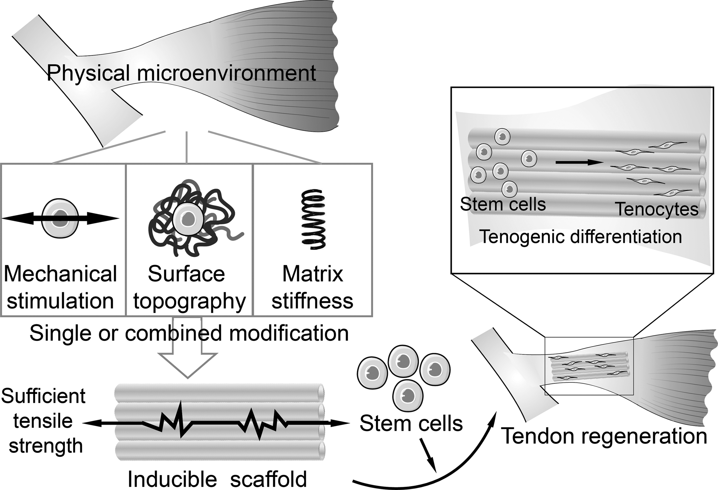
Inducible scaffolds with physical microenvironment-based modifications for stem cell tenogenic differentiation and tendon regeneration.
Mechanical stimulation
Mechanical stimulation is a vital component in the native tissue's physical microenvironment, and it exerts considerable influence on the determination of stem cells' fate; different degrees of mechanical stimulation drive stem cells to differentiate into distinct lineages. In addition, numerous studies have employed mechanical stimulation in scaffolds to obtain an inducible microenvironment for stem cell tenogenesis. Mechanical stimulation can induce stem cell maintenance and tenogenic differentiation. Under mechanical stimulations, stem cells, such as MSCs and TDSCs, exhibit good proliferation on scaffolds, with an upregulation of genes and proteins associated with the tendon ECM, including collagen type I, in vitro.54,59,61 Xu et al. also demonstrated that cyclic mechanical stimulation could promote TDSC differentiation into the tenolineage in a poly(
In addition, mechanical stimulation can promote the regeneration of tendon in vivo. For example, the transplanted TDSC–scaffold construct can promote rabbit patellar tendon defect regeneration under mechanical stimulation. 61 Furthermore, when culturing hESCs on a knitted silk–collagen sponge scaffold, a tenocyte-like morphology is induced, and tendon-related gene expressions are upregulated with mechanical stimulation, both in vitro and in vivo, thereby contributing to tendon regeneration. 58 Therefore, mechanical stimulation is a critical strategy for making a scaffold an inducible scaffold for stem cell tenogenesis and tendon regeneration.
Topography
In recent years, many scaffolds have been fabricated using nanoscale and microscale fabrication technologies for providing an inductive surface topography for tendon regeneration. The topography applied in scaffolds also modulates the differentiation of stem cells. For example, the surface nanopatterning of scaffolds can control the initial assembly of focal adhesions and then regulate the self-organization and tenogenic differentiation of hMSCs. 63 A recent study also showed that scaffolds with a parallel microgrooved topographical surface can enhance the expression of tendon-specific genes, such as tenomodulin and scleraxis, thereby enabling murine TDSC tenogenesis and inhibiting nontenogenic lineage differentiation. 64
Moreover, the fiber alignment of scaffolds plays a critical role in stem cell fate. For instance, MSCs seeded on the aligned and oriented fibrous scaffolds exhibit tenocyte-like morphology and improve tenogenic differentiation, whereas randomly oriented scaffolds display enhanced osteogenic differentiation.69,82,83 A study also showed that a three-dimensional (3D) collagen–silk scaffold with 3D alignment of collagen could enhance the cellular infiltration and tenogenesis of TSPCs in vitro and improve the outcome of tendon injury repair in a rotator cuff repair model. 66
Stiffness of materials can also influence tendon differentiation. For example, a moderately rigid collagen type I substrate can induce MSC differentiation into tenolineage, whereas MSCs differentiate into osteogenic cells on more rigid substrates. 84 Moreover, a recent study has shown that lower material stiffness is beneficial for Achilles tendon repair. 85 Fiber diameter and angles are other biophysical factors that promote tendon differentiation; fibers with larger diameters (>2 μm) enhance MSC tenogenesis and may be more suitable for the development of tendon tissue in vitro. 72 Moreover, braided submicron fibrous scaffolds with large braiding angles promote the tenogenic differentiation of hiPSC-MSCs. 73 However, some problems remain; for instance, polymer scaffolds with aligned fibers have the potential to cause chronic inflammation. 86 Thus, for successful tendon regeneration, artificially fabricated ECM-like materials with suitable physical properties and no inflammatory responses could be a promising scaffold for in vivo transplantation.
Combined modifications
The native tendons have a complex microenvironmental niche that comprises not only physical cues but also growth factors and other bioactive compositions, which exert effects on stem cell proliferation, tenogenic differentiation, and tendon regeneration.87–89 Therefore, in many cases, single factors, either physical cues or bioactive factors, cannot promote complete regeneration of the tendon, as they are unable to mimic the real tendon microenvironment.
For instance, imprinted substrates with parallel groove patterns influence cell density and tenocyte alignment, resulting in the deposition of the matrix; however, this does not have clear effects on the matrix gene expression and cell phenotype in vitro, 90 which indicates that single topographical modifications are unable to generate stem cell teno-differentiation. In one study, the researchers proposed a strategy to solve this issue; they incorporated a small-molecule Trichostatin A (TSA) into a PLLA scaffold with aligned fibers to develop a TSA-laden PLLA-aligned fiber (A-TSA) scaffold, and they showed that the A-TSA scaffold could significantly induce the tenogenesis of TSPCs in vitro, as well as improving the structural and mechanical properties of the regenerated Achilles tendon when implanted in rat models. 78
There is an obstacle to the employment of mechanical stimulation for successful tendon regeneration. Stem cells, even TSPCs, may differentiate into other lineages, such as bone and cartilage, when only mechanical stimulation is applied, resulting in tendon degeneration.13,91,92 To resolve this issue, many researchers consider the combined effects of two or more biological and mechanical factors on stem cell tenogenesis and tendon regeneration. One attempted method involves combining the mechanical force with the tendon-specific transcription factor, scleraxis, to induce hESC-MSC tenogenesis.10,92 Moreover, the combination of cyclical mechanical stimulation and tenocytic cell-free extract can increase ECM deposition with aligned dense fibrils, upregulate the tendon-related gene expression of MSCs, and improve the cellularity of the construct and ultimate tensile strength compared with the constructs with only mechanical stimulation. 76
Furthermore, another study investigated the combined effect of tensile strain, growth differentiation factors, and various oxygen tensions on equine ADSCs cultured in a collagen type I gel scaffold; the results demonstrated that these factors could improve the expression of tendon-relevant genes and enhance tenogenic differentiation of ADSCs. 74 Consequently, the incorporation of different physical cues or physical cues and biological factors into scaffolds enables the formation of an inductive microenvironment that is closer to that of native tendon than that emerging from treatment with a single element, thereby exerting superior effects on tendon regeneration.
Future directions of scaffolds in tendon regeneration
As already mentioned, several inducible scaffolds have been fabricated with suitable mechanical properties, and their merits for stem cell tenogenesis and tendon regeneration have been demonstrated. However, many current scaffolds do not simultaneously possess superior mechanical properties and inducible capacity to reconstruct the structure and function of the injured tendon. Some of them have insufficient mechanical properties to support native tendon regeneration,43,52 whereas others cannot better mimic the natural tendon microenvironment to induce stem cells' tenogenic differentiation,86,93 resulting in ectopic bone formation.13,93 Thus, it is imperative to design more suitable scaffolds for tendon regeneration. The design of scaffolds with superior mechanical properties and inducible capacity for stem cell tenogenesis and successful tendon regeneration remain a popular research topic.
Decellularized sliced tendon/matrices are promising scaffolds because they are derived from native tendon with tendon-like mechanical properties, and they can better mimic the native extracellular surroundings of tendon, providing an inducible microenvironment for stem cell tenogenesis and tendon regeneration.50,54,55 However, as the ECM biology of tendons remains in its infancy, the current decellularized ECM scaffolds have serious limitations for tendon regeneration. 94 Proteomics, a large-scale study of proteins, will be a useful tool for determining the composition and effects of ECM, allowing the evaluation of specific proteins that can promote stem cell proliferation and tenogenic differentiation in the ECM, as well as investigation of the interactions between scaffolds and biological systems.95,96 Based on the results, scaffolds can be more accurately designed to promote regenerative processes.
Furthermore, a recent study also designed a method of quantitatively predicting the changes in the tendon ECM, including key signaling, structural, and effector molecules, 97 which can provide a deeper understanding of the tendon and ECM to guide inducible scaffold design for tendon regeneration. Therefore, it is necessary to decode the native tendon ECM and determine the specific elements involved in stem cell proliferation and tenogenesis; it will be beneficial to employ these specific elements identified from the ECM into scaffolds for developing superior tendon regeneration scaffolds.
In recent years, composite scaffolds have attracted attention in the field of tendon regeneration. Because single scaffolds, such as collagen, exhibit poor mechanical properties, making them insufficient for tendon regeneration when implanted in vivo, 98 many researchers have developed composite scaffolds to address the disadvantages of single scaffolds for stem cell differentiation and tendon regeneration. For instance, in a recent study, the researchers developed a novel composite scaffold combining aligned poly-ɛ-caprolactone microfibers and methacrylate gelatin to obtain a tendon-like structure and mechanical anisotropy, generating seeded stem cells exhibiting a native tendon tissue phenotype. 99 Therefore, a composite scaffold with suitable physical properties and inducible ability is a promising strategy for tendon regeneration.
Novel responsive scaffolds have also been introduced in recent years. Magnetic responsive scaffolds with aligned structures have proven to be able to induce the tenogenic differentiation of ADSCs under magnetostimulation conditions. 100 These scaffolds also have the potential to be applied for inducing stem cell tenogenesis and promoting tendon tissue regeneration.
The stem cell is also a critical element in tendon injury repair; different types of stem cells need corresponding scaffolds to induce tenogenesis for tendon regeneration. TSPCs are ideal cells for tendon regeneration, but the current isolated TSPCs are considered to be heterogeneous cell populations; thus, different subtypes of TSPCs may exert unequal effects on tendon injuries. For example, a nestin+ TSPC subpopulation has been identified in the tendon cell population through single-cell analyses, and it exhibits superior tenogenic ability compared with nestin−TSPC. 101 Therefore, based on different TSPC subpopulations, designing stem cell-specific scaffolds will allow the advanced capability of inducing tenogenesis and promoting tendon regeneration.
Inducible scaffolds with superior mechanical properties, modified by specific factors that make the scaffolds inducible for stem cell tenogenesis, are promising for tendon tissue regeneration. Moreover, scaffolds designed for specific TSPC subpopulations will also arouse increasing interest among future researchers. Therefore, further efforts are needed to develop more efficient scaffolds for successful tendon regeneration.
Conclusion
The physical microenvironment, which includes, but is not limited to, mechanical force and topography, plays a vital role in stem cell tenogenesis and tendon regeneration. Based on the physical microenvironment of tendon, numerous inducible scaffolds have been developed with specific modifications, such as mechanical stimulation, surface topography, and stiffness; these scaffolds not only possess tendon-like mechanical properties, but they are also capable of inducing stem cell tenogenesis. However, they are still insufficient for tendon regeneration; further efforts need to be made to design better inducible scaffolds.
Decoding the tendon ECM will provide us with more information for guiding the development of inducible scaffolds. In addition, designing inducible scaffolds for specific TSPC subpopulations will emerge as a popular research topic in tendon tissue engineering. The increasing understanding of the material technology and tendon microenvironment represents a promising advancement for us to develop inducible scaffolds to bring us closer to regenerating native tendons for the treatment of tendon injury.
Footnotes
Acknowledgments
We are grateful to Ms. Ziwei Xia for illustration assistance, and we thank Ms. Varitsara Bunpetch for her help in grammar checking. This work was supported by the National Key Research and Development Program of China (2017YFA0104902, 2018YFC1105104), National Natural Science Foundation of China (NSFC) grants (81522029, 81772418, 31570987, 81330041, 81572157), and Fundamental Research Funds for the Central Universities.
Disclosure Statement
The authors declare no conflicts of interest.
References
Supplementary Material
Please find the following supplemental material available below.
For Open Access articles published under a Creative Commons License, all supplemental material carries the same license as the article it is associated with.
For non-Open Access articles published, all supplemental material carries a non-exclusive license, and permission requests for re-use of supplemental material or any part of supplemental material shall be sent directly to the copyright owner as specified in the copyright notice associated with the article.