
Abstract
Health care and medicine were revolutionized in recent years by the development of biomaterials, such as stents, implants, personalized drug delivery systems, engineered grafts, cell sheets, and other transplantable materials. These materials not only support the growth of cells before transplantation but also serve as replacements for damaged tissues in vivo. Among the various biomaterials available, those made from natural biological sources such as extracellular proteins (collagen, fibronectin, laminin) have shown significant benefits, and thus are widely used. However, routine biomaterial-based research requires copious quantities of proteins and the use of pure and intact extracellular proteins could be highly cost ineffective. Gelatin is a molecular derivative of collagen obtained through the irreversible denaturation of collagen proteins. Gelatin shares a very close molecular structure and function with collagen and thus is often used in cell and tissue culture to replace collagen for biomaterial purposes. Recent technological advancements such as additive manufacturing, rapid prototyping, and three-dimensional printing, in general, have resulted in great strides toward the generation of functional gelatin-based materials for medical purposes. In this review, the structural and molecular similarities of gelatin to other extracellular matrix proteins are compared and analyzed. Current strategies for gelatin crosslinking and production are described and recent applications of gelatin-based biomaterials in cell culture and tissue regeneration are discussed. Finally, recent improvements in gelatin-based biomaterials for medical applications and future directions are elaborated.
Impact statement
In this study, we described gelatin's biochemical properties and compared its advantages and drawbacks over other extracellular matrix proteins and polymers used for biomaterial application. We also described how gelatin can be used with other polymers in creating gelatin composite materials that have enhanced mechanical properties, increased biocompatibility, and boosted bioactivity, maximizing its benefits for biomedical purposes. The article is relevant, as it discussed not only the chemistry of gelatin, but also listed the current techniques in gelatin/biomaterial manufacturing and described the most recent trends in gelatin-based biomaterials for biomedical applications.
Introduction
For in vitro cell and tissue culture, the use of biomaterials provides several benefits for the proper growth and development of cells. Biomaterials not only function as a physical attachment for cells, but they also provide biochemical cues and activate the molecular signaling mechanisms that determine the fate of each individual cell in the culture. Generally, extracellular matrix (ECM) proteins such as collagen,1,2 fibronectin,3,4 and laminin 5 are widely utilized as biomimicking materials, as these proteins are the main interactors with cells in vivo and thus are valid candidates for use in vitro. 6
ECM proteins, which are generally produced and secreted by fibroblasts and other cell types such as epithelial cells, play significant roles in cellular growth and development in vivo. For instance, several types of collagen (the most abundant ECM protein) provide enormous tensile strength and structural integrity in connective tissues, tendons, and the skin. 7 When used as biomaterials in vitro, collagen not only provides structural integrity but also regulates the cells' proliferation and differentiation capacity. 8 In one study, it was found that collagen-coated tissue culture plates promoted the cellular adhesion and proliferation of mesenchymal cell even under stress conditions. 9 Moreover, mesenchymal stem cells (MSCs) showed increased osteogenic potential when cultured in collagen-infused polymer scaffolds. 10 Other important ECM proteins, such as fibronectin, laminins, tenascins, and vitronectins also contribute to a variety of cellular functions such as migration, growth, and differentiation.8,11,12 These proteins contain essential domains for the attachment of integrins that are present in cell membranes and are responsible for the induction and initiation of key molecular signaling pathways. Aside from the biochemical cues, the mechanophysical (stiffness, elasticity) properties of ECM also regulate the fate of the cells.13,14 In one study, human MSCs were found to differentiate into neurogenic, myogenic, and osteogenic precursor cells when cultured in soft, medium, and stiff microenvironments, respectively.15,16 Taken together, the response of cell to ECM depends on several factors such as the type of ECM present, the concentration, the physical and mechanical properties of ECM, adsorption procedure of the ECM on to the surface, and presence of other proteins.17–19
However, the use of pure ECM proteins for biomaterial studies could be cost ineffective. For instance, general protocols for coating cell culture dishes require about 0.1–1 mg/mL of ECM proteins (fibronectin, laminin, Matrigel) to induce better attachment of cells.20,21 In another study, it was found that a concentration of 3 mg/mL of ECM hydrogel was required to induce gelation in stroke cavities. 22 Thus, in routine studies, where a lot of protein is needed, the use of pure ECM proteins might be an expensive option. An intensive review of the types and functions of different ECM proteins can be found elsewhere. 23 The main purpose of this review is to discuss the use of gelatin as a replacement to ECM proteins for biomaterial purposes.
Gelatin polymer is a well-known biodegradable and biocompatible material that consists of 85–92% of proteins, mineral salts, and water.24,25 It is a molecular derivative of type I collagen and has a wide range of food, cosmetic, and pharmaceutical applications.26,27 It is generally produced by irreversible hydrolyzation of the triple helical structure of collagen through processes such as heat and enzymatic denaturation, producing random coiled domains. As such, gelatin has less organization but has a very similar molecular composition to collagen (Figure 1). 28 Because of this, gelatin has the capacity to replace and perform similar biomaterial functions as collagen for cellular development in vitro. Gelatin is readily available and can be extracted from several sources such as cattle bones, fish, pig skins, and some insects. Several studies on the biocompatibility of gelatin derived from various sources showed that gelatin, in general, does not induce toxicity, antigenicity, and other adverse effects in human cells. Toxicity, however, can arise depending on the reagent used to crosslink gelatin solutions.29–31
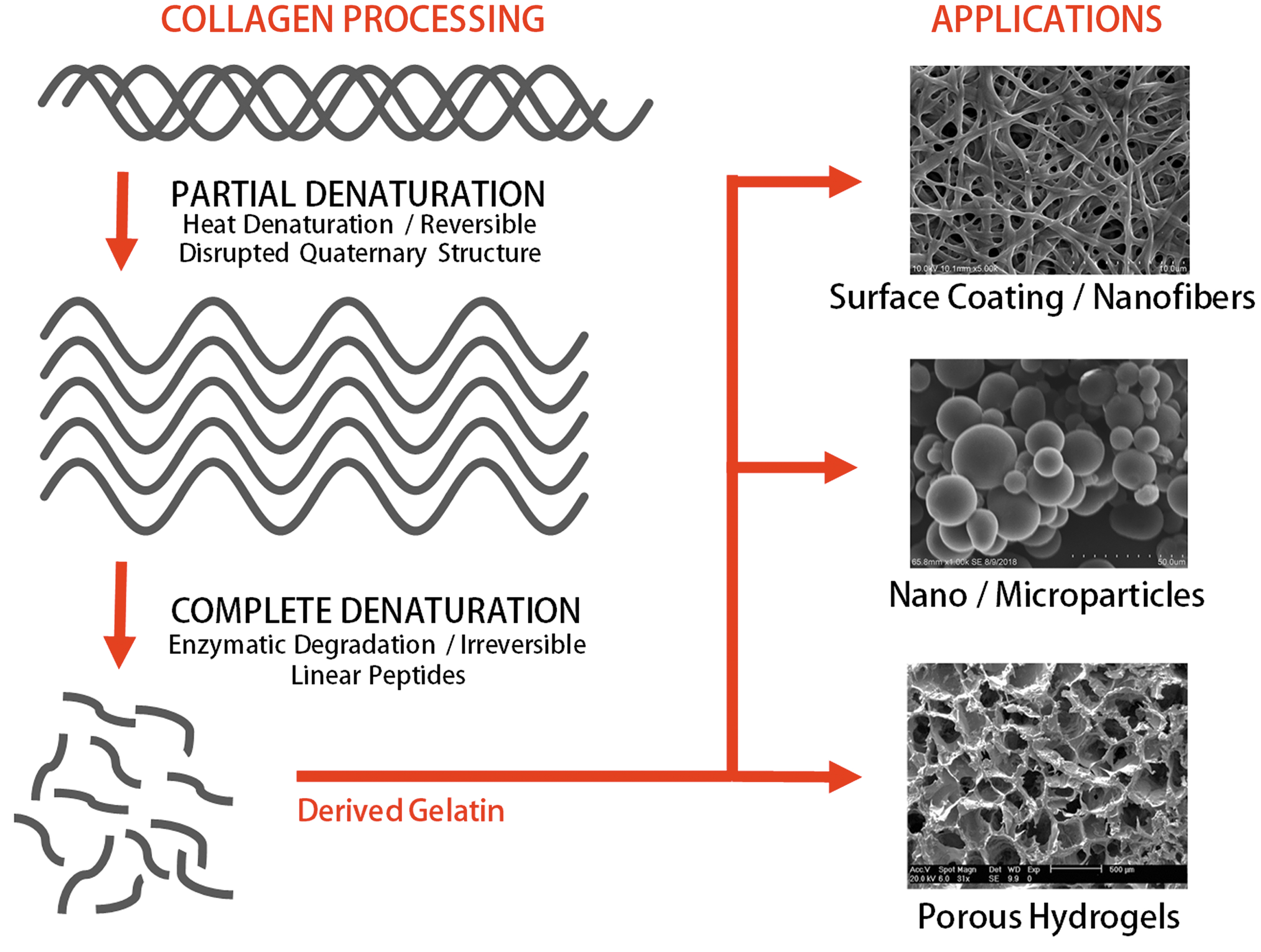
Gelatin is obtained through the irreversible denaturation of collagen type I through heat and enzymatic degradation. These processes produce linear peptides that can be reassembled in various forms, such as nanofibers, microparticles, and porous hydrogels for different biomaterial applications. Color images are available online.
Thus, the use of gelatin has gained popularity over pure and intact ECM proteins for the following reasons: (i) gelatin is more readily available and is much cheaper than ECM proteins, (ii) gelatin is highly soluble compared with other ECM proteins and thus is easier to use for biomedical purposes, (iii) only a few sections of the full ECM protein sequence is important for cell attachment and eliciting cellular response, thus the use of intact proteins may not be necessary, (iii) gelatin possesses a highly similar structure to collagen and contains important binding moieties for cell attachment, (iv) different gelatin sources are biocompatible, biodegradable, and do not induce antigenicity and toxicity in cells.
For biomaterial purposes however, gelatin also poses some disadvantages. The main drawback of using gelatin is that gelatin-based materials have poor mechanical properties, lack thermal stability, and have relatively shorter degradation rate. When used in studies that require longer period of time such as controlled drug release, cell differentiation, and wound healing; gelatin-based materials may not last. Moreover, compared with collagen, gelatin is highly susceptible to several proteases, and thus may lead to its faster degradation. 32 These disadvantages, however, can be easily overcome by modifying gelatin and making gelatin composites to increase the material's mechanical stability, biocompatibility, and bioactivity. The advancement of manufacturing technology and our knowledge of material chemistry have made these drawbacks less important compared with the limitless benefit of using gelatin for biomedical purposes.
In this review, the molecular properties of gelatin will be discussed and compared with those of other ECM proteins that are commonly used for biomaterial purposes. Moreover, recent two-dimensional (2D) and three-dimensional (3D) biomaterial applications of gelatin will be listed and compared. Current therapeutic applications and possible advancements of gelatin-based biomaterials will also be elaborated. Finally, the future direction of gelatin-based biomaterials, from cell culture and tissue engineering to material-based therapeutics, will be discussed.
Molecular Structure and Physical Properties of Gelatin
Molecular similarities between gelatin and collagen
Collagen is the most abundant protein, comprising almost one-third of the total protein concentration in the human body. 33 Collagen is an essential structural protein due to its rigidity, which is dictated by its structure. It is a triple-helical rod-shaped protein, composed of three left-handed helices twisted and intertwined to form a right-handed quaternary structure.32,34 In the body, there are several types of collagens, with collagen type I being the most abundant. Because of its structure, collagen is abundant in muscle and connective tissues. In vitro, collagen has been used in 2D and 3D biomimicking materials for a variety of research studies. For instance, culture plates coated with collagen type I increase the adhesion and proliferation rate of several cell types, including bone marrow mesenchymal cells, 9 smooth muscle cells, 35 and other cell lines used for vaccine production such as baby hamster kidney fibroblasts. 36 Collagen has also been used in 3D systems such as cell encapsulation to mimic in vivo microenvironments and study the behavior of cells.37,38
In terms of molecular composition, gelatin contains almost the same amino acid sequence as collagen. However, being denatured collagen, gelatin is a linear protein with molecular weights ranging from 15 to 250 kDa. Its amino acid composition, however, depends on the collagen raw material and extraction method but is mainly composed of a repetitive Gly-X-Y sequence, where X and Y are commonly proline and hydroxyproline, respectively. 39 Researchers have quantified the amino acid composition of gelatin to be ∼21% glycine (Gly), ∼12% hydroxyproline, ∼12% proline, and other amino acids such as alanine, arginine (Arg), aspartic acid, lysine, serine, leucine, and valine.32,40,41 The similarity of gelatin to collagen serves different purposes in cell and tissue culture. For instance, gelatin contains the linear tripeptide Arg, Gly, and Aspartate (Asp) or the Arg-Gly-Asp recognition sequences that bind to several integrin proteins and thus aid in cell attachment, migration, and survival.28,42 Another important similarity of collagen and gelatin is the lack or very low presence of the aromatic amino acids, tryptophan, tyrosine, and phenylalanine. The lack of these amino acids is one of the major contributing factors to the low antigenicity and toxicity of both gelatin and native collagen.32,43
General sources and extraction methods of gelatin
The demand for gelatin has drastically increased over the past decade. In 2015 alone, the global gelatin market reached 412.7 kilotons. 44 The ease of extraction and the countless uses of gelatin have made it a highly sought-after material. It has a wide range of applications in the food industry (emulsifier, gelling agent), cosmetics (components of cosmetic products), pharmaceutical (capsules, ointments), and specialized industries such as cell culture (surface coatings, hydrogels) and regenerative medicine, etc. 45
Gelatin is normally extracted from highly collagenous raw materials, such as pigskin, which accounted for about 40% of the global market in 2015. Cattle bones and bovine hides are also reliable sources of gelatin. 44 In recent years, several attempts have been made to also obtain gelatin from fish skin,46,47 chicken,48,49 and other materials. There are two general processes for extracting gelatin: through alkaline hydrolysis or acid hydrolysis. Depending on the extraction method, the obtained gelatin will have different properties. Type A gelatin (acid-based) and Type B (alkaline-based) gelatin have isoelectric points of ∼8.0 and ∼4.9, respectively. 50 This property affects the overall net charge of gelatin particles in solution and should be greatly considered when using gelatin for any biomaterial purposes. Another factor to be considered is the relative molecular weight of the extracted gelatin, commonly referred to as “bloom” and primarily dependent on what stage the gelatin was extracted. Gelatin obtained at the initial stages of extraction has higher bloom compared with those that undergo complete hydrolysis. Commercially available gelatins have blooms ranging from 50 to 300. The bloom value is directly proportional to the gelling capacity and thus the gel strength of gelatin. 51 To summarize, in using gelatin for biomaterial purposes, the following must be considered: raw material, type of gelatin, bloom value, the purpose of the experiment, and the crosslinking method. The last two factors will be discussed in the next sections.
Methods to crosslink gelatin
As previously mentioned, the method by which the gelatin is crosslinked is one of the most crucial factors in generating gelatin-based biomaterials. Like other proteins, gelatin moieties can be linked to each other to form a network of polymer chains under stable condition in aqueous solution. Changes in the environmental parameters and type of crosslinkers used determine the biophysical properties of the biomaterials such as the swelling properties, water absorption, elastic modulus, and transport of molecules. 52 In this study, we discuss several methods by which gelatin can be crosslinked for different purposes.
Physical gelation of gelatin (heat/pH)
One of gelatin's intrinsic properties is that its solution, under sufficiently high concentration, forms a semisolid gel at low temperatures. Gelatin solution is speculated to be in a random coil conformation when the temperature is about 40–50°C. 53 As the gelatin solution cools to below 30°C, a reverse coil to triple helix transition occurs and hydrogen bonds stabilize the conformation. 54 This physical gelation method can crosslink gelatin hydrogel in a facile and reversible process, modulated only by the concentration and the ambient temperature, without the aid of any enzymes and chemicals. 55 However, the transition of gelatin from solution to gel is unstable without the help of a stronger crosslinker. The gel strength of gelatin and the capacity to maintain its mechanical properties are highly dependent on several factors such as the bloom degree, the initial concentration of gelatin, and changes in the ambient temperature. 56 That is, gelatin monomers cannot coacervate with each other and solidify when the solution is either diluted or put at temperatures higher than 40°C. 57 A diagram of describing phase transition of gelatin with varying concentration and temperature is available elsewhere. 57
Aside from temperature, pH adjustments can also alter the physical gelation of gelatin. As pH value of the gelatin solution changes, the melting temperature adjusts, and thus affects the gel strength of the solution. Moreover, changes in pH affects ionic changes between gelatin monomers and consequently affects the hydrogen-bond crosslinking process. 58 It has been experimentally determined that visible changes occur in gelatin solution at pH 3.0–6.0. 57 In one study, it has been shown that gelatin forms helices more easily at pH 5.0, a pH closest to the isoionic point of the ossein, alkali-treated gelatin. 59
Chemical gelation (glutaraldehyde)
Since physical gelation is a transient method that fails to maintain a polymeric network system even with the slightest environmental changes, alternative methods have been developed. Among them, the use of chemical crosslinkers has been widely investigated. Formaldehyde, epoxy compounds, and dialdehyde are examples of chemical crosslinking compounds.60–62 Among them, glutaraldehyde is one of the most popular compounds used for crosslinking gelatin. It bridges gelatin molecules by forming a stable bond between the free amino groups of the lysine or hydroxylysine amino acid residues in gelatin and the aldehyde groups in glutaraldehyde. 63 Chemical crosslinking of gelatin using glutaraldehyde is highly preferred since the compound is an easily accessible and inexpensive reagent. 64 Moreover, glutaraldehyde is effective as it increases the strength of the crosslinked polymer network over a short period of time. 65 Because glutaraldehyde poses a potential threat due to its cytotoxicity and immunological responses,63,66 many studies have been carried out to minimize its toxicity by optimizing its concentration 67 or replacing it with other candidates, such as carbodiimides. 68
Natural compound gelation (genipin/phenols)
Chemical crosslinkers are depicted as toxic substances, causing researchers to shift their attention to naturally derived substances. Nature-derived chemical compounds include, but are not limited to, genipin, grape seed proanthocyanidin, epigallocatechin gallate, caffeic acid, and tannic acid. Genipin, an aglycone derivative from an iridoid glycoside called geniposide from the fruits of Gardenia jasminoides Ellis, is used to crosslink gelatin and other proteins by reacting with amine groups.69–71 The genipin crosslinking process is far less toxic than that of glutaraldehyde but the crosslinking mechanical properties are comparable.72,73 The use of genipin as a crosslinker, however, can be very expensive. Moreover, genipin/protein crosslinking often leads to the formation of dark blue pigment, thus limiting its applications for biomaterial purposes. Another natural compound that can be utilized as a gelatin crosslinker is phenol. Phenolic acid, along with its anti-inflammation effect, has shown the capacity to crosslink gelatin and other collagen-like proteins. Extracted from plants, phenolic acids react with the amine groups in gelatin chains or form dimers with other phenolic acids to create a network of polymer webs. 74 Several research studies suggest that the use of phenolic acids to crosslink gelatin is an irreversible yet highly controllable method that creates strong mechanical linkages between gelatin moieties.75–77
Enzymatic gelation (transglutaminase)
Enzymatic crosslinkers, such as tyrosinase or transglutaminase, have emerged as another solution to synthesize highly stable and biocompatible gelatin structures.78–80 Among these, transglutaminase is widely used as an enzymatic crosslinker. Transglutaminase catalyzes glutamine and lysine chains from gelatin to form N ε-(γ-glutamyl) lysine amide bonds, creating a permanent network of gelatin polymers.81,82 Studies on enzymatic crosslinkers are mainly performed with transglutaminase because it is abundant in nature, from plants to animals, and is mechanically stronger and more stable than other enzymes. 82 After the discovery of microbial transglutaminase, enzymatic crosslinking has become an economically competitive tool for protein crosslinking. 83
It is important to note, however, that the proper choice of a crosslinker depends on several factors such as availability of reagents, the nature of the research, the biophysical requirements of the synthesized material, and the overall purpose of the experiment.
Biomaterial Applications of Gelatin
Due to the biocompatibility and nontoxicity of gelatin, it has been used for different cell and tissue culture purposes. In this section, different biomaterial applications of gelatin will be discussed and both 2D and 3D gelatin systems will be elaborated. A list of studies about gelatin-based biomaterials is shown in Table 1.
Gelatin-Based Biomaterial Platforms for Cell and Tissue Engineering
Owing to its biocompatibility and biodegradability, gelatin has been widely used in both 2D (surface coatings, microsheets) and 3D (hydrogels, microparticles) biomaterial platforms for the enhanced proliferation and assisted differentiation of several types of cells and tissues in vitro.
2D, two-dimensional; 3D, three-dimensional; ASC, adipose-derived stem cell; BMP, bone morphogenic protein; BM MSC, bone marrow-derived mesenchymal stem cell; Dex-GMA, glycidyl-methacrylated dextran; FGF, fibroblast growth factor; hMSC, human mesenchymal stem cell; mTG, microbial transglutaminase; TMSC-GHH, tonsil derived-MSCs in gelatin-hydroxyphenyl propionic acid hydrogel.
Gelatin-based 2D biomaterials for cell and tissue cultures
Traditionally, cells are grown on glass and polystyrene culture plates in vitro. However, some cells, especially the primary cell cultures that are grown on regular, noncoated Petri dishes fail to adapt to the new environment and show altered cellular properties, such as cell proliferation, differentiation, and sensitivity to important proteins such as growth factors and hormones. 103 Essentially, cells grown on these platforms fail to efficiently attach and thus fail to survive. Cells require better substrates for proper attachment, growth, and proliferation. In this section, different gelatin-based 2D biomaterials will be discussed and their applications in tissue engineering and medicine will be elaborated.
Two-dimensional gelatin substrates
Probably the earliest research on the use of gelatin as a substrate for cell culture was recorded in 1982 when a group of Japanese scientists compared the adhesion of various cell types in culture plates coated with either fibronectin or gelatin. In this study, the authors reported that different cells have different degrees of attachment to both fibronectin and gelatin. The interaction between fibronectin and gelatin was also described. Cells that produce and release sufficient fibronectin into the medium were found to bind more effectively to gelatin-coated dishes. This highlights the role of fibronectin in bridging the cells to gelatin. 104 With the advent of innovative technologies for the extraction, purification, and crosslinking of gelatin, several researchers have since tried to investigate the use of gelatin as a substrate to support cell attachment and cell growth. For instance, gelatin crosslinked with various degrees of methacrylation was utilized as a 2D culture substrate for the growth of juvenile foreskin fibroblasts. The authors reported that methacrylated gelatin is biocompatible and nontoxic to fibroblasts; however, the metabolic activity decreased with increased substitution. 87 Increasing the degree of substitution increases the number of hydroxyl moieties (especially in lysine and hydroxyl lysine residues) in gelatin with attached methacrylic anhydride.105,106
However, it is worth noting that Arg-Gly-Asp sequences in gelatin do not contain reactive moieties for methacrylation and thus might have a very minimal effect on the adhesive functionality of methacrylated gelatin. 107 In another study, microbial transglutaminase-crosslinked gelatin with varying stiffness was tested as a substrate for the growth and differentiation of podocytes (epithelial cells). When podocytes were cultured in substrate with stiffness similar to that of in vivo glomeruli tissues (2–5 kPa), they exhibited phenotypes indicative of cellular differentiation. 86 This supports the theory that in general, the physical and mechanical properties of gelatin substrate whether it is because of the increased ECM concentration or the increase in the degree of crosslinking affects cell adhesion, proliferation, migration, and differentiation.
Nano and micropatterning of gelatin-based substrate
In vivo, the cellular morphology and shape are key determinants in the formation of highly organized, functioning tissue. These properties are dependent on the intrinsic properties of the cell and on the geometrical space available to them or their external boundaries.108,109 Cells are properly shaped according to their function in the body. For instance, the long, extended, and polarized morphology of nerve cells allows them to transmit signals effectively within the nervous system 110 and the cuboidal, tightly packed morphology of epithelial cells make them a strong barrier. 111 Thus, to elicit the same shape and functionalities in vitro, cells should be provided with the proper geometrical conditions similar to their native environment.
In vitro, cell morphology is not only dependent on the presence and the stiffness of the substrate but also on the ligand concentration, spatial presentation, and geometrical orientation. 112 Micromolding and nanopatterning are techniques used to modify substrate surfaces to accommodate various geometric patterns and configurations that control the behavior of the cells by imposing spatial restrictions to cell attachment, spreading, etc.113–115 Due to the advent of technology, surface modifications such as micromolding and nanopatterning can be achieved with various techniques such as photolithography, soft lithography, and microcontact printing. Extensive reviews on the different micro and nano surface patterning techniques are available elsewhere.113,116
Gelatin surface modifications have also been conducted for cell and tissue engineering purposes.115,117 In one study, μmolded gelatin crosslinked with bacterial glutaminase performed better compared with 2D-coated ECM matrices in maintaining cultures of skeletal myotubes in vitro for 3 weeks. The myotubes were found to have a longer morphology compared with other reported values. 85 In another study, nanoembossed gelatin substrates resulted in a significant increase in the proliferation and elongation of NIH3T3 fibroblasts. 118 Other studies include applications of micromolded gelatin in cardiomyocytes, 119 osteosarcoma cells, 113 and other cells. Micro/nanopatterning has immense potential in medicine and tissue regeneration. For instance, biofilms composed of a sheet of ECM proteins and cells may be nanopatterned to improve the viability and quality of the cultured cells before application to patients. Additionally, the behavior of a single cell may be studied in surfaces that contain microgrooves.
Gelatin-based 3D microenvironment for cell culture
The growth and directed differentiation of cells in vitro require more than a surface to attach to. Cells often need to interact with other cells in their surroundings for proper growth and development. It has been argued that the best way to reproduce the cells' natural growth and differentiation in vitro is to mimic their microenvironments in vivo.120,121 In this section, different 3D gelatin-based biomaterials will be discussed and compared. The techniques for creating these 3D gelatin biomaterials and their applications will be elaborated.
Gelatin microparticles for 3D cell spheroids
Over the past few years, the use of microparticles in cell and tissue culture has received a great deal of attention for the following reasons: (i) microparticles provide greater surface area for cell attachment; (ii) microparticle properties such as stiffness and biodegradability are tunable based on cell culture requirements; and (iii) microparticles can be loaded with virtually any protein, including the growth factors necessary for either cell proliferation, survival, and differentiation.122–124
Depending on the size and starting material, micro and nanoparticles serve different purposes. For instance, larger microparticles are used as microcarriers (Figure 2) which cells attach to. These microcarrier surfaces are modified to facilitate stronger attachment of cells. Microcarriers provide greater surface area for cell attachment and proliferation, and thus a scalable technique for the generation of clinically relevant cells.125,126 In one study, rat hepatocytes cultured on gelatin microparticles (GMPs) showed increased proliferation without compromising metabolic functions.98,127 Other gelatin-based microcarriers have been used for the delivery of embryonic stem cells for bone regeneration. 128

Gelatin and gelatin composites are good options in manufacturing microparticles for cell and tissue cultures. Depending on the size and starting material, micro and nanoparticles serve different purposes. For instance, larger microparticles are used as microcarriers, which cells attach to. Smaller gelatin microparticles can be used as microcapsules. Microcapsules (both solid and liquid) are loaded microparticles designed to deliver important growth factors and proteins when cocultured with cells. Color images are available online.
Smaller GMPs can be used as microcapsules. Microcapsules (both solid and liquid) are loaded microparticles designed to deliver important growth factors and proteins when cocultured with cells. For instance, in vivo microenvironments can be recreated in vitro through the formation of 3D cell aggregates called cell spheroids or embryonic bodies (for embryonic stem cells). However, ex vivo, the 3D conformation hinders the effective transfer of nutrients and the diffusivity of growth factors in different portions of the spheroid and thus results in the induction of a necrotic core.102,129,130 Formation of a necrotic core often leads to the failure of cell proliferation and further differentiation. Incorporation of growth factor-loaded microparticles in the developing cell spheroid prevents the formation of a necrotic core by effectively delivering nutrients at the center of the spheroids. 95 Additionally, GMPs can be loaded with specific cytokines for the directed differentiation of the cell spheroids. In one study, GMPs were utilized to deliver TGF-β1 into bone marrow-derived cell spheroids for the induction of chondrogenesis for cartilage tissue engineering. 131 Another study used TGF-β1-conjugated GMPs in conjunction with bone morphogenic protein-2 (BMP-2)-loaded hydroxyapatite microparticles with different properties to successively release growth factors into the bone marrow-derived MSC (BM MSC) spheroid for osteochondral ossification of MSCs. 94
Fabrication of gelatin nano and microparticles
Emulsion
Emulsion is probably the easiest and most efficient method of generating gelatin nano and microparticles. Generally, the process involves mixing at least two immiscible liquids (i.e., polar and nonpolar solutions) to create a heterogeneous mixture containing tiny droplets of one liquid (dispersed phase, smaller volume) in a solution with a higher volume (continuous phase).132,133 In literature, emulsions are often described as either water-in-oil or oil-in-water emulsions, although variations such as double emulsions (oil-in-water-in-oil) have also recently gained popularity among material and chemical scientists. 134 As for GMPs, the gelatin solution is the dispersed phase mixed in various oil selections.
Droplet microfluidics
Another method of generating gelatin micro/nanoparticles is through microfluidics. Like emulsion techniques, microfluidics generates monodispersed microdroplets with sizes depending on the diameter of the tube from which the liquid is constrained. The ejected microdroplets are released in an immiscible solution, stabilizing the particle's size and conformation. Finally, the microdroplets are crosslinked and washed to generate the desired microparticles. A schematic diagram of microfluidics-based generation of microparticles is shown below.
Compared with the emulsion technique, microfluidics offers a highly uniform and more homogenous population of microparticles.135,136 Moreover, the size and composition of the microparticles can be easily controlled. Finally, microfluidics allows variation of the microparticles by creating more advanced functionalities. For instance, two or three growth hormones may be combined in one particle by producing two inlets for the different solutions and one outlet for the microparticle. An extensive review on the generation of functional microparticles is available elsewhere. 137 GMPs have also been fabricated through the droplet microfluidic technology. In one study, capillary-based GMPs were generated for chemoembolization and were tested for cell cytotoxicity. 138 Other droplet microfluidic-based GMPs were fabricated for advanced drug delivery systems. 136
Spray methods
Another method of generating GMPs is through electrohydrodynamic spraying (electrospraying). In this method, the gelatin solution is loaded in a syringe and forced through a highly charged capillary system and into a small emitting tip such as the Taylor cone. This method produces tiny droplets whose size is dependent on several parameters, such as the viscosity of the solution, pump rate, and diameter of the capillary. The droplets immediately dry upon release and are collected below the needle tip.139–141 Provided the parameters are optimized, electrospraying is a fast and reproducible method for generating monodispersed microparticles for food, medicine, and tissue engineering purposes. A graphical representation of the different methods of gelatin microparticle formation is shown in Figure 3.

Various techniques in the generation of gelatin microparticles. Gelatin microparticles can be generated through different techniques for varying functionalities. Some of the commonly used techniques include emulsion, electrospraying, and microfluidics. Capillary microfluidics can be constructed with two or three-channel input and one output to create a hybrid microparticle that may contain different growth factors. Moreover, double or triple emulsion microfluidic systems can be constructed to create multilayered microparticles, with each layer bearing different properties or different growth factors. Color images are available online.
Composite microparticles overcome limitations of gelatin and other types of biomaterial
Other commonly used materials in generating microparticles include inorganic polymers (hydroxyapatite, calcium phosphate),142,143 organic polymers (poly-lactic-co-glycolic acid, poly-lactic acid, and poly-ethylene glycol),144–146 natural proteins (collagen, ECM, and gelatin),95,147,148 and polysaccharides (alginate, chitosan).148–150
The use of these materials for the generation of microparticles have their own advantages and drawbacks. For instance, hydroxyapatite-based microparticles have been widely used for bone regeneration for its natural osteoinductive capacity. 151 However, hydroxyapatite have weak and brittle mechanical property, which limits its use for biomaterial purposes. 152 Gelatin on the other hand, has a more tunable mechanical property but with less osteoinductive capacity. In one study, gelatin/hydroxyapatite composite showed enhanced biocompatibility and bioactivity. Moreover, the composite microspheres showed enhanced proliferation and differentiation of osteoblast-like cells both in vitro and in vivo. 152 In another study, collagen/gelatin composite microparticles showed higher swelling capacity, better mechanical stability, and higher resistance to dissolution compared with pure collagen and pure gelatin alone. 153 Advantages and drawbacks of other types of other materials are listed in Table 2. It is important to note also that the benefits of making gelatin composites are also applicable to other types of biomaterial platforms such as 2D coating, 3D hydrogel, and others.
Gelatin Composite Materials Overcome Drawbacks of Gelatin and Other Types of Biomaterials
Like gelatin, other materials also have their own advantages and drawbacks. These drawbacks however can be overcome by producing gelatin composites for specific purposes such as enhanced mechanical strength, increased biocompatibility, or boosted bioactivity (proliferation, differentiation).
HA, hydroxyapatite; PEG, poly-ethylene glycol; PLA, poly-lactic acid; PLGA, poly-lactic-co-glycolic acid.
Three-dimensional gelatin hydrogels and cell encapsulation
Hydrogels are polymeric materials that can accommodate large volumes of water. 154 These hydrogels are made up of either synthetic or natural starting materials that are crosslinked to stable polymeric networks for the growth of cells. However, natural materials such as ECM proteins, gelatin, and hyaluronic acid are preferred as these materials not only support a true 3D microenvironment for the cells but also provide sites for cell attachment which are needed for several cellular processes such as cell proliferation, differentiation, and mechanosensing.
Among the natural materials, gelatin is a popular choice for making hydrogels for the same reasons previously mentioned: it is natural, easy to obtain, cheap, and has tunable biophysical properties. Moreover, gelatin hydrogels are bioresorbable materials that are biocompatible with almost all cell types. These properties have made gelatin hydrogels a popular choice in biomedical and tissue engineering applications. For instance, porous gelatin hydrogels have been fabricated to mimic the epidermis. It was found that methacrylated gelatin hydrogel not only supports the growth of immortalized human keratinocytes, but also showed increased proliferation with an increase in the hydrogel concentration. 155 In another study, photo-crosslinkable furfurylamine-conjugated gelatin hydrogels (gelatin-FAs) were developed to study osteochondral development. When a modified BMP-4 containing a collagen-binding domain was incorporated into the gelatin/FA hydrogel, BMP-4 was retained more often in the hydrogel network and supported the osteochondral differentiation of BM MSCs, thus leading to improved tissue regeneration in osteochondral defect rabbit models. 99 These results are consistent with those from other studies that utilized gelatin hydrogels for bone tissue repair using MSCs and preosteoblasts.100,101
Three-dimensional printed gelatin hydrogels for enhanced functionality and spatial control
One exciting advancement in hydrogel-based research is the ability to create a 3D structure that contains biological materials such as cells and growth factors. Three-dimensional bioprinting provides researchers with spatial control of materials to enhance their function and better mimic in vivo microenvironments. 156 Due to the advent of modern technologies, 3D bioprinting for tissue engineering has become relatively easy. The technique involves the computer-based and MRI-assisted generation of a tissue blueprint, preparation of a suitable bioink with cells and growth factors, and the precise deposition of the cell-laden bioink.156,157 Three-dimensional bioprinting has been utilized to create tissues such as bone, 158 skin, 159 and heart tissues. 160
In 3D bioprinting, gelatin is a popular choice for the bioink, mostly due to its tunable chemical composition and physical strength. The viscosity of the bioink is crucial in creating a defined structure and gelatin solutions can be modified to have various degrees of viscosity and thus stiffness. For instance, the correct ratio of alginate and gelatin can be used to produce highly viscous bioinks and thus create materials with high resolution. 161 Gelatin-based hydrogel bioink has been used for different cell culture applications162,163 and for the regeneration of various tissues such as bone, 164 skin, 165 and heart tissues. 166
Biomedical Applications of Gelatin-Based Biomaterials
The goal of biomaterial-based research is to mimic in vivo microenvironments to maximize the therapeutic potential of the cells for repair and to develop biomimicking substitutes for effective tissue engineering. In this study, we outline different gelatin-based biomaterials and their uses for biomedical applications such as cell codrug delivery systems, the enhancement of cell functions, and tissue substitutes. The use of gelatin as a microcapsule for drug delivery was not included in this section as numerous publications have been released describing advancements in gelatin-based drug delivery. In this study, we will discuss advancements in the use of gelatin biomaterials for cell support, scaffolds, and tissue replacement (Figure 4).
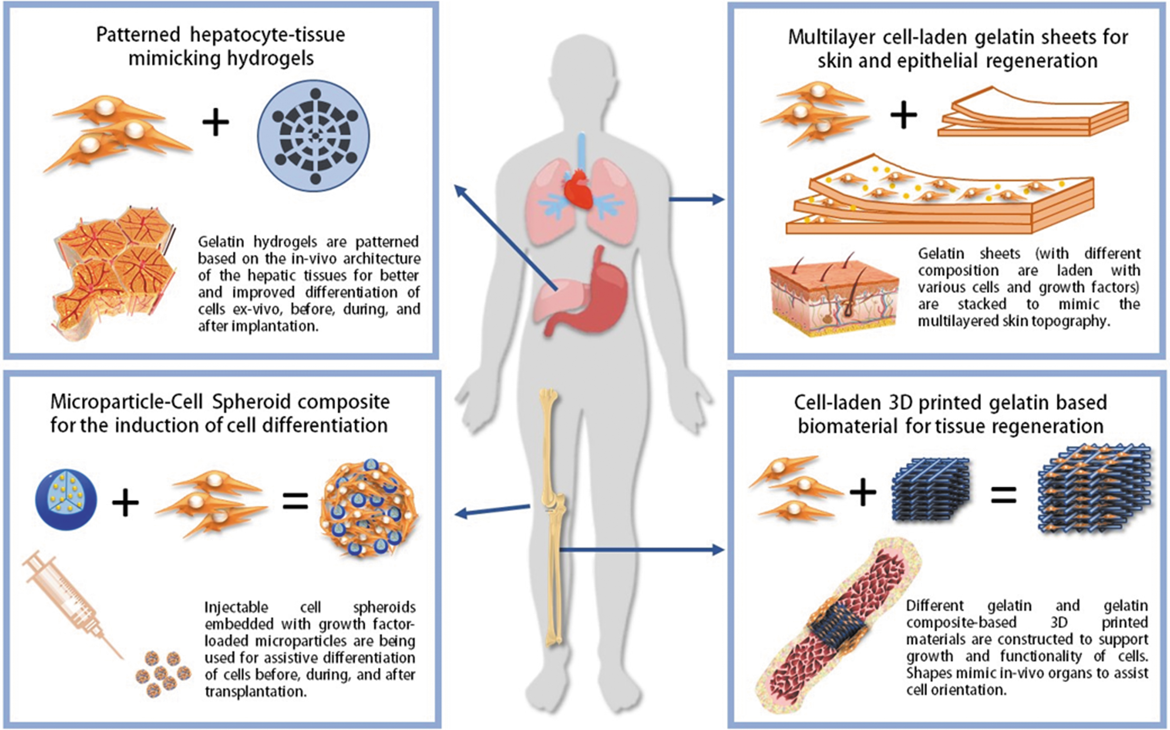
Representative medical application of gelatin-based biomaterials. Recent studies on gelatin-based biomaterials include the development of multilayered cell-laden sheets for skin and corneal regeneration, macro and micropatterned gelatin hydrogels mimicking in vivo microenvironments for the spatially assisted differentiation of cells, generation of injectable microparticle-incorporated cell spheroids for supported transplantation of exogenous cells, and 3D-printed bioscaffolds for the replacement of damaged tissues such as the bone and cartilage. 3D, three-dimensional. Color images are available online.
Gelatin sheets for cell support
Gelatin can be made into carrier sheets and serve as an attachment support for cells during different medical procedures. This form of gelatin biomaterial has been utilized in several medical applications and gelatin sheets are widely utilized for wound dressing. Several studies have shown that gelatin and gelatin composites are efficient in inducing the natural healing process of the body by supporting the recruitment, attachment, and growth of endogenous cells in the damaged area. 167 When infused with growth factors like epidermal growth factor and fibroblast growth factor, the healing functionality of these gelatin wound dressings is shown to be greatly improved.165,168–170
In an interesting study, gelatin films were fabricated to transport and deliver corneal endothelial cells for transplantation. Corneal endothelial cells are important for controlling the water content of the corneal stroma and thus, their replacement is essential in the event of tissue damage. In the study, corneal endothelial cells were shown to have a normal growth in gelatin films, as indicated by the normal expression of several marker genes. Moreover, the cells were shown to have a regular, mosaic, and polygonal arrangement similar to their physiological counterparts. These results demonstrated that gelatin films can be good materials for supporting and delivering cells for tissue repair. 171 One advancement in gelatin films is the production of multilayer biomaterials for enhanced functionality. In one study, a multilayered polycaprolactone/gelatin composite fiber was seeded with adipose-derived stem cells between layers. The layered conformation increased the scaffold's tensile strength and generated a cell-laden construct for enhanced differentiation of cells into native tendon cell phenotypes. 172
Gelatin-cell composites for cartilage and bone tissue regeneration
Cells encapsulated in gelatin scaffold have long been utilized to replace the damaged portions of certain tissues such as cartilage and bone. It is important, however, that materials used to replace tissues possess similar physical and biochemical properties as the physiological tissue. For instance, bone graft substitutes should both be rigid and porous, mimicking the outer cortical layer and the inner cancellous/trabecular layers, respectively. 173 Moreover, bone grafts should at least contain the inorganic (mainly hydroxyapatite crystals) and organic components of the bone. Since the organic component of the bone is rich in collagen type I, it is therefore valid to hypothesize that gelatin is a good candidate for designing bone biomimicking scaffolds. 174
In one study, gelatin/chitosan/nanosilica composite scaffolds were created and tested for the in vitro growth and proliferation of the osteosarcoma cell line. The composite scaffold was found to be better in terms of cytocompatibility, cell attachment, and alkaline phosphate activity compared with the conventional gelatin/chitosan scaffolds, indicating that nanosilica might be a good additive in the generation of gelatin-based bone graft substitutes. 175 In a similar study, the nanofibrous gelatin/apatite scaffold showed enhanced osteogenic differentiation of loaded osteoblasts in vitro. 176 In another study, gelatin/nanohydroxyapatite/chitosan microscaffolds (HaCGM) were fabricated and used for in vivo tissue regeneration in a subchondral bone lesion model in the rabbit. The results showed that animals treated with HaCGM microscaffold showed improved tissue regeneration compared with other setups. 177 A similar result was obtained when photo-crosslinkable gelatin/furfurylamine scaffolds were tested for the regeneration of subchondral bone in rabbit osteochondral defect models. 99 In all these studies, gelatin has been one of the most essential contributing factors in bone regeneration, attributed to its close biocompatibility to the physiological bone composition. An extensive review of gelatin-based biomaterials for bone regeneration is available elsewhere. 178
Gelatin as a substitute for tissue engineering
Aside from bone and cartilage tissue regeneration, gelatin and gelatin composite scaffolds have also been utilized in engineering other tissues. With the advent of 3D printing and other advanced techniques, the generation of scaffolds for more complex tissues and organs has become possible. For instance, several gelatin-based scaffolds have been tailored to support hepatic tissues.179–181 In an interesting study, a chitosan/gelatin composite scaffold with an organized microstructure analogous to the physiological hepatic tissue was shown to support the growth and proper development of hepatocytes in vitro. 182 This indicates that mimicking the topography of the native tissue is essential to generate scaffolds for tissue replacement.
Gelatin biomaterials have also been used for cardiac and vascular tissue regeneration. A 3D printed gelatin/hyaluronic acid patch infused with human cardiomyocyte progenitor cells (hCMPCs) was created to serve as a tissue patch for mouse models of cardiac infarction. In this study, the transplanted gelatin/hyaluronic acid/hCMPC patch in mice supported the proliferation and differentiation of the cells, leading to the preservation of cardiac performance in the mice. 166 In another study, aortic valves were also recreated in vitro using 3D printed gelatin/alginate composite scaffold. The printed scaffold was found to support the growth of two essential cardiac cell types, namely aortic root sinus smooth muscle cells and aortic valve leaflet interstitial cells. The cells were viable and showed a cellular phenotype similar to the physiological conditions such as elevated alpha-smooth muscle actin and elevated vimentin expression. 183 Other gelatin-based scaffolds for tissue engineering created through 3D and 4D printing are available in a review elsewhere. 184
Concluding Remarks
Although our understanding of gelatin and its uses for stem cell research and tissue engineering has skyrocketed over the past decades, many more studies are needed to maximize its full therapeutic potential in medicine. Among the natural sources for biomaterials, gelatin reigns supreme due to its intrinsic biocompatibility, guaranteed biodegradability, and abundance. Moreover, gelatin is relatively cheaper and does not induce an immune response in the body. Finally, its molecular simplicity makes it more susceptible to modifications for enhanced functionality. Given proper modifications such as the generation of gelatin/ECM composites or combination with other chemicals or growth factors, gelatin-based biomaterials can provide a great solution in current material-based therapeutics. With the advent of 3D printing technologies such as additive manufacturing, rapid prototyping, and free-form fabrication, rapid developments have been made in the biomaterial field. Gelatin is now being utilized as a base material in bioink production for various functional scaffolds that offer a solution for replacing damaged tissues and organs. Indeed, gelatin might be underrated, but its potential for developing fully functional biomaterial-based therapeutics is limitless.
Footnotes
Disclosure Statement
No competing financial interests exist.
Funding Information
This work was supported by the National Research Foundation of Korea (NRF) grant funded by the Korea government (MSIT) (NRF-2019R1A2B5B03069690 and NRF-2019M3A9H1032376), the Korea Health Technology R&D Project, in the Korea Health Industry Development Institute (KHIDI) and the Ministry of Health & Welfare, Republic of Korea (Grant No. HI14C3484).