
Abstract
Several imaging modalities are available for investigation of the morphological, functional, and molecular features of engineered tissues in small animal models. While research in tissue engineering and regenerative medicine (TERM) would benefit from a comprehensive longitudinal analysis of new strategies, researchers have not always applied the most advanced methods. Photoacoustic imaging (PAI) is a rapidly emerging modality that has received significant attention due to its ability to exploit the strong endogenous contrast of optical methods with the high spatial resolution of ultrasound methods. Exogenous contrast agents can also be used in PAI for targeted imaging. Applications of PAI relevant to TERM include stem cell tracking, longitudinal monitoring of scaffolds in vivo, and evaluation of vascularization. In addition, the emerging capabilities of PAI applied to the detection and monitoring of cancer and other inflammatory diseases could be exploited by tissue engineers. This article provides an overview of the operating principles of PAI and its broad potential for application in TERM.
Impact statement
Photoacoustic imaging, a new hybrid imaging technique, has demonstrated high potential in the clinical diagnostic applications. The optical and acoustic aspect of the photoacoustic imaging system works in harmony to provide better resolution at greater tissue depth. Label-free imaging of vasculature with this imaging can be used to track and monitor disease, as well as the therapeutic progression of treatment. Photoacoustic imaging has been utilized in tissue engineering to some extent; however, the full benefit of this technique is yet to be explored. The increasing availability of commercial photoacoustic systems will make application as an imaging tool for tissue engineering application more feasible. This review first provides a brief description of photoacoustic imaging and summarizes its current and potential application in tissue engineering.
Introduction
The fields of tissue engineering and regenerative medicine (TERM) are primarily focused on the development of new sources of tissue or organs for replacement, regeneration, or reconstruction. 1 New approaches for the design of engineered tissues are continually under development and investigation. 2 However, tissue regeneration is a complex process modulated by multiple cues in the physiological environment. The better understanding of the interactions between engineered and host tissues is required for the design of new systems, but the tools available for evaluating this process are limited even in preclinical models. There is a significant need for new tools that enable monitoring and assessment of engineered tissues in vivo in animal models to enable the design and evaluation of next-generation tissue engineering strategies. 3
Histological analysis is the gold standard for tissue evaluation. While histology provides crucial information, it is limited particularly when faced with unique aspects of tissue engineering. Histological analysis is generally an end point experiment precluding longitudinal monitoring or assessment. Besides, the processing required for histology can alter biomaterial structure in unpredictable ways, and the limited volume assessment can provide misleading results. A broad range of biomedical imaging technologies are available that may allow long-term monitoring of the morphological, functional, and molecular properties of engineered tissues. In addition, noninvasive imaging modalities may ultimately allow monitoring of therapeutic progress clinically as a potential guide for intervention if necessary.
The ideal characteristics of an imaging system depend, in part, on the application under evaluation. Desired characteristics for any imaging technique include (1) safe, (2) noninvasive, (3) high spatial resolution, (4) high tissue penetration, (5) quantitative, (6) functional, (7) allow longitudinal monitoring, (8) enable cell tracking, (9) provide information of biomaterial properties, and (10) free of exogenous labeling agents. Common imaging modalities such as magnetic resonance imaging (MRI), computed tomography (CT), and ultrasound (US) can achieve some of the characteristics.
Regardless of modality used, there is a natural give and take based on relative advantages and limitations in TERM applications (Table 1). For instance, MRI may have high spatial resolution and high tissue penetration, but it requires long scan times, is expensive, and may not enable material contrast. X-ray based imaging modalities (e.g., X-ray CT) provide superior resolution and imaging depth, but uses ionization radiation and provides limited soft-tissue contrast when used at a standard X-ray dose. US, in contrast, is safe and provides higher imaging depth, but generally has low spatial resolution for the TERM application. Therefore, the continued development and application of new imaging tools would benefit the TERM fields.
Summary of Imaging Modalities in Tissue Engineering and Regenerative Medicine
CT, computed tomography; MFM, multi-photon fluorescence microscopy; MRI, magnetic resonance imaging; OCT, optical coherence tomography; PAI, photoacoustic imaging; PET/SPECT, positron emission tomography/single photon emission computed tomography; US, ultrasound.
Photoacoustic imaging (PAI) system has emerged as a promising alternative for biomedical imaging for TERM applications.4–9 PAI has been extensively investigated in biomedical imaging such as cancer diagnosis, cardiovascular diseases, and neurology to name a few. While the PA effect was first discovered by Alexander Bell in the 18th century, 10 its rapid progress in the past few decades is fueled by fast-paced advancement in laser technology and emerging fundamental research in PAI. PAI, also known as optoacoustic imaging, is a hybrid technique meaning it exploits two different phenomena, optical and acoustic, to generate images. When an optical pulse from a laser is used to excite a tissue, optical absorption results in a transient local thermal expansion of tissues. Thermal expansion leads to a change in pressure distribution in the tissues and the generation of broadband ultrasonic waves. The resultant acoustic signals can then be detected outside the tissue for reconstruction.5,6 The reconstructed image depicts a map of initial pressure distribution that is proportional to absorbed optical energy. 11 These images provide anatomical, functional, and molecular information of biological tissues.
PAI generally offers a high spatial resolution, increased imaging depth relative to most optical methods, label-free vascular imaging, and retrieval of functional and molecular information. In addition, extrinsic contrast agents can be used to generate or enhance PA contrast. Moreover, PAI does not use ionizing radiation, and therefore, PAI demonstrates high potential for clinical applications where safety is an important concern. As there are a number of excellent review articles on PAI,4,5,6–9,11 we provide only a brief overview of three major modalities of PAI: PA computed tomography (PACT), PA microscopy (PAM), and multispectral PAI. The bulk of the review focuses on current and potential applications of PAI in TERM.
Basic Image Formation Principles of PAI
Optical imaging plays an important role in preclinical and clinical studies. However, it suffers from poor spatial resolution at depth due to light diffusion in highly scattering tissues.
12
Conventional optical imaging techniques, including confocal microscopy and two-photon fluorescence microscopy, have an imaging depth typically limited to <1 mm.
13
In contrast, US images can be obtained at a much deeper tissue level (a few centimeters). However, US imaging does not provide molecular specificity and its image quality typically has low contrast limited by the US contrast mechanism that depends on the mechanical and elastic properties of the tissue rather than individual molecules.8,9 PAI has emerged as a promising modality to address many of the most challenging issues in conventional optical and US imaging methods. It is based on a hybrid technology that combines rich optical contrast mechanisms and superior ultrasonic penetration depth and resolution.5–8,14–16 For PAI, photons from a nanosecond laser pulse are absorbed by certain endogenous chromophores or exogenous contrast agents in a tissue sample, causing impulsive heating and acoustic stress. The acoustic stress relaxes by launching broadband US pressure waves (i.e., PA emission), which propagate to the outside of the tissue and are detected by a mechanically scanned US receiver or an array of US receivers to form PA images.5,17,18 For effective PA signal generation, it is necessary to reach the so-called thermal and stress confinement conditions using a pulsed laser with its pulse duration normally within several nanoseconds, so that the thermal diffusion and stress relaxation can be neglected during the laser illumination period.
4
The initial pressure, P0, generated by an optical absorber is determined by the expression
Since acoustic waves scatter much less than optical waves in tissue, PAI allows for deep-tissue imaging by leveraging the acoustic signals originated from optical absorption and offers advantages of rich endogenous and exogenous optical contrast agents for functional molecular imaging without the use of ionizing radiation. 20
Photoacoustic Computed Tomography
In a canonical PACT experiment, an expanded laser beam is used to irradiate the whole tissue region of interest. PA waves generated from the whole volume of the targeted tissue are detected by an US detection system that completely or partially surrounds the object. From the detected acoustic signals, a reconstruction algorithm is utilized to form an image that depicts the distribution of the initial PA-induced pressure distribution
Similar to other computed imaging modalities such as X-ray CT and MRI, image quality in PACT will be influenced by the quality of measurement data and the choice of image reconstruction algorithm used. An extensive effort from many research groups has been devoted to the development of different approaches for image reconstruction in PACT. 11 Multiple reported approaches, including filtered back-projection,23,24 model-based inversion algorithms,25–27 and wavelet-based approaches,28,29 have been demonstrated to provide a successful mapping of the distributions of targeted optical absorbers in a 3D tissue volume. Most of the reported implementations of PACT sought to reconstruct estimates of the PA-induced pressure distribution. Active research for developing new image reconstruction algorithms is undergoing to correct the artifacts and distortions in PACT images due to tissue heterogeneity.30–32 For example, researchers are currently developing a transcranial image reconstruction algorithm to compensate for the effects of wave propagation through the skull to better image the brain.33,34 Quantitative PACT methods seek to obtain accurate estimates of the medium's optical parameters. Obtaining absolute chromophore concentrations from PA images obtained at multiple wavelengths is a nontrivial aspect of PAI but is essential for accurate functional and molecular imaging. 7 However, such methods are still in a stage of development and have not been widely validated. 35
Photoacoustic microscopy
In contrast to PACT for deep tissue imaging, PAM is used to achieve superior high-resolution imaging, although its imaging depth is limited. PAM typically utilizes a confocal configuration by having overlapped foci of both the optical excitation and ultrasonic detection to optimize its sensitivity and resolution. 36 As the time-of-flight information of the PA signal provides the axial resolution, each focused laser beam position produces a depth-resolved one-dimension (line) image into the tissue without mechanical scanning. Thus, a 2D transverse scanning of the laser beam (together with the US receiver) generates a 3D image. The lateral resolution is determined by the product of the point spread functions of the laser focus and acoustic detection focus. 37 Optical-resolution PAM (OR-PAM) can be achieved when the optical focus is significantly tighter than the acoustic focus. 38 Even though other optical imaging techniques, such as confocal and two-photon fluorescence microscopy, also provide high-resolution imaging (spatial resolution of 1–2 μm) capability at an imaging depth similar to that of PAM (0.2–1 mm depth), PAM possesses a unique feature for label-free imaging with optical absorption as a contrast. For example, in vivo label-free functional imaging of hemoglobin oxygen saturation (sO2) in single blood capillaries can be imaged clearly with OR-PAM (Fig. 1). 38 Different from PACT, image formation for PAM is relatively straightforward and does not require the use of complicated image reconstruction algorithms, as it utilizes direct image formation principles.
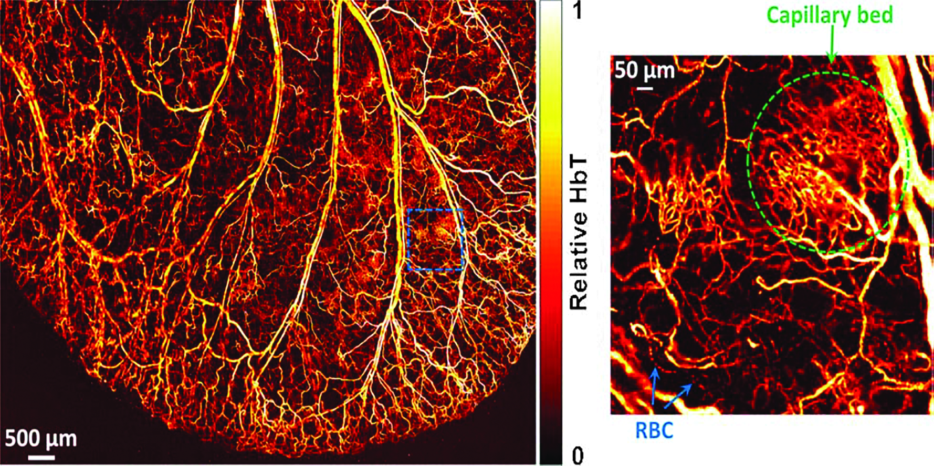
OR-PAM of relative total hemoglobin concentration (HbT) in a living mouse ear, revealing the vascular anatomy. Insert shows a densely packed capillary bed and individual red blood cells traveling along a capillary. © 2011 Reprinted with permission of Optical Society of America. 38 OR-PAM, optical-resolution PAM. Color images are available online.
Multispectral imaging
In this section, we will discuss the principle of multispectral PAI and its pertinent applications in tissue engineering. In addition, we will briefly cover the use of multispectral imaging for quantitative PACT.
PAI at a single optical excitation wavelength can spatially resolve certain photoabsorbers with a distinct absorption spectrum. However, for samples containing multiple absorbers with overlapped absorption spectra, the different sources of absorption cannot be distinguished from one another if PAI is conducted at a single excitation wavelength. This situation is not uncommon especially for in vivo studies, as the spectra of the targeted photoabsorber may overlap with other endogenous photoabsorbers or other exogenous contrast agents used to label different tissues. 39 In such a situation, multispectral PAI is required to identify the photoabsorbers of interest through their unique absorption spectra through spectral unmixing.
Spectral unmixing algorithms are applied to the resulting images obtained at multiple excitation wavelengths to distinguish targeted photoabsorbers (whether endogenous and/or exogenous) with overlapped absorption spectra.
40
In short, 2D spectral unmixing commonly adopts a linear mixture model, which assumes that the measured spectrum at each image location M(r, λ) (function of position, r, and wavelength,
Quantitative PAI is a technique that aims at obtaining/estimating the absolute concentrations of the present chromophores from PA images. 7 As the attenuation of light, even at a fixed depth, can vary with the excitation wavelength used, it is challenging to accurately quantify the concentration of the targeted photoabsorber. Therefore, it is of great interest to develop novel methods to correct/overcome this challenge to obtain quantitative results in deep tissue. There are multiple ongoing studies on this topic as reported recently.7,42,43 The most common method hinges on multispectral imaging to estimate the optical absorption and scattering coefficients. Once determined, estimates of local concentrations of absorbers can be calculated, with the most common one being hemoglobin oxygen saturation (sO2).
To summarize, by leveraging the use of multiple excitation wavelengths in conjugation with either PACT or PAM, multispectral imaging can offer an analysis of multiple endogenous and exogenous contrast agents concurrently in real time. Currently, these multispectral imaging techniques have been used to image blood vessels, 44 quantify oxygen saturation levels, 45 identify/monitor melanoma, 46 detect lipids in vessels, 47 and to detect and characterize glioblastoma, 48 to name a few of the multitude of applications capable with this powerful technique.
Contrast in PAI
Several contrast mechanisms can be exploited in PAI to provide insight into engineered tissues. Contrast agents can be categorized as endogenous and exogenous agents. One of the exciting aspects of PAI is the differential optical absorption of endogenous chromophores that provides intrinsic contrast. Intrinsic optical absorbers such as blood, lipid, melanin, and collagen can be imaged with PAI without the need for additional contrast agents (Fig. 2). Multispectral analysis can allow identification and separation of distinct optical absorbers. In addition, exogenous contrast agents can be introduced to enhance detection sensitivity and to target specific structures, molecules, or cells.
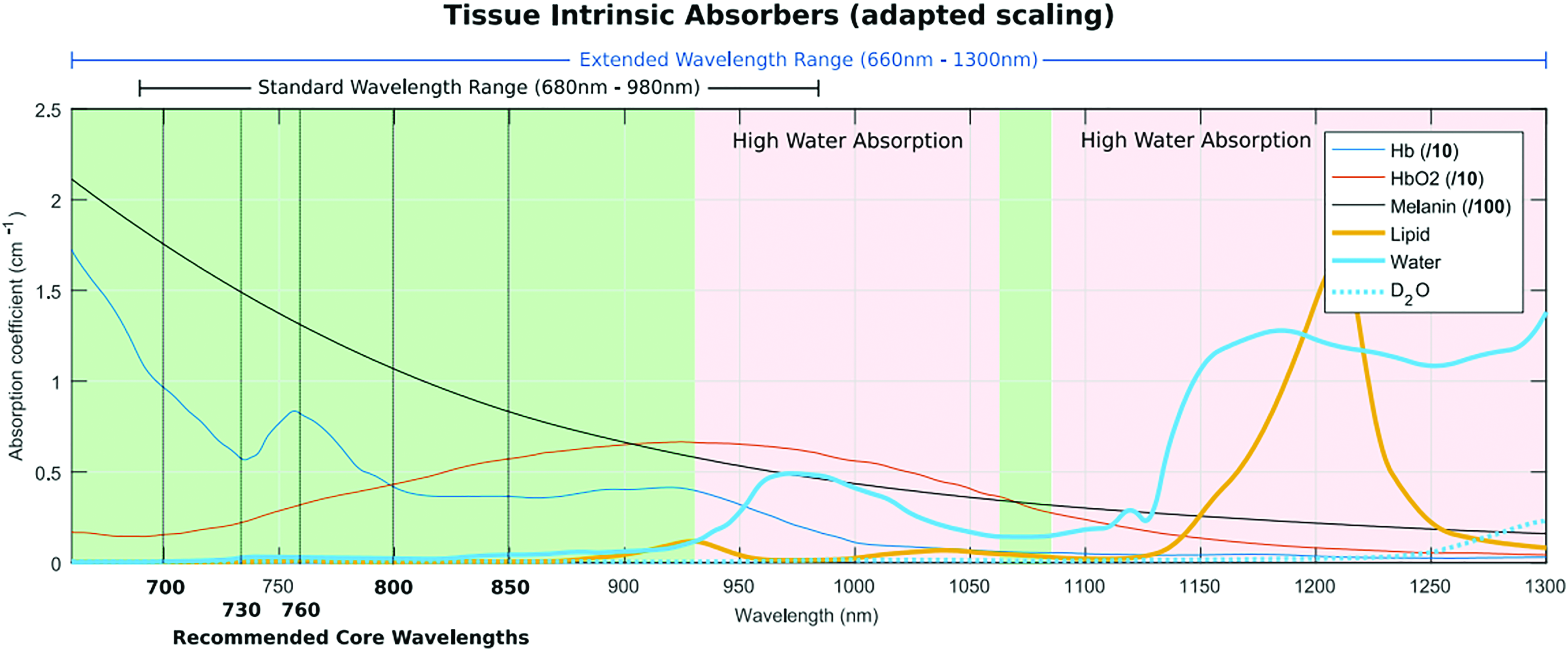
Absorbance spectra of endogenous contrast agents. © 2012 Reprinted with permission from iThera Medical, Inc. Color images are available online.
Endogenous contrast agent
Common endogenous contrast agents, including blood, lipid, and melanin, have been widely investigated to predict, detect, or monitor a variety of disease conditions ranging from cancer to inflammatory diseases, such as atherosclerosis. The sections below provide examples of how endogenous contrast has been exploited to provide insight into tissue features relevant to TERM applications.
Blood
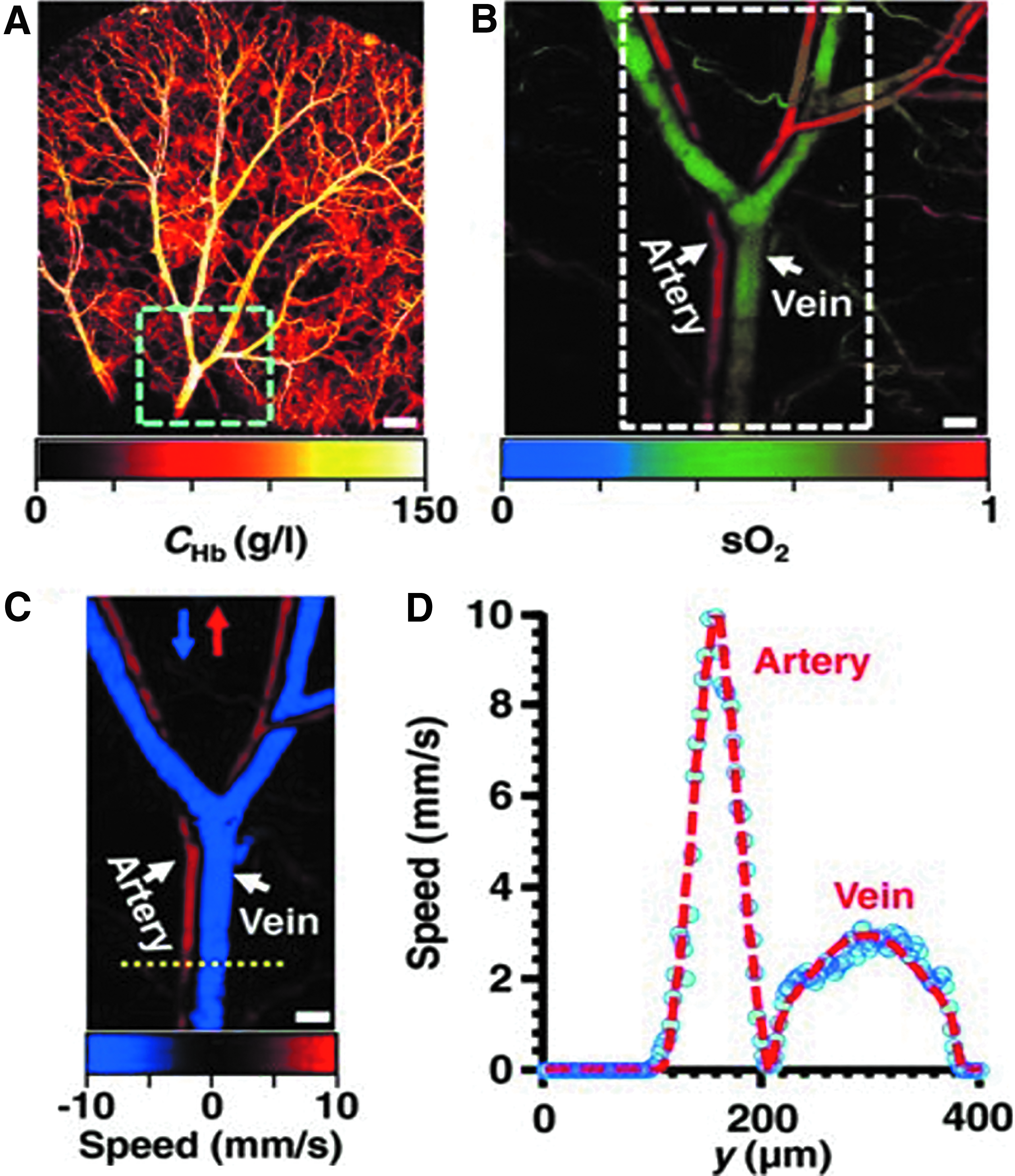
Blood is the most studied endogenous contrast agent for PAI. The absorbance of blood is in a broad spectral range and is significantly higher relative to other common chromophores, at least by an order of magnitude. As a result, functional vasculature (i.e., vasculature containing blood) generates significant contrast with PAI. This contrast can be used to quantify the microvascular density and evaluate vascular network structure. The optical absorption of blood is primarily due to hemoglobin. The absorption properties of hemoglobin are dependent on its oxygenated (HbO2) or deoxygenated (Hb) state. This optical property of blood allows PAI to determine total hemoglobin concentration, oxygen saturation level of blood, differentiate between veins from arteries, and blood flow speed (Fig. 3). 49 These features result in extensive applications of PAI in diagnosis, monitoring, or evaluation of conditions that are typically characterized by alterations in vasculature such as cancer and cardiovascular diseases. 50 Vascularization is also critical for the clinical success of engineered tissues, making this an important feature of PAI. 51 In PA, signal generation is independent of the movement of the absorber (i.e., hemoglobin). Other commonly used tools, such as power doppler imaging, require movement of blood and which can result in false positive or undetected vessels.52,53
Metabolic PAM images.
Lipid/fat
Lipids exhibit absorbance in the range between 1150 and 1310 nm. 54 The strong absorption of lipids has enabled intravascular lipid deposition to be detected with PAI. 55 Lipid deposition inside the arterial wall is increased in atherosclerosis, a harbinger of many cardiovascular diseases. In addition to diagnosis, PAI of lipids can also be used to characterize the lipid content and its spatial distribution within plaques and thus can be used to monitor disease progression. 56 Wang et al. reported the presence of a “valley” in the water absorption spectra between 1450 and 1900 nm. Based on this result, they reported a new optical window for deep tissue imaging from 1600 to 1850 nm, where the absorption of pure water is comparable to the absorption coefficient of heme protein at 800 nm. 57 Spectral features of C-H overtones within this range demonstrated label-free detection of arterial plaques. Lipids are rich in CH2 groups, and therefore, CH2 overtones can distinguish the presence of lipids at 1.7 μm wavelength through the layer of blood.58–60
Melanin
Melanin is a group of natural pigments that determine skin color. Melanocytes are the cells that play a crucial role in the production and distribution of melanin. Melanin exhibits a strong absorbance (1000 times than water) in the visible range of 700–730 nm. At these wavelengths, melanin demonstrates high contrast relative to other endogenous contrast agents. Melanin PAI has mostly been investigated to diagnose or monitor the progression of Melanoma, a skin cancer that arises from mutation of melanocytes.61,62
Collagen
Collagen is the most abundant protein in the body and a key component of the extracellular matrix in many tissues and organs. Collagen imaging may allow disease diagnosis and monitoring of disease progression.63,64 For example, Kruizinga et al. showed that lipid and collagen could be differentiated in the arterial wall for the detection of atherosclerotic lesions. 65 However, the absolute spectrum of collagen is not well established, partly due to the high scattering and fluorescence properties of collagen in the visible range. In addition, a tunable laser system that operates in the range of above 1000 nm is required to evaluate the absorbance of collagen. 66 Recently, Sekar et al. compared collagen absorption in the visible and short wave infrared region (SWIR). They reported higher absorption of collagen in the range of 1.1–1.7 μm than in the visible range of the spectrum. On further comparison within the SWIR range, they observed collagen absorption to be 5 times higher than lipids at 1700 nm and 1.5 times higher at 1100 nm. 67 Similar to lipids, Wang et al. have reported that vibrational overtone absorption of CH3 group can distinguish collagen in PAI. 60
Exogenous contrast agent
PAI without the introduction of exogenous contrast has been widely studied and shown to be useful in a variety of applications. Nevertheless, there are many limitations with relying solely on label-free imaging. Contrast agents have played an important role in molecular imaging, due to their versatility, customizability (for specific applications) and ease of use. There are various principles, materials, shapes, and sizes of contrast agents suitable for different imaging modalities. Ideal contrast agents could significantly increase contrasts, effectively improve imaging depth or accuracy, and provide pertinent molecular specific information.68–70 In this section, a short review about various exogenous contrast agents for PA molecular imaging is presented.
Synthetic near infrared dyes
There are many biocompatible dyes that absorb in the near infrared (NIR) optical window, 71 including heptamethine cyanine dyes (i.e., Indocyanine Green [ICG], and the IRDye family [range from 680 to 800 nm]), borondipyrromethene dyes (i.e., the AlexaFluoro family [range from 650 to 790 nm]), squaraine dyes (i.e., D172,73), rhodamine dyes, and azo dyes (i.e., methylene blue), which have all been used extensively in PAI. These dyes are typically small molecules, on the order of 1 nm, and are able to quickly clear the body through the renal system. Many of these dyes are fluorescent and are commonly used in purely optical imaging. For PAI, the PA emission comes from the energy due to the optical absorption. A low fluorescence quantum yield of the dye results in more efficient PA signal generation, as more of the absorbed energy is converted into the PA signal rather than being emitted as fluorescence. 68
It has been shown that to obtain static contrast in PAI, a NIR dye such as ICG can be detected at nanomolar concentrations in tissue.8,74 However, with respect to responsive imaging probes for PAI, very few reports are currently available. One such report by Mishra et al., demonstrated the development of a metallochromic sensor utilizing a heptamethine cyanine dye, IR780, to measure calcium concentrations. 75 Quantifying calcium ion concentrations pertain to many biological processes/responses. Specifically, pertaining to bone tissue regeneration, calcium is a fundamental building block of the native extracellular matrix, 76 as well as being shown to have a significant role in osteogenic differentiation, 77 making it an ideal candidate for molecular PAI in tissue engineering.
NIR protein dyes
With the steady rise in applications capable with PAI, synthetic dyes (which require methods for target and delivery) present many hindrances for more longitudinally relevant applications. Protein dyes, as conventionally used in optical imaging, can be genetically encoded, thus ever present in a targeted sample in vivo, allowing for a multitude of imaging applications. One particular family of the dyes that shows great promise for PAI is the iRFP family.78–81 With optical absorptions in the range of 650–750 nm and a low quantum yield (for better PA signal generation), they can be coupled with gene-delivery technologies (i.e., CRISPR) to be a powerful tool for the targeting, labeling, and longitudinal monitoring of specific cell groups in vivo. 82
Plasmon nanoparticles
Noble metal (i.e., gold and silver) nanoparticles (NPs) have been widely used as PA contrast agents. This is primarily due to their strong and tunable optical absorption that results from the surface plasmon resonance (SPR) effect. The SPR effect occurs when free charges on the surface of noble metal NPs oscillate in concert with the electromagnetic field, resulting in an optical absorption that is five orders of magnitude greater than dyes (on a per-particle basis). When the size and shape of the NP change, so does its resonant frequency. For example, as a gold nanorod grows longer, its peak absorption shifts to longer wavelengths. 83 The ideal wavelength for absorption depends on the other endogenous and exogenous absorbers in the region and the excitation wavelengths available for imaging. In addition to being highly tunable, gold NPs (GNPs) form strong gold-thiolate bonds that enable covalent surface modification, such as polyethylene glycol functionalization, for increasing biocompatibility 84 and active targeting (i.e., antibody conjugations). 68 Furthermore, utilizing a silica coating around GNPs can both increase PA stability, 85 allowing for longer image times without damaging the contrast agent, while also increasing the PA signal generation, 86 for better image quality.
The field of stem cell therapy is currently advancing toward clinical trials in a variety of biomedical applications, most notably in the field of TERM.87–91 This advancement leads to the new requirements of medical imaging to monitor stem cell therapies (i.e., stem cell tracking). One example of utilizing plasmonic NPs to meet the demands of monitoring novel stem cell therapies was reported by Ricles et al. They showed the use of a dual GNP system in conjunction with PACT for the monitoring of both implanted mesenchymal stem cells, loaded with gold nanorods, and infiltrating macrophages, loaded with gold nanospheres, to the wounded area. 92
Carbon nanostructures
Carbon forms different allotropic structures as follows: carbon nanotubes, graphene-based nanomaterials, and nanodiamonds. All three classes can be synthesized to possess an intrinsic absorption in the NIR range and thus play an important role for PAI. Despite possessing a lower molar extinction coefficient than gold, carbon NPs (CNPs) hold significant potential for molecular PAI due to their flexibility of synthesis and functionalization.93,94 CNPs can be also conjugated with plasmonic NPs to further enhance their PA emission properties. For example, fluorescent nanodiamonds (FNDs), a novel nanomaterial that has been utilized for biomedical imaging applications due to its excellent photostability, high biocompatibility, and extended far-red fluorescence emission, 95 may also be utilized as a PA contrast agent by conjugating them with GNPs. 96 It has been demonstrated that their PA signals can be significantly enhanced by a factor of 30 when the FNDs are conjugated with GNPs. The large increase in the PA signal has been attributed to the local field enhancement of the GNPs and the energy transfer between FNDs and GNPs, which increases the nonradiative decay processes of FNDs through fluorescence quenching, thus enhancing the conversion efficiency of the absorbed laser energy into heat for enhanced thermoelastic effects (i.e., PA signal generation). 96
One example of utilizing carbon nanostructures in tissue engineering was reported by Cai et al., where single-walled carbon-nanotubes (SWNTs) were incorporated in poly(lactic-co-glycolic acid) (PLGA) polymer scaffolds to both image and characterize the scaffold through PAM. 97 Characterization and longitudinal monitoring of porous polymeric scaffolds are critical, as these structures are vital in the development of neo-tissue formation, as they provide structural support, and can be integrated to house implanted cells and elude pertinent growth factors. 98
Polymer NPs
There are many different polymer NPs, in which their strength comes from their ease of customizability (i.e., conjugations with other NPs or dyes, encapsulations of other NPs or dyes, and functionalization for molecular targeting). Organic semiconducting polymer NPs prepared from semiconducting polymers are promising nanoagents with excellent optical properties for imaging and therapy and have been demonstrated in PAI. 99 Conjugated polymers are macromolecular structures with a highly delocalized conjugated backbone. The conjugated system is spread through the entire polymer backbone, and the polymers are densely packed into NPs, yielding much higher extinction coefficients and photostability than possible with small molecule dyes. One can tune the optical properties (which are strongly dependent on the conjugated core) by adopting different backbone structures, combining different conjugated polymers, and controlling aggregation and surface functionalization. 100
Wound healing and regenerative medicine go hand in hand. Whether implanting an engineered biomaterial or transplanting stem cells, the wound healing process follows the required invasive procedure. 101 pH is a recognized indicator of the state of the wound, providing information about bacterial contamination and the stage of healing. 102 Pu et al. have reported a novel PA pH-dependent contrast agent, made of semiconducting polymer NPs in response to reactive oxygen species. 103 This can potentially be implemented to work in conjugation with PACT to monitor pH in vivo, thus wound healing, following biomaterial or stem cell therapies.
Table 2 summarizes the different types of exogenous contrast agents used in PAI, its application in tissue engineering, and general limitations.
Summary of Exogenous Photoacoustic Contrast Agents Used in Tissue Engineering and Regenerative Medicine
MWNTs, multi-walled carbon nanotubes; NIR, near infrared; NPs, nanoparticles; OSPNs, organic semiconducting polymer NPs; PA, photoacoustic; PAI, photoacoustic imaging; SWNT, single-walled carbon-nanotube; TERM, tissue engineering and regenerative medicine.
PAI in TERM
Tissue engineering scaffolds
Biomaterial scaffolds, both natural and synthetic, are often a fundamental component of a successful tissue engineering strategy. The scaffolds provide physical support for tissue growth but also can directly modulate cell activity and function. 104 The physical, chemical, and mechanical properties of tissue engineering scaffolds play a vital role in cell migration, proliferation, and differentiation, 105 and changes in these properties due to degradation (intentional or not) can facilitate the success or failure of the tissue. Substantial scaffold degradation studies are often performed in vitro before implantation, but the characterization of degradation in vivo is a significant challenge in tissue engineering. 3 Longitudinal quantitative analysis of the scaffolds can provide information that is critical to understanding the behavior of the materials in vivo. In addition, tools that allow evaluation of cell-scaffold interactions in vivo may provide important cues in understanding cell behavior in tissue engineering systems.
Common imaging modalities, including CT and scanning electron microscope (SEM), are used for preimplantation characterization of scaffold structure. However, SEM cannot be applied to image implanted samples and most biomaterials do not generate sufficient contrast for imaging with CT under in vivo conditions. Besides, imaging of cells within scaffolds is often performed by staining with osmium tetroxide, which requires harvest and postprocessing.106,107 PAI has been explored as an alternative technique for imaging polymeric scaffolds and the cells cultured in these scaffolds. Usually polymer itself does not generate enough PA signal for PA contrast; therefore, contrast agents are incorporated into biomaterial scaffolds to enhance PA contrast. In the absence of intrinsic cellular contrast, exogenous contrast agents such as dyes or NPs can be incorporated into cells108–110 or biomaterial scaffolds to enhance PA contrast. 111 For instance, SWNTs were entrapped in PLGA scaffolds for mechanical reinforcement and to provide PAI contrast. 97 Both OR-PAM and acoustic resolution PAM (AR-PAM) were used to characterize average porosity and pore size of PLGA scaffolds immersed in a biological buffer. Micro CT images of dry and wet scaffolds were taken for the comparisons. Micro-CT images of dry samples clearly depicted well resolved porous structure. However, wet scaffolds (immersed in fetal bovine serum) failed to provide any structural details. The structural details from PAM images agreed well with measurements obtained by micro CT of dry scaffolds. To evaluate scaffolds in an environment mimicking in vivo imaging, the scaffolds were embedded in chicken breast tissue. With ∼660 μm tissue penetration depth and lateral resolution of 2.6 μm, OR-PAM images depicted detailed structures of the scaffold that was comparable to that of an optical microscope such as one-photon fluorescence microscope. AR-PAM, in contrast, was able to image scaffold through 1.7 mm of soft chicken tissue. The lateral resolution of AR-PAM is less (45 μm), but able to achieve greater tissue depth. As resolution depends on the penetration of US signals, the deeper tissue penetration typically corresponds with a sacrifice in lateral resolution.
PA has received significant attention largely due to its advantages for noninvasive imaging in people or animal models. 112 Talukdar et al. used PA and US imaging concomitantly to image SWNT-PLGA scaffolds ex vivo, as well as in vivo. 113 In this study, US images provided the anatomical location of the scaffold, whereas PA images provided the blood oxygen saturation maps around and within the scaffolds which is difficult to achieve with other imaging modalities. PLGA and PLGA-SWNT scaffolds were first embedded in the chicken breast tissue of varying thickness for ex-vivo testing (Fig. 4). For in vivo imaging, the scaffolds were implanted subcutaneously in rats at ∼2 mm depth. The PA and spectroscopic images (blood oxygen saturation maps) of the PLGA scaffolds and SWNT-PLGA scaffolds were obtained at day 7 and 14, which suggest that noninvasive long-term monitoring of scaffolds postimplantation is possible with PAI. The rate of scaffold degradation plays an important role in tissue engineering. While in vitro degradation studies are routine, evaluation of in vivo degradation is difficult. Recently, PAI has been used for noninvasive structural assessment of degradable polymers in vivo in parallel with US shear wave imaging for analysis of polymer mechanical properties. 114 The PAI signal from the scaffolds agreed with histological images obtained from the explanted tissue. This result indicates that PAI can be utilized to monitor structural changes that may occur in the scaffolds in vivo. PAI technique may provide vital information about tissue engineering scaffolds postimplantation and hence fill the gaps in understanding between in vitro and in vivo degradation behavior.

PA imaging of scaffolds. US-PA images of PLGA
Vascular imaging
One of the most established applications of PAI is the analysis of vasculature in tissue.115–120 This noninvasive and label-free vasculature imaging approach has been utilized to study existing disease conditions, monitor therapeutic progress, and diagnose clinical conditions. Vascular imaging with PA has been successfully used in diagnosis and assessment of cancer,121–123 cardiovascular diseases, 124 microvascular abnormalities,120,125,126 and superficial soft tissue damages. 127 Vascularization is crucial for engineering tissues of sufficient volume for clinical impact.128–130 In the absence of adequate nutrients and oxygen, tissues can become hypoxic hindering tissue development and function. Assessing vascular development and function in engineered tissues in vivo is vital. Early detection of relevant abnormal vascularization condition may provide the opportunity to intervene to improve therapeutic outcomes or avoid catastrophic events.
Vascular imaging with PAI has been performed to evaluate wound healing. Wound healing involves three phases: Inflammation phase, proliferative phase, and remodeling and scar formation. The structural changes that occur following the inflammation phase include extracellular matrix formation, angiogenesis, granulation tissue formation, and reepithelialization. 131 PAI of angiogenesis and oxygen saturation level can be used to evaluate wound healing. A higher oxygen saturation level together with angiogenesis has been used as an indicator of wound healing and tissue regeneration. 132 In one study, PAI was used to monitor perfusion and hemodynamic changes in the burn healing process. PA images taken postburn at different interval demonstrated increased PA amplitude indicating neovascularization. The distribution and the density of neovascularization were qualitatively confirmed using histological analysis. 133 In another interesting application, PAI in combination with US imaging was first used to diagnose the severity of the burn and later used to track stem cells to monitor vascularization and skin tissue regeneration. 127 Three different temperatures (87°C, 100°C, and 133°C) for various durations were applied to mimic varying burn degrees. The PA images of the wound caused by 100°C and 113°C for 30 s indicated significant subcutaneous bleeding similar to third degree burns. No PA signal was observed in case of 87°C for 30 s and 113°C for 10 s indicating less severe burn injury. Skin incision verified PA images to be in well accordance with subcutaneous bleeding level suggesting that this technique can be used to determine the severity of burn injury. In a follow-up to this initial injury, PAI was used to monitor the therapeutic progress after treating burn injury with adipose-derived stem cells (ASCs). These ASCs were labeled with gold nanorods for imaging contrast. PA images were used to evaluate blood vessel density and blood perfusion at 2 weeks postinjury. Spectral PA images of ASCs and vascularization provided critical information regarding the morphological changes that occurred along the wound healing process (Fig. 5).

PA imaging in cell tracking and monitoring. PA images of burn tissue treated with ASCs. ASCs were labeled with gold nanorods for PA contrast. Spectroscopic PA imaging allowed to identify and track ASCs over the period of 14 days. Oxygen saturation (oxygenated-red and deoxygenated-blue) indicated wound healing or tissue regeneration process. © Reprinted with permission of Mary Ann Liebert. 127 ASCs, adipose-derived stem cells. Color images are available online.
Recently, PAI was used to detect thrombosis in the carotid artery of a mouse model of ferric-chloride induced endothelial injury and arterial occlusive thrombus formation. Within thrombi, the hemoglobin concentration decreases. By measuring the optical absorption of hemoglobin, Li et al. reported the detection of artery thrombosis in mice. 134 Red blood cells (RBCs) in fresh clot formation generate higher PA signal than old clots. As the clot ages, it retracts forcing RBCs and other content out of the clot 135 resulting in a reduced PA signal. Based on this phenomenon, Karpiouk et al. used PAI to visualize thrombosis and estimate the age of the blood clot to differentiate the acute and the chronic stage of thrombosis. 136 While this application is not tissue engineering per se, the ability to evaluate clot formation could be useful in evaluating tissue-engineered vascular grafts. One recent study demonstrated PAI as a technique to monitor neovascularization and integration of decellularized human scaffold over an extended period of time postimplantation. 17 Decellularized scaffolds were implanted subcutaneously and monitored for neovascularization and integration over the period of 15 weeks. The higher PA signal intensity at 5 weeks indicated increased neovascularization, which plateaued at 12 and 15 weeks. The initial increase in PA signal intensity usually results from inflammatory effects; however, this effect lasts for a couple of days. A stable increase in PA intensity over the extended period could be attributed to neovascularization. Physically, the scaffolds retrieved after 15 weeks postimplantation were reported to integrate well. This study shows that we can leverage label-free imaging of vasculature to explore or test novel strategies to evaluate or improve the vascularization of bioengineered scaffolds or even organs using PAI.
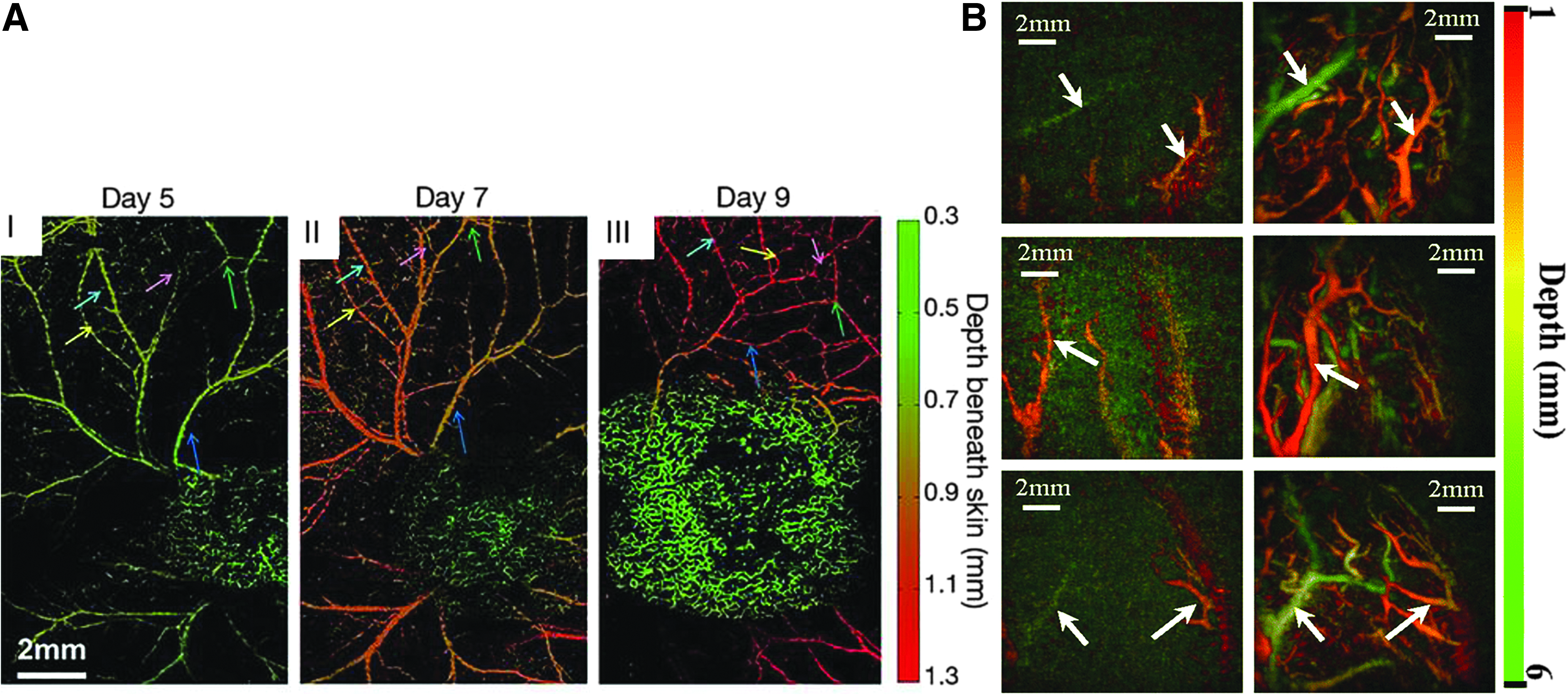
PAI is a promising approach for vascular imaging and characterization due to its ability to achieve resolution in the range of 0.1–800 μm in the tissue thickness up to 8 cm all based on intrinsic contrast (Fig. 6A). 137 Furthermore, PAI of vascular function has been used to assess peripheral arterial disease (PAD). 138 PAD involves both large and small vessels. While large vessels are easily assessed using ultrasonography and CT angiography or MRI angiography, assessment of smaller vessels is hampered due to limited spatial resolution. PAI has been reported as a potential technique to evaluate small vessels-PAD. Detecting vascular changes induced in normal or diseased physiological conditions can provide crucial information relevant to PAD. To show changes in microvasculature circulation, the human index finger was subjected to the hot and cold stimulus to induce constriction and dilation of blood vessels (Fig. 6B). Stimulus induced changes in microcirculation were clearly visible in PA. This experiment represents a proof of concept for evaluating arterial diseases, including small vessels-PAD. The same technique can be used to test the results of strategies focused on increasing blood flow in peripheral vascular disease through the assessment of changes in vasculature circulation in response to a given stimulus.
Cell tracking and monitoring
Like polymers, cells also lack intrinsic cellular contrast except for melanoma cells. Melanin, a characteristic of melanoma cells, generates intrinsic PA contrast. Due to this intrinsic contrast of melanoma cells, Zhang et al. reported cell migration, distribution, and quantification of melanoma cells in PLGA scaffold for the first time. 139 PLGA scaffold did not generate PA contrast; however, spatial distribution and temporal proliferation of cells inside the 3D scaffold of thickness over 1 mm were visible with PAI. In the absence of intrinsic absorbance, external agents are used to generate PA contrast. One of the major external contrast applications in tissue engineering is cell tracking and monitoring.
Cells are often implanted as a critical component of a tissue regeneration strategy. One of the major challenges in stem cell therapy is tracking and monitoring of cells following implantation. This may provide vital information about the therapeutic outcome of the treatment, mechanism of the therapeutic effect, and potential risks of ectopic effects. It is crucial to understand cell fate, engraftment, and survival in vivo to understand the underlying mechanism of the stem cell function. PAI has been used as a noninvasive measure to track stem cells following implantation. To date, various contrast agents have been investigated for stem cell tracking, including dyes,36,140 reporter genes, 141 fluorescent probes, 142 and NP.143,144 Recently, bone mesenchymal stem cells (BMSCs) were investigated as a treatment in a mice brain injury model and were labeled with Prussian blue particles to generate PA image contrast. 36 PA tomography images following BMSC treatment provided pathophysiological status of brain injury, vascularization information, and recovery process. PA images indicated injury location and change in the bleeding area starting at 3 min following injury. There was dramatic increase in PA signal until 30 min postinjury; later, PA signal gradually decreased due to blood coagulation and spreading of blood pool and could no longer be detected on day 13 indicating the natural recovery process of about 15 days. Another study applied PAI to image and monitor cells implanted 6–8 mm deep in mice. In this study, a tyrosine based reporter gene was used to selectively label cells for long-term monitoring of cells. 145 With this technique, cells could be engineered to constitutively express tyrosinase (Tyr), an enzyme that catalyzes the formation of eumelanin, to provide PA contrast. Expression of the tyrosinase reporter allowed for monitoring of MSCs post transplantation in an acute myocardial infarction rat model. 146
Gold labeled NPs have also been used to track cells. GNPs generate PA contrast that can be tuned based on NP size and structure. Nam et al. demonstrated in vivo imaging of GNP-labeled MSCs using PAI. 147 MSCs were prelabeled with GNPs and mixed with a PEGylated fibrin gel in 1:1 volume ratio and then injected intramuscularly at the tissue depth of ∼5 mm. They reported that GNP-labeled MSCs could be monitored for 10 days following injection. The same group previously reported that GNPs did not alter the cell viability, cell proliferation, or differentiation of MSCs suggesting that GNPs could be used for long-term monitoring of cells in vivo.148,149 PAI has been used to evaluate endothelial cell distribution in decellularized scaffold during reendothelialization. 150 In another similar study, PAI was used to track and monitor GNP labeled ASCs for dermal tissue engineering. In this study, GNP labeled ASCs were detected up to 2 weeks in a PEG-based scaffold. 151 Furthermore, silica coating on GNPs as a contrast agent demonstrated enhanced PA in comparison with uncoated GNPs to track and monitor MSCs.152,153 Similar to PEGylated GNPs, silica coating did not evoke any significant effect in the therapeutic effectiveness of stem cells. Jokerst et al. showed that silica coated GNPs did not affect the pluripotency of stem cells. 153 Further research is needed for assessing cell behavior and the immune response toward the silica coated GNPs. 154 Recently, PAI was used to guide the delivery of stem cells into the rodent spinal cord. GNP labeled MSCs were imaged in real time while being injected into the spinal cord of the rat. They report detecting cell number as low as 1000 cells with this imaging system. 155 In another study, human embryonic stem cell derived cardiomyocytes were delivered to the heart of the mouse for cardiac regenerative therapy. These cells were tagged with PA NPs that consist of semiconducting polymers, poly [2,6-(4,4-bis-(2-ethylhexyl)-4H-cyclopenta [2,1-b;3,4-b′]dithiophene)-alt-4,7(2,1,3 benzothiadia-zole)], for cell tracking and monitoring purpose. Strong PAI signals from these NPs easily differentiated implanted cells from background tissues. 156
Current Status of PAI in Diagnostic Applications
Label-free detection of vascularization is one of the most appealing applications of PAI. Based on this and other advantages of multispectral PAI techniques have been developed for the evaluation and assessment of many disease states. Generally, these studies have exploited the ability of PAI to evaluate hemodynamic changes due to abnormal vessel assembly or function in these conditions. For example, hypervascularization and increased oxygen saturation are hallmarks of inflammation. Extensive vascularization co-occurring with low oxygen saturation may indicate tumor stage or progression. 157 As blood vessels are easily visualized with PAI system without the aid of external contrast agent, PAI holds great potential for clinical diagnostic applications. This includes cancer, cardiovascular disorders, inflammatory diseases, rheumatoid arthritis (RA), myocardial infarction, and diabetic retinopathy. 158 The research described in this section provides examples of PAI used in other medical applications using approaches that have not been fully exploited by researchers in the fields of TERM.
Inflammatory diseases
Increased vascularization and oxygen saturation often mark an inflammatory state. 159 In many instances, clinical outcomes of inflammatory disease treatments depend on early diagnosis and intervention. 160 Increased vascularization in arthritic joints of RA patients exhibited up to a 10-fold increase in the PA signal compared to noninflamed joint. 161 In addition to diagnosis, PAI reportedly monitored the escalation of inflammation and the therapeutic effect of the treatment.162,163 For example, in Crohn's disease (CD) chronic relapse of inflammation is common. A tool that allows quick detection of inflammation based on tissue perfusion and oxygenation allows for intervention or adjusting the treatment therapy. Although a detailed study is required, preliminary results performed in 108 patients suggest that PAI can be used to noninvasively evaluate CD based on hemoglobin levels in the intestinal wall. 164 In addition, CD may be characterized by fibrosis in addition to inflammation that is attributed by the irreversible deposition of collagen. CD fibrotic stricture may require surgical intervention, and therefore, accurate characterization of stricture is critical. Due to the optical absorbance of collagen around 1300 nm, PAI has shown to detect the presence of fibrotic stricture noninvasively in CD. 165
Atherosclerosis is characterized by prolonged inflammation. The complex structure of vascular plaques correlates with disease progression and risk. 166 The plaque composition is important to access its rupture vulnerability and to determine the course of treatment. Intravascular PA (IVPA) imaging has been reported to allow differentiation between the various components within an atherosclerotic lesion.56,167–169 IVPA utilizes a catheter or probe-based system for intravascular delivery of light to generate PA signal. The basis for detecting atherosclerotic plaque using PAI is the difference in optical absorbance of blood, collagen, and lipid. 170 The vulnerability of plaques can be evaluated in an atherosclerosis rabbit model by targeted macrophages with NPs. 171 However, the inherent challenge in IVPA is to design a flexible catheter or probe with clinically relevant diameter that has high sensitivity and chemical specificity. 58 Furthermore, these catheters or probes should have sufficient resolution to provide structural and functional information of the vascular wall. Many design considerations are currently being investigated to optimize IVPA catheter designs.58,172,173 Nevertheless, PAI is a powerful tool for evaluating inflammation in a variety of disease states suggesting that it could be utilized to assess the response of a tissue to an implanted tissue engineering or biomaterial-based therapeutic.
Cancer
PAI in oncology is rapidly evolving. PAI is already in clinical trials for the detection of some types of cancers, while for many others it is in preclinical or investigational phase.123,174–178 While a standardized technique is required for PAI before its transition to routine clinical use, the current in-house prototypes have demonstrated promising results for a wide range of cancers such as breast,179–181 cervical, 182 prostate, 183 skin, 184 thyroid, 185 and ovarian cancer. 157 PAI leverages vasculature imaging and functional information such as oxygen saturation for cancer detection, 178 progression, 186 characterization,187–191 prognosis, and monitoring therapeutic efficiency of treatment.192–194 Delivery vehicles for cancer therapy are equipped with different PA contrast agents for multimodal imaging for better diagnosis and staging of cancer. 195 Neovascularization is commonly observed in tumors, triggered by the hypoxic state within the tissue. 196 Oxygen saturation level and extent of vascularization can be an indication of cancer progression. Longitudinal monitoring of vascular parameters gives insight into the therapeutic progress of a given treatment. In addition to the detection and characterization of cancer, PAI has also been used to stage cancer. When a primary tumor metastasizes, they first localize to a regional sentinel lymph node. With the aid of an external contrast agent, PAI has been used to noninvasively locate a sentinel lymph node to guide biopsy with minimal surgical procedures. 197
Future Prospects
PAI offers several potential advantages of other imaging modalities when real-time longitudinal monitoring of functional imaging is needed. PAI does not use ionization energy allowing long term monitoring over the course of weeks or months. PAI allows tracking and monitoring of cells, as well as biomaterials, following implantation with the aid of external contrast agents. While PAI has seen heavy investigation in stem cell tracking, biomaterial monitoring is not well explored yet. One of the challenges in tissue engineering is monitoring in vivo degradation of scaffold. 3 Extrinsic contrast agents can be incorporated in the scaffolds or tissue engineering constructs to monitor and evaluate degradation behavior in vivo. Concomitant vasculature development can be imaged following scaffold implantation. Often after the surgical biomedical therapeutic intervention, there exists a risk of certain complication. Based on the amount of hemoglobin present at the injury site, it can be monitored to evaluate whether the injury is progressing toward wound healing route or inflammation route which would allow to better manage the clinical outcome of the therapeutic procedure.
There has been a recent upsurge in interest in PAI based on its strong potential for clinical imaging. However, several challenges still need to be addressed. The depth of tissue imaging can be increased but typically at the cost of resolution. Catheter or endoscopic PA probes are under development, which enable localized evaluation of specific internal organs or tissues while overcoming depth-related resolution issues. These tools may not be broadly applicable in TERM but certainly can be an important tool in specific applications such as tissue engineered vascular grafts. While there are many PA contrast agents under investigation, longitudinal monitoring could be improved through the development of contrast agents that provide sustained PA contrast. Most PA systems allow qualitative evaluation, and the development of more quantitative PA techniques will have a significant impact on biomedical research. Finally, the development of standardized, easy-to-use commercial systems will increase the impact of PAI.
Conclusions
There is a significant need for tools that allow monitoring and tracking of tissue engineering therapies. While there are many imaging strategies that are currently used to evaluate these processes, PAI offers an attractive alternative. The endogenous contrast resulting from vascularization, collagen, and lipids enables insight in the absence of any additional factors. In addition, given the wide range of available external contrast agents, PAI has immense potential to monitor specific characteristics such as cells or scaffolds by incorporating or targeting with exogenous agents. Monitoring cell behavior or processes in vivo real time, therefore, can provide better understanding of how the given stimulus or biological process affects the cellular or therapeutic response in general. In addition, this information can be used as a feedback to design improved tissue engineering constructs for reconstruction, repair, or regenerative purpose.
Footnotes
Disclosure Statement
No competing financial interests exist.
Funding Information
Our work was supported by National Institute of Health (NIH) grants 5R01EB020604, 1R21EB008765, and R01NS102213, Cancer prevention and Research Institute of Texas (CPRIT) RP120558, and U.S. Department of Defense (DoD) W911NF-17-1-0488. This work was also supported by Research Initiative for Scientific Enhancement (RISE) R25GM060655.