
Abstract
The recent advances in the field of cell-based therapeutics open promising perspectives for oral tissue regeneration. The development of large animal models, which overcome the limits of the rodent models and allow to emulate clinical situations, is crucial for the validation of regenerative strategies to move toward clinical application. Currently, porcine, canine, and ovine models are mainly developed for oral regeneration and their specific characteristics have an impact on the outcomes of the studies. Thus, this systematic review investigates the application of porcine, canine, and ovine models in present cell-based oral regeneration, according to the species characteristics and the targeted tissue to regenerate. A customized search of PubMed, EMBASE, Scopus, and Web of Science databases from January 2015 to March 2020 was conducted. Relevant articles about cell-based oral tissues engineering in porcine, canine, and ovine models were evaluated. Among the evaluated articles, 58 relevant studies about cell-based oral regeneration in porcine, canine, and ovine models matched the eligibility criteria and were selected for full analysis. Porcine models, the most similar species with humans, were mostly used for bone and periodontium regeneration; tooth regeneration was reported only in pig, except for one study in dog. Canine models were the most transversal models, successfully involved for all oral tissue regeneration and notably in implantology. However, differences with humans and ethical concerns affect the use of these models. Ovine models, alternative to porcine and canine ones, were mainly used for bone and, scarcely, periodontium regeneration. The anatomy and physiology of these animals restrain their involvement. If consistency was found in defect specificities and cell trends among different species animal models of bone, dentin-pulp complex, or tooth regeneration, variability appeared in periodontium. Regeneration assessment methods were more elaborate in porcines and canines than in ovines. Risk of bias was low for selection, attrition and reporting, but unclear for performance and detection. Overall, if none of the large animal models can be considered an ideal one, they are of deemed importance for oral cell-based tissue engineering and researchers should consider their relevance to establish favorable conditions for a given preclinical cell-based therapeutics.
Impact statement
This systematic review investigates porcine, canine, and ovine models for current oral cell-based regeneration procedures, and researchers could refer to it for the choice of the most pertinent preclinical model for a given cell-based therapeutics.
Introduction
Injuries and pathologies affecting the oral region, as well as lesions resulting from invasive and destructing therapeutic approaches can determine extensive loss of tissues and function. Moreover, due to the heterogeneity of the tissues of this area, their reconstruction is particularly complex. 1
In recent years, thanks to the exponential growth of tissue engineering, new perspectives have been opened in cell-based oral regeneration. 2 In fact, the use of new sources of stem cells,2,3 the development of high-performance biomaterials, and promising biotechnologies, such as tridimensional (3D) bioprinting,3,4 allowed a considerable progress toward human application of cell-based oral tissue regeneration procedures. Before clinical trials, validation in animal models is required.
It has been established that rodents' substantial anatomic and physiologic dissimilarities with humans affect extrapolation of results from murine studies to patients. Thus, the development of large animal models, overcoming the limits of the rodent ones and allowing the reproduction of near-to-real clinical situations, plays a crucial role in the translation of cell-based regenerative procedures from bench to bedside.5,6
The identification of the most relevant animal model is a crucial step of the study conception. However, this selection is far from being a simple process, since multiple factors are at stake. Literature on oral regeneration reports large animal models that significantly contributed to the current knowledge on the field. In particular, nonhuman primates, porcines, and canines have been involved, over decades, to investigate surgical procedures, pathogenesis of periodontal and endodontic diseases, guided-tissue regeneration, and implantology.7–9
The identification and characterization of dental stem cells, in 2000, allowed a significant development of cell-based approaches of oral tissue regeneration.10–12 Therefore, the above-mentioned animal species have been used to validate key models of bone,3,9,13 periodontium,14,15 dentin-pulp complex,16–18 and tooth organ regeneration, 19 which opened the way to current research and still represent the benchmarks in the field.
More recently, animal research faced considerable changes. Indeed, the introduction of the Animal Research: Reporting of In Vivo Experiments (ARRIVE) guidelines supported transparency and systematization in reporting on animal studies, addressing the problem of poor reproducibility of scientific findings. 20 Moreover, an emerging debate on the protection of animals used for scientific purposes led to substantial revisions of the existing regulation, notably introducing the 3Rs principle (Replacement, Reduction, and Refinement) and restricting the use of specific species, with an actual full application by different countries only in the last few years.
This results in the exclusion of nonhuman primates' models for cell-based oral tissue regeneration, leaving the choice of porcines, canines, and ovines.21–23
Porcines are widely used due to their similitudes with humans in terms of anatomy (Table 1 and Fig. 1), physiology, and pathology, as well as for ethical reasons. Nevertheless, their temperament can be difficult to manage. 5
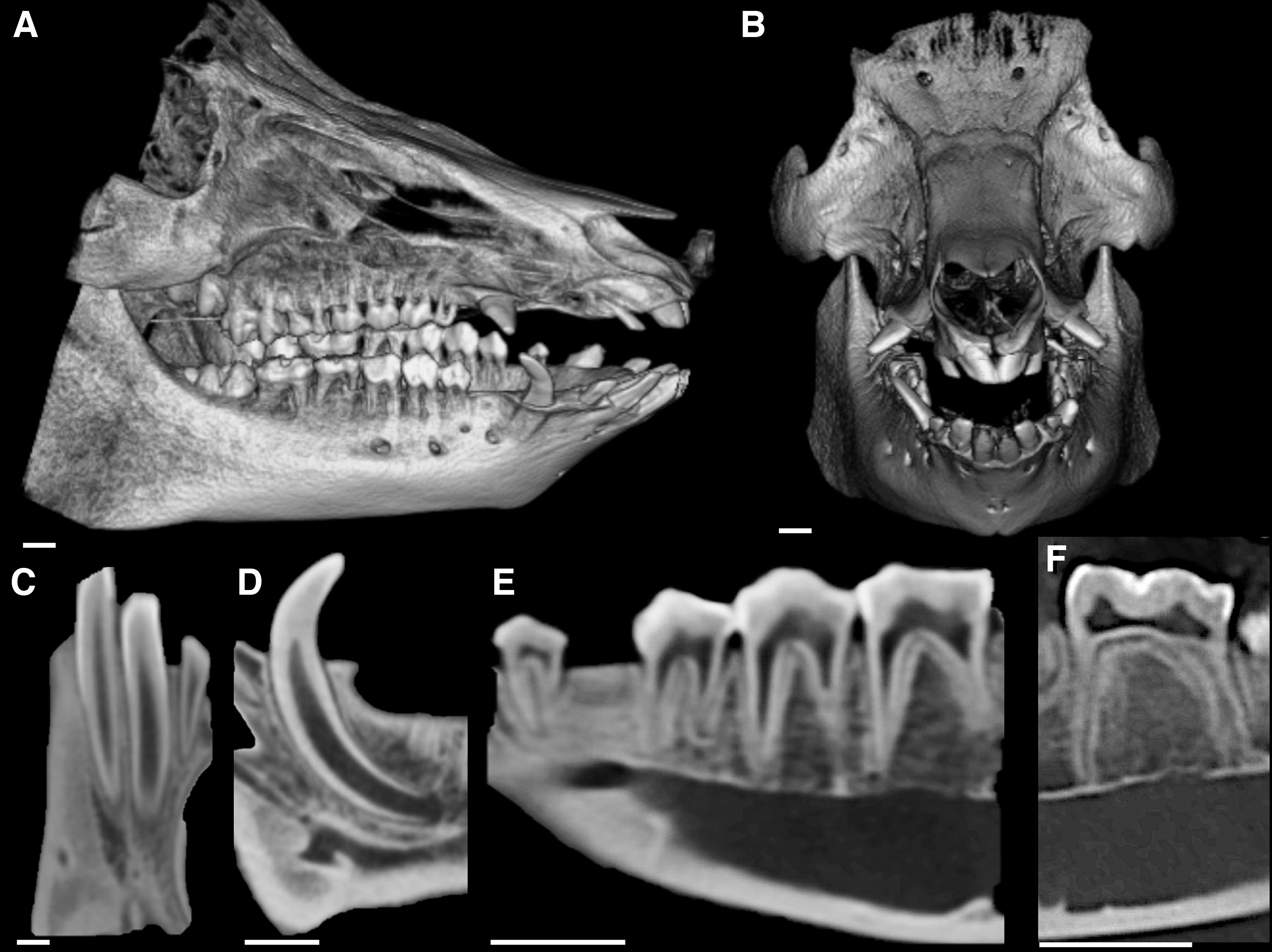
Pig anatomy—Cone beam CT.
Characteristics of the Large Animal Models
Canines are one of the most common large animal models for oral and dental regeneration, notably due to their familiar behavior and the comparable growth, physiology and pathology with humans. However, dogs are considered companion animals and their use in medical research is negatively perceived by society 7 (Table 1 and Fig. 2).

Dog anatomy—Cone beam CT.
As an alternative, the use of ovines increased over the last decade, but the ruminant nature leads to substantial anatomical differences in comparison with humans 7 (Table 1 and Fig. 3).

Sheep anatomy—Cone beam CT.
Hence, this review aimed to investigate porcine, canine, and ovine models for current cell-based oral tissue regeneration procedures, to (1) provide an exhaustive analysis of the present potential application of each model and (2) support researchers in the choice of the most pertinent one for a given study, according to the animal characteristics and the tissue to regenerate.
Methods
Search strategy
The review process followed the Preferred Reporting Items for Systematic Reviews and Meta-Analyses (PRISMA) guidelines 24 and the protocol was registered in PROSPERO (International Prospective Register of Systematic Reviews) under the number CRD42020201550.
The peer-reviewed literature reporting about large animal models on most recent cell-based oral regeneration procedures was systematically searched in PubMed (National Library of Medicine, NCBI), Embase, Web of Science and Scopus databases, from January 2015 to March 2020. The following combination of key words was used: Oral AND (regeneration OR tissue engineering) AND stem cells AND (pig OR dog OR sheep). A manual review of articles' references was also performed.
Eligibility criteria
Inclusion criteria were as follows: (1) cell-based oral tissues regeneration studies, (2) large animal studies, (3) English language available full text, and (4) publication between 2015 and 2020.
Exclusion criteria were as follows: (1) murine studies, (2) in vitro studies, (3) noncell-based studies, (4) ectopic and semiorthotopic regeneration models, and (5) literature reviews.
Two independent reviewers (F.M. and S.V.) screened all relevant titles and abstracts against eligibility criteria. If the abstract did not provide sufficient information, the full text article was analyzed. A third reviewer (B.S.) was involved to resolve disagreements.
Data extraction and analysis
The selected articles were assigned, depending on the regenerated tissue, as follows: (1) bone regeneration, (2) periodontium regeneration, (3) dentin-pulp complex regeneration, and (4) tooth/tooth-root regeneration studies.
For each tissue, articles were subsequently classified according to the involved animal; reproduced clinical context/pathology, source of stem cells, scaffolds, follow-up, and assessment techniques were considered.
Assessment of quality of the studies
Risk of bias for the included studies was evaluated by SYstematic Review Centre for Laboratory animal Experimentation (SYRCLE) risk of bias tool. The following criteria were used: (1) selection bias, (2) performance bias, (3) detection bias, (4) attrition, and (5) reporting bias. Studies were scored with a “yes” for low risk of bias, “no” for high risk of bias, and “?” for unclear risk of bias by the two reviewers independently. Disagreements were resolved by a third reviewer (B.S.).
Results
As presented in the flowchart based on PRISMA (Fig. 4), the initial search resulted in a total of 148 articles. Eight relevant publications were manually added from reference lists of the articles identified. After duplicate removal, a total of 123 articles was identified. The review and selection procedure resulted in the exclusion of 43 articles at title screening stage and 11 articles based on the content of the abstract. Of the remaining 69 articles, 11 were excluded at the full-text reading stage for the following reasons: (1) 3 articles were not in vivo studies, (2) 1 article reported ectopic cell-based regeneration, (3) 3 articles reported studies not developed on large animal models, and (4) 4 studies presented noncell-based regeneration. The entire selection process therefore resulted in a total of 58 articles, included in this systematic review.

Flowchart of the article selection process.
Characteristics of included studies
Results are summarized in Tables 2–5. Considering the regenerated tissues, 24 articles focused on bone,25–48 17 on periodontium,49–65 10 on dentin-pulp complex,66–75 and 7 on tooth or tooth root.76–82 Regarding bone regeneration, 8 studies developed a porcine model,25–32 14 a canine,33–46 and 2 an ovine one.47,48 Eight studies reported periodontium regeneration in pig,49–56 eight in dog,57–64 and one in sheep. 65 Among the articles about dentin pulp-complex regeneration, two studies were developed in pig66,67 and eight in dog.68–75 Except for one canine model, 82 in all the studies, tooth or tooth root regeneration was reported in porcines.76–81
Bone Regeneration
Acute defect model.
Chronic defect model.
μCT, micro-CT; 2D, bidimensional; 3D, tridimensional; BIC, bone to implant contact; BMM, bovine bone mineral; BMP2, bone morphogenetic protein 2; b-TCP, beta tricalcium phosphate; cADSCs, canine adipose-derived stem cells; cBMSCs, canine bone marrow stem cells; cEPCs, canine endothelial progenitor cells; CLSM, confocal laser scanning microscopy; cPDLSCs, canine periodontal ligament stem cells; CT, computed tomography; DBM, demineralized bone matrix; dPDLSCs, dog periodontal ligament stem cells; EPCs, endothelial progenitor cells; FDB, freeze-dried bone; GFP, green fluorescence protein; HA, hydroxyapatite; hADCs, human adipose-derived cells; hADSCs, human adipose-derived stem cells; hDPSCs, human dental pulp stem cells; oBMSCs, ovine bone marrow stem cells; oMSCs, ovine mesenchymal stem cells; pASCs, porcine adipose-derived stromal/stem cells; pBMPCs, porcine bone marrow progenitor cells; pBMSCs, porcine bone marrow stem cells; PLGA, poly-co-glycolic acid; pMSCs, porcine mesenchymal stem cells; PRP, platelet-rich plasma; pUC-MSCS, porcine umbilical cord-mesenchymal stem cells; rhPDGF, recombinant human platelet-derived growth factor; TCP, tricalcium phosphate; VEGF, vascular endothelial growth factor.
Periodontium Regeneration
Acute-chronic defect model.
Acute defect model.
Chronic defect model.
BCP, biphasic calcium phosphate; CaP, calcium phosphate; cBMMSC, canine bone marrow mesenchymal stem cells; cPDLCs, canine periodontal ligament cells; ESEHT, extraction socket early healing tissue stem cells; HGF, hepatocyte growth factor; hMSCs, human mesenchymal stem cells; hPDLCs, human periodontal ligament cells; hPDLSCs, human periodontal ligament stem cells; hSCAPs, human stem cells from apical papilla; HyA-sECM, hyaluronic acid-synthetic extracellular matrix; IGFBP5, insulin-like growth factor binding protein 5; oPDLSCs, ovine periodontal ligament stem cells; PAB, proper alveolar bone; pADMPC, adipose-derived multilineage progenitor cells; pMSCs, porcine mesenchymal stem cells; pPDLSCs, porcine periodontal ligament stem cells; SFRP2, secreted frizzled-related protein 2; TRAP, tartrate-resistant acid phosphatase; TRL2, toll-like receptor 2.
Dentin-Pulp Complex Regeneration
Acute defect model.
Chronic defect model.
cDPSCs; (G-CSF)cMADSCs, granulocyte colony-stimulating factor canine-mobilized adipose-derived stem cells; (G-CSF)cMBMSCs, granulocyte colony-stimulating factor canine-mobilized bone marrow stem cells; (G-CSF)cMDPSCs, granulocyte colony-stimulating factor mobilized dental pulp stem cells; BMP7, bone morphogenetic protein-7; DLA, dog leukocyte antigen; HyA, hyaluronic acid; MRI, magnetic resonance imaging; NGF, nerve growth factor; PDGF, platelet-derived growth factor; pDPC, porcine dental pulp cells; pDPSCs, porcine dental pulp stem cells; PRF, platelet-rich fibrin; SIM, simvastatin; TDM, treated dentin matrix; VEGF-2, vascular endothelial growth factor-2.
Tooth/Tooth Root Regeneration
CBCT, cone beam computed tomography; dTBs, decellularized tooth buds; HA-TCP, hydroxyapatite- tricalcium phosphate; hBMMSCs, human bone marrow mesenchymal stem cells; hDPCs, human dental pulp cells; hUVECs, human umbilical vein endothelial cells; pDFCs, porcine dental follicles cells; pECs, porcine epithelial cells; SEM, scanning electron microscopy.
Bone regeneration (Table 2)
Porcine models
The clinical situations were exclusively acute defects such as mandible noncritical 26 and critical size bone defects,27,29,31 mandible extraction socket, 32 alveolar cleft,25,30 as well as ramus and condyle defects. 28
Employed cells were mostly porcine cells such as porcine mesenchymal stem cells (pMSCs),25,30,32 porcine bone marrow stem cells (pBMSCs),26,29,31 and porcine adipose-derived stem cells (pADSCs), 28 except in one study in which human dental pulp stem cells (hDPSCs) were used. 27 The used scaffolds were poly-co-glycolic acid (PLGA),25,29,30 beta tricalcium phosphate (b-TCP),26,27 and decellularized bone scaffolds.28,31 In one article, cell sheets were involved. 32
Follow-up was performed at 4 weeks (or 30 days),25,31 6 weeks, 32 8 weeks,26,27 12 weeks (or 90 days),29,30 and 6 months. 28
Regenerated tissues were assessed by histology in all studies,25–32 in association with computed tomography (CT) and/or microcomputed tomography (μCT) evaluation,25,28–31 immunohistochemistry,25,26,30 histomorphometry,26,27,32 mechanical tests, 30 and fluorescence microscopy. 32
Canine models
The studies focused on acute models of mandible peri-implant bone defects,33,39,43,45,46 mandible noncritical 34 and critical size bone defects,37,42,44 alveolar cleft, 35 bilateral sinus lift, 38 and mandible segmental defect. 41 Two studies developed chronic mandible peri-implant bone defects.36,40
The employed cells were canine bone marrow stem cells (cBMSCs),34,35,38,41,45 recombinant human platelet-derived growth factor (rhPDGF)-cBMSCs, 39 canine adipose-derived stem cells (cADSCs),37,43 canine epithelial progenitor cells, 42 bone morphogenetic protein 2 (BMP2)-cADSCs, 40 BMP2-canine periodontal ligament stem cells (cPDLSCs), 36 and cPDLSCs. 46 Only two studies used human adipose-derived cells (hADCs).33,44 Cell sheets were used in two articles.41,46
Cells were seeded into various scaffolds such as b-TCP,35,39,45 tricalcium phosphate (TCP), 40 hydroxyapatite-tricalcium phosphate (HA-TCP),33,44 TCP-fibronectin,37,43 PLGA, 44 b-TCP coated with PLGA releasing vascular endothelial growth factor (VEGF), 42 freeze-dried bone, 41 and hydroxyapatite (HA) collagen. 36
The follow-up was performed during 4 weeks, 33 8 weeks,41,42,44 11 weeks, 46 12 weeks or 3 months,35–37,39,43,45 16 weeks, 34 6 months, 38 and 10 months. 40
In 13 out of 14 studies, assessment was made by histology and histomorphometry33–40,42–46; these techniques were also associated with bidimensional (2D) and/or 3D (μCT) radiographic analysis,38,40,41,44,45 bone to implant contact evaluation,33,36,39,40,43,45,46 hardness mechanical tests,34,38 and fluorescence microscopy. 39 Only one study evaluated regenerated tissues by combining histology, immunohistochemistry, and radiographies. 41
Ovine models
One study focused on sinus lift 47 and the other one on acute mandible segmental bone defect. 48 Ovine mesenchymal stem cells (oMSCs) 47 and ovine bone marrow stem cells (oBMSCs) 48 were used, respectively, associated with autologous serum and BBM scaffold. Follow-up was made for 1647 and 32 weeks. 48
Histology and histomorphometry were performed in both studies,47,48 one case also combined with CT and μCT evaluations. 48
Periodontium regeneration (Table 3)
Porcine models
The experimental models of periodontitis reproduced acute or acute-chronic mandibular class II furcation defects53,56 and acute-chronic maxillary and/or mandibular alveolar three walls bone defects.49–52,54,55
Porcine cells such as pMSCs, 50 porcine adipose-derived cells (pADCs), 56 and porcine periodontal ligament stem cells (pPDLSCs) 53 were used in three studies. Human cells like human periodontal ligament stem cells (hPDLSCs) transfected with hepatocyte growth factor, 49 hDPSCs, 52 insulin like growth factor binding protein 5 (IGFBP5), human mesenchymal stem cells (hMSCs), 51 and human stem cells from apical papilla (hSCAPs) 54 transfected or not with secreted frizzled-related protein 2 (SFRP2) 55 were employed in the other articles.
Cells were seeded into fibrin gel complex, 56 IL1-hyaluronic acid (HyA)-synthetic extracellular matrix 50 hydrogel, and collagen 53 scaffolds. In the other studies cells were injected49,51,52,54,55 and in one case also associated with cell sheets. 52
The follow-up was performed during 4 weeks, 56 12 weeks or 3 months,49,51–55 and 16 weeks. 50
Assessment was made by clinical, radiographic, and histological evaluations in five studies,49,50,52,54,55 three out of which also performed histomorphometry.49,54,55
One article presented clinical, photographic, and histological, but no radiographic analysis. 51 Two articles reported histology and histomorphometry, respectively, associated with μCT and immunohistochemistry, 56 as well as fluorescence microscopy, 53 but no clinical evaluation.
Canine models
The experimental models of periodontitis reproduced acute mandible alveolar bone dehiscence 59 and maxillary-mandibular class II furcation defects, 63 acute-chronic mandible class III or II furcation defects,58,60 mandibular alveolar bone dehiscence,62,64 as well as chronic maxilla and mandible alveolar bone defects. 61 One model of tooth reimplantation was reported. 57
The employed cells were cBMSCs58,59,63 also transfected with green fluorescence protein (GFP) 63 or toll-like receptor 2 (TRL2), 64 canine extraction socket early healing tissue stem cells with proper alveolar bone, 60 cPDLSCs, 57 and b-defensin-3-canine periodontal ligament cells. 61 Human cells (human periodontal ligament cells) were used once. 62
One study used cell sheets. 61 Each article reported a different scaffold material such as atelocollagen combined with b-TCP, 58 HA collagen, 59 collagen, 64 BCP, 62 platelet-rich plasma, and fibrin glue, 63 decellularized dental root coated with calcium phosphate-fibronectin. 57 In one study, grafting materials were not specified. 60
The follow-up was performed at 2 weeks, 64 8 weeks,57,58,60,61,63 12 weeks, 62 and 6 months. 59
The assessment was performed in half of the studies by histology and histomorphometry,58–60,63 also associated with μCT analysis, 59 immunohistochemistry, and tartrate-resistant acid phosphatase. 58 Histology and μCT analysis,57,62,64 in one case also associated with fluorescence microscopy, 62 were reported. In one study, assessment consisted of histology and immunohistochemistry. 61
Ovine model
The periodontal defect was an acute dehiscence in the mandibular premolar-molar area. Ovine periodontal ligament stem cells (PDLSCs) and BMSC sheets were used, associated with polycaprolactone biphasic scaffold.
The follow-up was 10 weeks. The assessment of regenerated tissues was made by histology, histomorphometry, and μCT. 65
Dentin-pulp complex regeneration (Table 4)
Porcine models
One study focused on partial pulp regeneration, 66 the other one investigated total pulp regeneration. 67 In both cases, upper and lower mature multirooted teeth were involved and the pulp defects were acutely induced.
Porcine dental pulp cells (pDPCs) and porcine dental pulp stem cells (pDPSCs) were used. The cells were seeded in a nanopeptide 66 and in HyA or collagen hydrogels, 67 respectively.
The follow-up was performed at 21 days 66 and 4 months. 67
In one study, assessment was made by histology, immunohistochemistry, μCT, and histomorphometry 66 ; in the other one, histological, immunohistochemical, and 2D radiographic analysis were performed. 67
Canine models
Models of partial pulp regeneration involving upper and lower multirooted teeth were reported in two studies.71,75 Authors used immature 71 and mature 75 teeth, respectively. Five studies focused on total pulp regeneration.68–70,73,74 The involved teeth were upper and lower mature incisors,69,70,74 upper immature incisors, 73 as well as upper and lower mature multirooted teeth. 68 Only one study evaluated a model of pulp chamber floor perforation in upper and lower mature premolars. 72 Expect for one article, 73 dentin-pulp complex defects were acutely generated.
Regarding the source of cells, canine dental pulp stem cells (cDPSCs),68,71–73 granulocyte-colony stimulating factor mobilized dental pulp stem cells,69,70,74 as well cBMSCs 75 were involved.
Cells were implanted with atelocollagen scaffold,69,70,74 platelet-rich fibrin, 68 treated dentin matrix (TDM) or TCP, 72 gelatin sponge associated with simvastatin 71 and chitosan hydrogel releasing VEGF-2, PDGF, nerve growth factor, and bone morphogenetic protein-7 (BMP7). 73 In one study, cells were injected. 75
Follow-up was performed at 2 weeks, 69 8 weeks, 68 9 weeks, 75 10 weeks, 71 3 months or 12 weeks,72,74 4 months, 73 and 6 months. 70
Histological assessment was reported in all studies,68–75 associated with histomorphometric analysis,68,69,72 2D radiographies,71,73 magnetic resonance imaging (MRI), 70 in situ hybridization, 69 immunohistochemistry, 69 as well as blood and urine tests. 75
Tooth or tooth root regeneration (Table 5)
Porcine models
Three studies focused on tooth root regeneration in the mandibular incisor 78 and premolar area.76,77 Three articles described whole tooth regeneration79–81 in the upper incisor and premolar region, 81 in lower canine and premolar region, 80 as well as in lower premolar and premolar region. 79
Porcine dental follicles cells (DFCs) transfected or not with GFP were used in two studies.76,77 One article described re-associated tooth germ implantation associated with BMSCs systemically infused. 81 In two cases, pDPSCs were combined with pPDLSC sheets 78 or epithelial cells from gingiva. 79 In one study, porcine epithelial cells were associated with human dental pulp cells (hDPCs) and hUVEC1. 80
The scaffolds in which cells were seeded were TDM,76,77 HA-TCP, 78 gelatin-chondroitin-hyaluronan scaffold, 79 and decellularized tooth bud (dTB). 80
The follow-up was performed at 12 weeks, 76 150 days, 81 6 months,77,78,80 and 13.5 months. 79
Histology, radiography (2D, μCT, CT, and cone beam computed tomography [CBCT]), and immunohistochemistry analysis were reported in five studies,76,77,79–81 in one of which clinical oral assessment was performed too. 77
In only one study, clinical, radiographic (CT, μCT), biomechanical, and elemental analysis were combined. 78
Canine model
Whole tooth regeneration was performed in lower premolar area. The regenerated teeth underwent orthodontic traction to test periodontal ligament remodeling.
Canine tooth bud cells were used. The follow-up consisted of 6 months plus 1 month for orthodontic treatment evaluation. The assessment was made by histology, 3D radiography (CBCT and μCT), scanning electron microscopy, and energy dispersive X-ray spectroscopy. 82
Assessment of quality of studies
In 98% of the studies, a low risk of selection bias (baseline characteristics) was found.25–42,44–64,66–82 Performance bias was considered unclear in 98% of cases,25–47,49–82 because no information about random housing was given. Random outcome assessment was scored as low risk for 24% of the studies25–29,33,34,36–40,42,43,46–48,50,52,54,57–60,62–74,76–82 and unclear for the rest of them. In none of studies, blinding was described and the risk was rated as unclear.25–82 A low risk of attrition and reporting bias was estimated for all studies25–82 (Fig. 5 and Supplementary Figs. S1–S4).
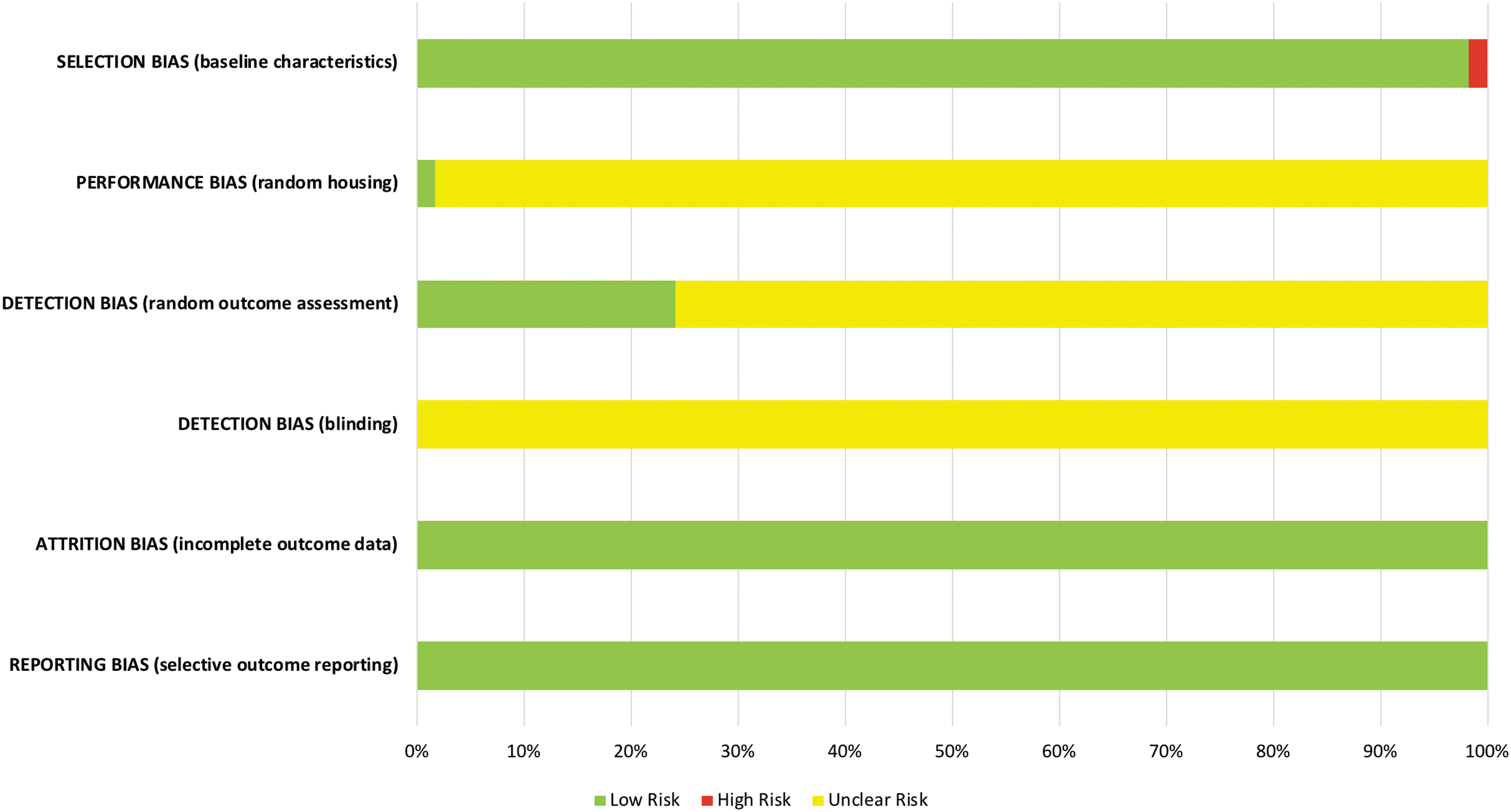
Risk of bias assessment evaluated according to the SYRCLE. Selection bias: baseline characteristics. Performance bias: random housing. Detection bias: random outcome assessment; blinding. Attrition bias: incomplete outcome data. Reporting bias: selective outcome reporting. SYRCLE, SYstematic Review Centre for Laboratory animal Experimentation.
Discussion
Large animal models according to regenerated tissues
Bone regeneration
Our research highlights that porcine, canine, and ovine models were developed for cell-based regeneration of acute critical maxillary and mandible jawbone defects, mimicking congenital lack of tissues as well as traumatic or postsurgical sequels, such as clefts or segmental osteotomies.25–48 Moreover, procedures requiring long healing process were possible, since reported follow-up went from 1 month even up to 10 months.25–48
As supported by broad literature on cell-based bone regeneration in large animals, 83 these models make up for multiple limitations of widespread murine calvarial defects, such as the impossibility to perform long-term studies, the lack of biomechanical loading, and faster tissue healing than in humans.5,84,85
In accordance with previous studies,86–88 it appears that porcine models are preferred to the other models for higher challenge bone regeneration procedures, because of their similarities with humans, in terms of anatomy, morphology, healing, remodeling, and mechanical properties.25,28,30
For instance, Bhumiratana et al. demonstrated regeneration of ramus-condyle unit by using an autologous, anatomically shaped, living graft, made by decellularized bovine trabecular bone and pADSCs. 28
Furthermore, since congenital clefts occurring in pig resemble those in humans, Caballero et al. reported porcine alveolar cleft regeneration using porcine umbilical cord mesenchymal stem cell (MSC) sheets associated with nano-microfiber PLGA scaffold.25,30
In implantology, canines play a key role.14,89 In this review, numerous peri-implant bone defects and re-osteointegration models were identified33,36,39,40,43,45,46; in fact, dogs' bone turnover, composition, and mechanical properties are the most similar to humans among large animal models, even if jaws show a denser and more resistant bone.7,89
Even if ovine models of cell-based bone regeneration are reported in literature,90,91 in our research, only two studies developed maxillary sinus lift and mandible segmental osteotomy. Indeed, ovine bone dissimilarities with humans such as higher density and mechanical resistance, as well as age-related changes in structure and remodeling can limit the relevance of ovine models for follow-up studies.7,14,25,89,92
Periodontium regeneration
Since murine periodontium and bacterial resistance sensibly differ from humans, porcine, canine, and ovine models of periodontitis are developed.14,89
In this review, similar types of periodontal lesions were reported in pig and dog. However, variability in defects' standardization and follow-up was encountered between the two species.
Only one study developed an acute ovine model of mandible dehiscence. 65 Sheep periodontium displays constant cement apposition as a compensation response to teeth ware, which is typical in ruminants. 14 Hence, this periodontal physiology is likely to have an impact on regeneration mechanisms, which represents a non-neglecting bias for the potential extrapolation to patients.14,92,93
Dentin-pulp complex regeneration
Regenerative endodontics opens up the perspective of an alternative to millions of endodontic treatments each year.94–96
Validated in murine ectopic models, dentin-pulp regeneration is hardly performed orthotopically due to frequent dental fractures and differences with human pulp reparation process. Thus, large animals are required to address these limitations.97,98
Interestingly, despite a more important similarity of pig dental anatomy and physiology with humans in comparison with dogs, mostly canine and only two porcine models were developed.66–75
Besides, in both animals, partial and total dentin-pulp complex regeneration procedures, involving upper and lower mature or immature single-rooted or multirooted teeth, were evaluated, meaning that several clinical situations can be reproduced in these models. In addition, in dog also, a pulp chamber floor perforation model was reported. 72
However, regarding partial pulp regeneration, contrasting findings were reported. In 2017, our team demonstrated, after 3 weeks of follow-up, no pulp regeneration, but reparative osteodentinogenesis in minipig mature multirooted permanent teeth by implanting pDPCs into a self-assembling injectable hydrogel scaffold in a pulpotomy model. 66 In dog, after 9 to 10 weeks, normally organized pulp tissue with a complete dentin bridge was found in single-rooted immature teeth as well as in multirooted mature teeth, using cDPSCs seeded in a gelatin sponge scaffold releasing simvastatin and injected cBMSCs, respectively.71,75
In line with previous studies,99–102 canine models as well as the only porcine model of total pulp regeneration used autologous dental pulp stem cells (DPSCs)67–70,73,74 mostly combined with collagen-based scaffolds67,69,73,74 in mature single-rooted upper and lower teeth.69,70,73,74 However, the role of neoangiogenesis was solely investigated in dog, since cells were constantly conditioned or associated with angiogenetic factors. One could assume that such a difference in regenerative environments between animal models influenced the duration of the regenerative process 84 since functional dentin-pulp complex was obtained after 4 months in pig 67 and in 2 weeks to 6 months in dog.68–70,73,74
Tooth regeneration
The challenging regeneration of tooth organ, which depends on the recombination of dental mesenchymal and epithelial stem cells, has been demonstrated in several animal models.19,103,104 However, it has been shown that the dental functionality can only be assessed in large animals.19,103
In this review, tooth regeneration was reported in numerous porcine models76–81 and in only one canine model. 82 In particular, in pig, consistent with literature,87,105,106 two procedures were studied: tooth root and whole tooth regeneration. Functional bioroot formation was reported after implantation of HA/TCP/DPSC/PDLSC sheets. 78 Positives outcomes were obtained, also combining TDM with minipig DFCs.76,77 Whole single-rooted tooth regeneration was achieved by allotransplanted re-associated tooth germs into minipigs jaws associated with systemic infusion of porcine bone marrow mesenchymal stem cells as well as recellularized dTBs seeded with porcine dental epithelial cells, hDPCs, and human umbilical vein endothelial cells, with an average follow-up of 6 months.80,81
In line with porcine models, premolar regeneration was achieved, in dog, 6 months after transplantation of bioengineered tooth germs made with autologous germs cells, in the lower jaw. Periodontal functionality was eventually confirmed by 4-week-long orthodontic traction. 82
Defect characteristics
The reported oral tissue defects were mostly acute.25–35,37–39,41–48,53,56,57,59,62,63,65–72,74,75 Indeed, surgically made lesions imply standardized configuration, clear understanding of the regenerative process, and reduced experimental time. However, these models reproduce simplified regenerative environments. 89
The bacterial component of oral pathologies was considered in few chronic36,40,61,64,73 or acute-chronic49–52,54,55,58,60 models of peri-implantitis, periodontitis, and pulp necrosis. Certainly, these models, requiring time-consuming procedures, result in a variable degree of standardization of the defect, complicating the comparisons between studies. 89
Stem cell trends
The pertinence of an animal model for oral tissue engineering also relies on the potential to study different stem cell populations/sources. Thus, the accessibility to autologous stem cells as well as the feasibility of allogenic grafts are crucial criteria of choice.
Similar trends of cell-based bone regeneration were reported in all large animal models with comparable results. In fact, most of the studies demonstrated increased bone formation in critical size defects using autologous BMSCs, ADCs, or MSCs principally seeded into b-TCP, PLGA, or demineralized bone matrix scaffolds.25,26,28–32,34,35,37–45,47,48
Unlike the ovine models, in pig and dog, large jaw reconstruction and peri-implant defects were also successfully treated by implanting autologous or human ADSCs and/or MSCs co-seeded with endothelial progenitor cells associated with b-TCP and/or PLGA,25,28–30,32,33,40,42–44 as well as autologous or human stem cells from dental tissues alone or combined with b-TCP or HA/collagen.27,36,46
A substantial discrepancy between trends of periodontal defect regeneration was found; in fact, in pig, hMSCs, hPDLSCs, and hSCAPs49,51,52,54,55 were mostly involved, while in dog and sheep, mostly autologous MSCs or PDLSCs and no stem cells from apical papilla were used.57–61,63–65 Moreover, porcine stem cells were mostly injected or used as sheets,49,51,52,54,55 whereas in dog and sheep, they were implanted combined with a large variety of grafting materials,57–61,63–65 which even more complicate comparisons within studies.28,89
Regarding pulp and root or whole tooth regeneration, in both porcine and canine models, mostly autologous dental stem cells66–71,73–79,82 were used, which is coherent with the manageable accessibility to this source of cells in animals showing similar locoregional anatomy and dental eruption physiology with humans.6,7,25
Regeneration assessment
Regeneration assessment is essential for the validation of tissue engineering procedures. A pertinent large animal model should allow an appropriate follow-up duration for a given procedure as well as the quali-quantification of the newly formed tissues and their relation with the surrounding structures.7,25
In the porcine, canine, and ovine studies included in this systematic review, mineralized as well as nonmineralized oral tissue regeneration was assessed within periods even up to 13.5 months and the analysis was performed by similar approaches in the three models.
Overall, most of the studies reported histological, histomorphometric, and/or 2D/3D imaging analysis.25–40,42–60,62–73 Regardless for the μCT, which is an ex-vivo technique, imaging assessment was made by technologies currently used in patients such as intraoral 2D radiography, CT, CBCT, and MRI. Furthermore, reiterative blood and urine tests, impossible in murine, were reported, 74 which highlights the importance of large animals for mimicking clinical conditions.5,25
However, specific animals' characteristics, data/means unavailability, and the necessity to contain the number of samples, according to the 3Rs principle, give rise to some technical boundaries restraining tissue assessment.5,25,66,107
For example, immunohistochemistry was not constantly performed in pig or dog and not reported at all in sheep models; indeed, some tissue-specific markers cannot be revealed, because of the lack of suitable antibodies. Moreover, due to their size, specimens require even up to several months for the demineralization before histological analysis. Thus, aggressive acids or techniques used to accelerate the process can impair antigenic sites and limit antibody bond.66,107
Risk of bias
Overall, the included studies presented a low risk of bias in terms of animal selection (notably ARRIVE guidelines were respected), attrition, and reporting. However, poor reporting in terms of performance and detection affected evaluations and synthesis of results. Thus, SYRCLE guidelines should be followed, especially for randomization protocols, animal housing facilities, and blinding, which could improve homogeneity of large animal models' trials in oral cell-based regeneration.
Conclusion
The development of large animal models for oral tissue engineering is crucial for human application. Pig, dog, and sheep are the most relevant species allowed by current regulation, but they can have significant drawbacks, including functional dissimilarities when compared to the human craniofacial and dental anatomy.
Porcine models, the most similar with humans, were successfully developed for bone and periodontium regeneration, but very little was demonstrated about dentin-pulp complex. Interestingly, tooth/tooth root regeneration was reported only in pig, except for one canine study.
Canines are indeed the most transversal models as they showed positive outcomes for the regeneration of bone, in particular in implantology, as well as periodontium and dentin-pulp complex; however, canines substantially differ from humans and ethical concerns arise from their involvement.
Ovines are the least developed models, as they emerged as an alternative to dog and pig. Besides the economic and ethical advantages, these animals display essential dissimilarities with humans. Hence, ovines were mainly used for bone and very little for periodontium regeneration.
If a consistency was found in defect specificities and cells trends among different species, animal models of bone, dentin-pulp complex, or tooth regeneration, a variability appeared in periodontium.
Indeed, methods of regeneration assessment were more elaborate in porcines and canines than in ovines.
Overall, preclinical models display specific properties to take into account for oral tissue engineering. Thus, studies of different regenerative procedures should be related to the choice of the most pertinent large animal model for a given cell-based therapeutics.
Footnotes
Authors' Contribution
Conceptualization, F.M. and S.V.; writing/original draft preparation, F.M.; writing/review and editing, F.M., S.V., M.E., B.S., C.C., and R.J.
Acknowledgments
We would like to thank Elisabeth Dursun for her support as well as Fondation des Maladies Rares.
Disclosure Statement
No competing financial interests exist.
Funding Information
No funding was received for this article.
References
Supplementary Material
Please find the following supplemental material available below.
For Open Access articles published under a Creative Commons License, all supplemental material carries the same license as the article it is associated with.
For non-Open Access articles published, all supplemental material carries a non-exclusive license, and permission requests for re-use of supplemental material or any part of supplemental material shall be sent directly to the copyright owner as specified in the copyright notice associated with the article.