
Abstract
Neurological disorders such as Alzheimer’s disease, Parkinson’s disease, and stroke pose significant challenges for conventional therapy due to the complexities of the blood–brain barrier (BBB) and the restricted delivery of drugs to the central nervous system. Exosomes, a type of small extracellular vesicle secreted by nearly all cell types, hold substantial promise as delivery vehicles for therapeutic agents in treating these conditions. Notably, stem cell-secreted exosomes have emerged as particularly effective due to their regenerative potential and natural ability to cross the BBB. Similarly, hydrogels have gained recognition as versatile biomaterials capable of supporting sustained release and targeted delivery of therapeutics. The combination of the regenerative properties of stem cell-derived exosomes (SC-Exos) with the structural and functional benefits of hydrogels offers a promising approach for enhancing neurogenesis, modulating neuroinflammation, and facilitating tissue repair. This review explores the origin, structure, and modifications of exosomes as well as the synthesis and incorporation methods of hydrogels in the therapeutic context for debilitating neurological disorders. It highlights recent advancements in using SC-Exos and hydrogels for therapeutic delivery, addressing both current challenges and future applications. Improving our understanding of hydrogels loaded with SC-Exos for cargo transportation and neural tissue regeneration may pave the way for novel therapeutic strategies.
Impact Statement
In this review, we critically appraise the innovative use of stem cell-derived exosomes (SC-Exos) combined with hydrogels for treating neurological disorders, focusing on their dual role in therapeutic cargo delivery and tissue regeneration. We provide a comprehensive overview of the current methods for exosome isolation, stem cell sources, cargo loading techniques, type of hydrogels and their synthesis, exosome-hydrogel incorporation methods, and preclinical applications. This article also offers insights into the current advancements, future trends, and challenges in SC-Exos-loaded hydrogels for neural regeneration and suggests improvements for future research.
Introduction
Enhancing the delivery of therapeutic agents for treating neurological disorders has received increased attention and extensive research in recent years. Overcoming neurological diseases is challenging because of the presence of protective barriers, such as the blood–brain barrier (BBB) and blood–cerebrospinal fluid. The BBB is composed of several elements, that is, brain capillary endothelial cells, basal lamina, pericytes, and astrocytes. Its main function is to protect the brain by preventing potentially dangerous substances in the bloodstream from entering. 1 Although the barrier is essential for safeguarding the brain, it also presents a significant obstacle by impeding the entry of most medications into the central nervous system (CNS). As a result, the BBB leads to a major challenge in the direct delivery of therapeutic molecules to the brain, which hampers the efficacy of possible treatments for CNS disorders such as Alzheimer’s disease (AD), Parkinson’s disease (PD), multiple sclerosis (MS), spinal cord injury (SCI), traumatic brain injury (TBI), stroke, and brain tumors. 2
One potentially effective strategy being investigated is the use of extracellular vesicles (EVs) secreted by the stem cells as carriers for medication transportation. Stem cell-derived EVs provide great benefits, such as their capacity to cross biological barriers and transport therapeutic cargo directly to specific cells in the CNS. Stem cells, known for their regenerative potentials and capacity to transform into diverse cell types, have become promising candidates for generating EVs with improved therapeutic abilities. 3 Stem cells release EVs that have been utilized to transport bioactive substances, including proteins, nucleic acids (deoxyribonucleic acid [DNA] and ribonucleic acid [RNA]), and lipids, to other cells. 4 The nonhematopoietic multipotent stem cells that are most readily available are mesenchymal stem cells (MSCs). Bone marrow, umbilical cord vein, Wharton’s jelly, adipose tissue, placental tissues, peripheral blood, menstrual blood, liver, spleen, and deciduous tooth pulp are some of the various adult and perinatal tissues from which these cells can be harvested.5,6 EVs include various types of plasma membrane-derived microvesicles, with exosomes being a remarkable subset. 7 Exosomes, formerly thought of as waste disposal units for cells, have recently gained great interest for their function in communication between cells 8 and their structure in cargo delivery. 9 These lipid bilayer nanovesicles promote information transfer and material exchange between cells by carrying nucleic acids, proteins, and lipids, which mirror the composition of their parent cells. 10 Exosomal cargo, such as nucleic acids, especially miR-21 and miR-124, regulates neuronal differentiation, whereas miR-133b promotes axonal sprouting and synaptogenesis to regenerate damaged neuronal networks. 11
Hydrogels serve various purposes in regenerative medicine, including scaffolds, carriers, drug delivery systems, and cell encapsulation matrices. Hydrogels are hydrophilic, three-dimensional polymer networks that can be physically or chemically crosslinked. By varying the type and degree of crosslinking, molecular weight, and chemical reaction of the network polymers, their properties can be easily altered to meet different applications. In aqueous solutions, hydrogels absorb a large amount of water while maintaining their structural integrity without dissolving. 12 It has been demonstrated that embedded cells or bioactive compounds can retain their structure and function longer when incorporated into a hydrogel compared with those without it. 13 Key criteria for an ideal hydrogel include adjustable factors in the controlled release of encapsulated cells, molecules, or particles, such as biocompatibility, biodegradability, porosity, swelling, and proper mechanical properties that match the surrounding CNS tissues and support the regeneration rate. 14 Therefore, combining the use of stem cell-derived exosomes (SC-Exos) and hydrogels as scaffolding for delivering exosomes into the targeted site of neurological disorders could further enhance neural regeneration and therapeutic effects. 15
This review attempts to present existing information regarding the background of exosomes and stem cells, the source of exosomes, exosome modification, and engineering approaches. An overview of hydrogels is provided, emphasizing their role in enhancing the stability and targeted delivery of therapeutic cargo. Integrating SC-Exos with hydrogels for delivery and neural regeneration in neurological diseases is further analyzed to showcase their potential in treating conditions such as AD, stroke, and SCI. This review explores the therapeutic advantages and challenges of using SC-Exos combined with hydrogels as innovative delivery systems for major neurological disorders.
Characteristics of EVs and Exosomes
EVs are generated and released from various cellular environments, where they play a significant role in many biological processes. It is difficult to classify EVs into subtypes because of their vast heterogeneity, and their size and markers are similar. 16 Nevertheless, they can be classified into distinct categories—microvesicles, exosomes, and apoptotic bodies dictated by several factors, including their size, mode of formation, and functional attributes.17,18 The term “extracellular vesicles” refers to a varied range of membrane-bound structures that can transport a wide variety of bioactive cargo. This cargo includes DNA, RNA, proteins, lipids, and metabolites. 19 Among the EVs, microvesicles (200–1000 nm) are produced through the process of direct outward budding and subsequent cleavage of the cell membrane. The entrapment of membrane proteins and cargo within microvesicles is facilitated more conveniently by this distinctive regulatory mechanism. 20 Apoptotic bodies (800–5000 nm) are formed from the cellular fragmentation and cytoskeletal breakdown that occurs during the process of programmed cell death. The presence of fragmented nuclear components or organelles within these vesicles, which are ultimately removed by macrophages through phagocytosis, underscores their importance in maintaining cellular homeostasis and regulating the cell cycle. 21 In contrast, exosomes (30–150 nm) are enclosed by a phospholipid bilayer membrane and are a ubiquitous feature across mammalian cellular systems. 22 The diversity of exosomes from different cell types reflects their varied molecular profiles, which contribute to their functional roles in intercellular communication, immune modulation, and disease progression.23,24 The composition of exosomes is influenced by the cellular phenotype and can also be further regulated by environmental factors or external interventions (Fig. 1). For example, exosomes derived from MSCs and neural stem cells (NSCs) demonstrate the ability to decrease neuroinflammation, thereby creating a more conducive environment for neuronal repair.25,26 By releasing specific neurotrophic factors, microRNAs, and proteins that regulate neuroinflammation and promote neuronal survival, synaptic plasticity, synaptic remodeling, neural differentiation, and axonal growth, the SC-Exos have been found to significantly contribute to neuronal regeneration and functional recovery, subsequently leading to improved treatment outcomes for neurological disorders.27,28 Exosomes enhance neuroprotection and tissue restoration by triggering and mediating intracellular signaling pathways, such as PI3K/Akt (induce cell survival and antiapoptosis) and Wnt/β-catenin (promote neurogenesis and neurite outgrowth). 29
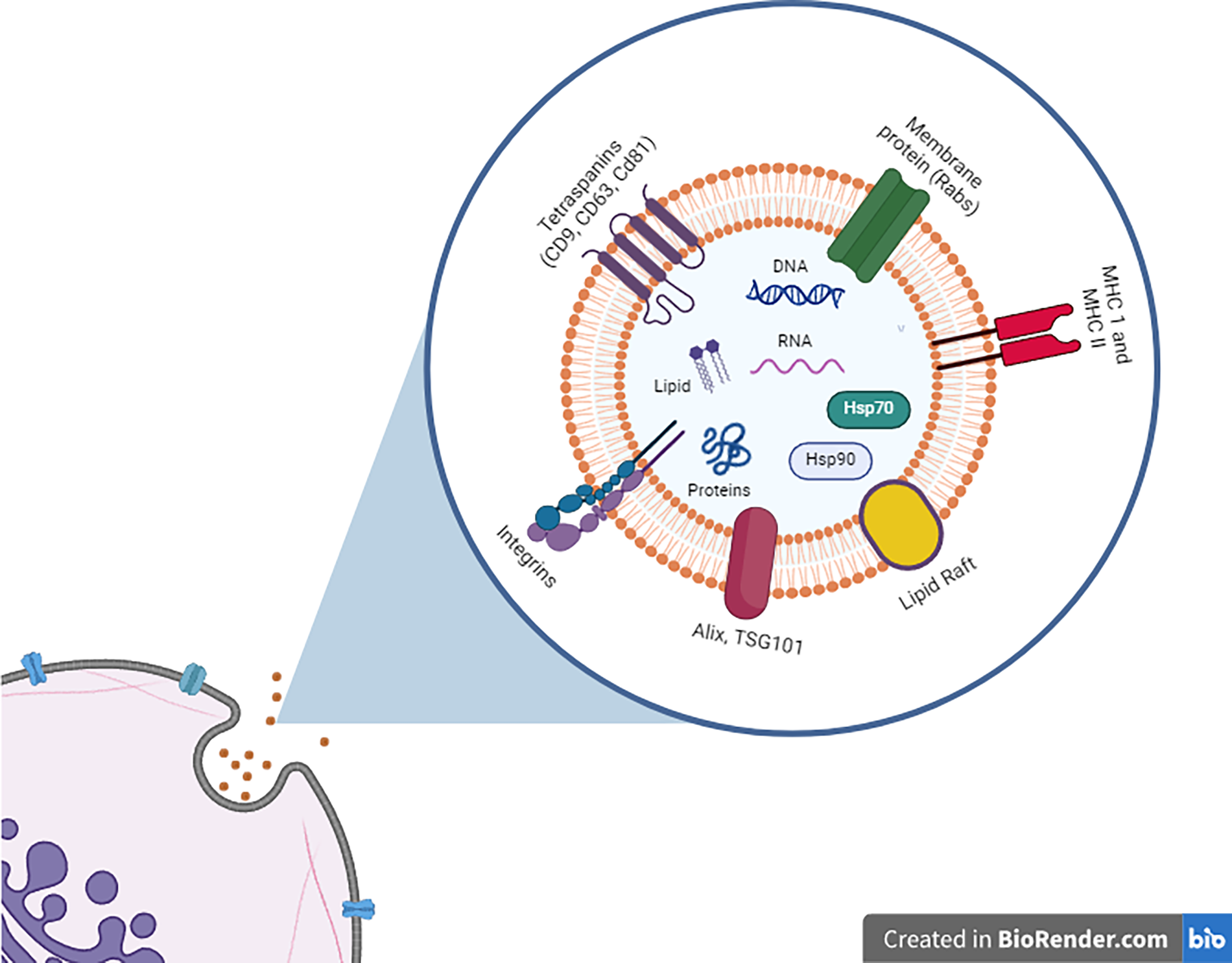
The illustration of the structure and composition of exosomes released from parent cells into the extracellular matrix.
Type of SC-Exos
Mesenchymal SC-Exos
MSCs are self-renewing adult multipotent progenitor cells that can differentiate into several cell lineages, specifically mesodermal lineages to generate osteoblasts, chondrocytes, and adipocytes. 30 MSCs can be obtained from various tissues and organs, including bone marrow, adipose tissue, and various types of blood, such as peripheral and umbilical cord blood. They are growing in interest as potential therapeutic components because they can be easily harvested from different adult tissues and multiplied ex vivo. 30 They can also give rise to many types of brain cells, including neurons, astrocytes, and oligodendrocytes.31,32 Interestingly, MSC derived-exosomes (MSC-Exos) have been reported to exhibit similar therapeutic advantages as their parent cells (natural MSCs) or even better. 33 Their intrinsic structure and size allow them to stimulate low immune reactions when introduced into the body. 34
The beneficial impact of MSCs primarily relies on their paracrine actions rather than their ability to directly differentiate. 35 Of utmost importance are the processes of initiating neuroprotection, neurogenesis, angiogenesis, and synaptic plasticity, as well as regulating neuroinflammation, boosting BBB integrity, and degrading aberrant protein aggregates. 36 These effects are primarily driven by the secretion and delivery of neurotrophic factors. 37 For example, glial cell-derived neurotrophic factor stimulates neuroprotective effects and regenerative responses in PD models 38 by shifting astrocytes and microglia toward a neuroprotective phenotype, which promotes axonal regeneration, supports cell survival, limits secondary damage, and partially restores neuronal functions. Nerve growth factor is found to interact with Schwann cells, induce Schwann cell activation and proliferation, promote myelin formation, and facilitate nerve regeneration for damaged peripheral nerves. 39 Brain-derived neurotrophic factor regulates neurogenesis, neuroprotection, and synaptic plasticity. It plays a synergistic role in promoting neuronal differentiation and enhancing axonal regrowth when combined with neurotropic 4/5, increasing the expression of phosphorylated extracellular signal-regulated kinase. 40 In animal models of TBI, MSC-Exos promote neuronal survival and inhibit inflammation, where miR-181b plays a key role that regulates the interlukin-10/signal transducer and activator of transcription 3 pathway. 41 In addition, MSC-Exos containing components with anti-apoptotic (Bcl-2 and miR-21), anti-inflammatory (IL-10 and TGF-β), and antioxidant (catalase and superoxide dismutase) properties help improve neurological outcomes in AD and PD studies.42,43
MSCs have been studied for many years in clinical trials for neurological disorders, with some MSC-based therapies having further received Food and Drug Administration designations, such as “breakthrough therapy” or “expanded access” for specific conditions.44–46 Furthermore, current clinical trials have investigated the potential of MSC-Exos as a therapeutic agent in various types of diseases, such as eye diseases (NCT05738629, NCT05413148), heart and blood diseases (NCT05669144), and orthopedic diseases (NCT05060107). 42 Only two clinical trials have been conducted for neurological disorders studies and have advanced to the early testing stage, both of which are in phase 1/2. 47 These trials focus on AD (NCT04388982) and acute ischemic stroke (NCT03384433), underscoring the preliminary phase of MSC-Exos research in neurology. 47
Neural SC-Exos
NSCs are a distinct type of stem cell found naturally in the CNS. Neural stem/progenitor cells can self-renew and are multipotent, they can generate neurons, astrocytes, and oligodendrocytes during the development of the CNS. 48 They differentiate into specific neural cells for preservation and restoration of injured brain and spinal cord tissues. 49 Native NSCs are found to increase neuronal growth, proliferation, and survival in endogenous neurogenic niches, such as in AD cellular models, through paracrine effects. 50 In mouse AD models, NSCs transplantation reduces amyloid deposition, induces neurogenesis, and improves memory and spatial learning. 50 Given the neurodegenerative nature of AD, regenerating neural circuits with exogenous NSCs presents a promising treatment strategy. 50 Compared with MSCs, NSCs have been involved in relatively few clinical trials related to neurological diseases. 51 These trials have mostly explored treatments for disorders such as amyotrophic lateral sclerosis (ALS), Huntington’s disease (HD), MS, AD, PD, and others. 51 The limited application of NSCs in clinical trials is mainly attributable to (1) ethical considerations because certain sources of NSCs involve fetal tissues, 52 (2) the possibility of tumor formation, 52 and (3) the increased risk of autoimmune responses due to the allogeneic transplantation. 53 Recent stem cell technology enabled researchers to differentiate NSCs into specific types of neurons, including oligodendrocytes, cholinergic motor neurons, dopaminergic, GABAergic, and glutamatergic neurons, in a controlled environment using chemically defined systems or the overexpression of lineage-specific transcription factors.54–56
Exhibiting biological capabilities similar to native NSCs, NSC-exosomes (NSC-Exos) have been discovered to improve the brain’s neuroplasticity, support the regeneration of nerve cells, and facilitate the recovery of motor function after brain injury. They achieve therapeutic effects by altering the activity of both neurons and glial cells in nearby and far-reaching regions. 57 To date, no clinical trials have explored the use of NSC-Exos for treating neurological diseases. 42 This gap may be caused by the difficulty in obtaining a sufficient and consistent supply of NSCs. 56 Currently, NSCs can be acquired from three primary sources: directly from CNS tissues (adult or fetal brain), through the differentiation of pluripotent stem cells (embryonic stem cells or induced pluripotent stem cells), or by reprogramming somatic cells (fibroblasts or blood cells) into induced NSCs (iNSCs).58,59 The iNSCs-derived exosomes (iNSC-Exos) have been shown to promote cell survival and proliferation effectively, comparable with NSC-Exos, while demonstrating therapeutic potential in enhancing recovery from ischemic stroke 60 and reducing AD-like symptoms in preclinical models. 61
Modification and Engineering of SC-Exos for Cargo Delivery
Isolation of exosomes
Effective methods of isolating exosomes are essential due to their varied origins and uses. Among the methods are commercial kits, immunological affinity capture, size exclusion chromatography, differential centrifugation, and microfluidic technology 62 (Fig. 2). Differential ultracentrifugation (dUC) is a method for sorting particles based on their size and density using repeated cycles of centrifugation. By exposing samples to varied centrifugal strengths and durations, dUC can effectively extract exosomes, generating high-purity populations. dUC is widely recognized as the gold standard for exosome isolation, providing a dependable method of obtaining purified exosomes. 63 Immunological affinity capture is achieved by coating magnetic beads with specific antibodies, which allows for the capture of exosomes that display those surface antigens. 64 Size exclusion chromatography separates molecules by size using a column packed with porous beads (Sephadex, Sepharose, Sephacryl, or BioGel P), where larger particles elute faster because they cannot enter the pores. 65 The advantages and disadvantages of each isolation approach result in varying degrees of exosome yield, which may comprise different forms of EVs or aggregated proteins found in body fluids.

A schematic diagram illustrating the various methods utilized for isolating exosomes and preparing drug-loaded exosomes from fluid samples.
Cargo loading
Cargo loading can be accomplished via either endogenous or exogenous pathways. Endogenous loading involves engineering parent cells to create exosomes with specified chemicals. Target molecules or cargo such as RNAs, proteins, and small-molecule drugs are coincubated or transfected into donor cells to alter them. 66 Coincubation refers to the process of combining exosomes and cargo in a controlled environment. 67 The cargo-loaded exosomes are then extracted and purified by ultracentrifugation or immunoaffinity capture after activation. For example, paclitaxel (PTX)-loaded MSC-Exos have been developed with strong antiproliferative activity and better biocompatibility than PTX alone.68,69 Transfection is another typical approach for delivering nucleic acids or other substances into donor cells, accomplished by chemical methods, electroporation, or viral vector-mediated techniques. 58
In exogenous loading, therapeutic cargo is introduced into preseparated exosomes through methods that penetrate their membranes. Nanomaterials, small-molecule pharmaceuticals, and proteins are molecules that can be loaded using this technology. The technical complexity and number of options available with exogenous loading are smaller than those with endogenous loading. Exogenous loading has been extensively used, including coincubation, electroporation, freeze–thaw cycles, extrusion, transfection, saponin treatment, and sonication 70 (Fig. 2). In the coincubation method, pure exosomes are incubated with drugs at room temperature for a set duration to facilitate the incorporation of the drug into the exosomes. However, ultrasonic incubation uses pulsed-focused ultrasound to enlarge holes in the exosome membrane temporarily. By adopting this technology, therapeutic agent delivery can be improved to target specific brain regions, such as the areas affected by a stroke, without inflicting any damage to the normal structures of the brain. 71 Transfection involves introducing exogenous DNA or RNA into exosomes to enable novel functionalities using techniques such as electroporation, chemical transfection, and cationic polymer-based transfection. Electroporation, in particular, creates pores in the exosome membrane by applying short current pulses, allowing cargo molecules to enter and making it an effective delivery system. 70
Hydrogels Loaded with SC-Exos for Delivery and Neural Regeneration
Exosomes may still encounter a challenge in being eliminated by the immune system, which hinders their clinical application. 72 There is a need for the continued development of delivery methods to ensure targeted delivery and maximize therapeutic efficacy. Despite serving as delivery vehicles, exosomes themselves may necessitate a biomaterial carrier. Hydrogels appear as a potential carrier for the controlled release of exosomes and as interaction matrices for improving the retention of exosomes in tissues. 73 Recently, hydrogels have also been reported for prolonged and localized delivery of engineered SC-Exos, including their application in tissue engineering and regeneration. 74
Characteristics and synthesis of hydrogels
Hydrogels are synthesized by cross-linking polymer chains, which may originate from natural, synthetic, semisynthetic, or hybrid polymers. Natural hydrogels are biocompatible, bioactive, and biodegradable; however, they lack stability with low biomechanical properties. For example, collagen, fibrin, gelatin, agarose, alginate, cellulose, chitosan, and hyaluronic acid are among the common types of natural hydrogels with inherent properties that are usually safe or well tolerated.75,76 Conversely, synthetic hydrogels are produced from man-made polymers by polymerization of the monomer, such as polylactide acid, poly lactic-co-glycolic acid (PLGA), polyethylene glycol (PEG), poly (ethylene oxide), poly (vinyl alcohol) (PVA), poly-N-isopropylacrylamide, Pluronic F-127, poly (acrylic acid), and polyacrylamide. 77 Most of them are less biocompatible or are incompatible, but they provide stability and better mechanical strength than natural hydrogels. To enhance their original properties, both natural and synthetic polymers can be chemically modified and combined with each other to produce semisynthetic and hybrid hydrogels, respectively. Studies have shown that mixing different types of polymers or combining different chemical structures can achieve synergic properties or specific properties not found in either component alone. For example, methacryloyl-modified gelatin (GelMA), 78 acrylate-modified hyaluronic acid, 79 PVA-mixed chitosan and PEG, 80 and PEG-conjugated fibrinogen or gelatin 81 have combined the bioactivity of natural hydrogels with the alterability of their chemical parameters. 82 This makes them ideal for encapsulating, delivering, and gradually releasing exosomes in the application of tissue regeneration.
Categories of hydrogels
Hydrogels are categorized into nanohydrogels, microgels or hydrogel microspheres, self-healing hydrogels, and 3D-bioprinted hydrogels. Nanohydrogels (1–100 nm) have a high surface area-to-volume ratio that increases their interaction with biological systems. Unlike conventional macro and micro hydrogels, nanohydrogels involve intramolecular cross-linking, resulting in environmental stability, reduced swelling and solubility, and improved mechanical properties and controlled release. These features and their diminutive size enable them to more efficiently retain drugs or nanoparticles loaded within them compared with larger hydrogels. Nanohydrogels can efficiently penetrate and distribute throughout tissues, making them suitable for the accurate administration of exosomes in the field of regenerative medicine. 83 In contrast, microgels, known as hydrogel microspheres, are produced in the micrometer size range. They can be injected directly and provide a greater proportionate surface area than macro hydrogels, enabling the natural removal of substances and enhancing tissue penetration. Despite the previous success of microgels in targeted delivery and long-lasting treatment for osteochondral joint injuries, 84 alginate hydrogel microspheres loaded with MSC-EVs have recently demonstrated promising therapeutic outcomes in acute colitis treatment. 85
Self-healing hydrogels, also known as stimulative hydrogels, can autonomously repair and regenerate themselves following injury when triggered by external stimuli, including light, heat, pH changes, or interactions among functional groups within the hydrogels. 86 By incorporating both covalent and noncovalent bonds, these hydrogels are engineered with deliberately weak sacrificial bonds, allowing for energy dissipation during deformation while enabling self-reformation without requiring external energy input. 87 Self-healing hydrogels loaded with SC-Exos may further improve spontaneous recovery from injuries.
In addition, 3D-bioprinted hydrogels can be designed to imitate the intricate structures and microenvironment of tissues or organs, making them highly promising for tissue regeneration. The incorporation of SC-Exos into these hydrogels further enhances their ability to promote tissue regeneration and improve treatment efficiency. 3D bioprinting techniques such as inject bioprinting, extrusion-based bioprinting, laser-assisted bioprinting, and stereolithography are utilized to deposit bioink(s) in a layer-by-layer hydrogel structure together with living cells, bioactive molecules, drugs, EVs, or any substances of interest. 88 For example, Li et al. bioprinted a highly complex, spatially heterogeneous bilayer hydrogel scaffold composed of methacrylated gelatin (GelMA), oxidative hyaluronic acid (HA), dopamine-conjugated HA, and decellularized extracellular matrix of cartilage and bone, along with exosomes isolated from human adipose-derived mesenchymal stem cells (ADMSC-Exos), for the regeneration of osteochondral defects. 89
Methods of exosomes incorporation in hydrogels
There are three methods to incorporate exosomes into hydrogels: physical approach, chemical approach, and direct mixing approach (Fig. 3). The physical approach is simple, where the hydrogels will be immersed in exosomes containing solutions, allowing absorption that is regulated by the porosity, swelling ability, pH levels, temperature, surface charge, and hydrophobic–hydrophilic nature of the hydrogels. This approach, also known as the “breathing” or “swelling” method, enables the hydrogels to expand and absorb the exosomes, while noncovalent bonds are utilized for exosome attachment. Thus, the pore size of the hydrogels must be larger than the size of exosomes and stem cells. In contrast, the chemical approach incorporates exosomes with polymers before the addition of crosslinkers or precursors, which then induce gelation through covalent bonds. This approach provides significant flexibility in controlling degradation rates and mechanical properties. 90 However, some of the active precursors or crosslinkers used may potentially be cytotoxic. For example, MSC-Exos have been loaded into GelMA/lithium phenyl-2,4,6-trimethylbenzoylphosphinate hydrogel and HA-bioprinted hydrogel for axon regeneration 91 and bone regeneration, 92 respectively. The third approach involves the direct mixing of exosomes, polymers, and crosslinkers simultaneously to achieve efficient in situ gelation for injectable hydrogels. The mechanisms of in situ gelation are ultraviolet radiation, temperature change, pH change, and ion exchange. 93 A dual-chamber syringe is typically used to inject the exosome-loaded hydrogels directly into the targeted site. This method is suitable for filling critical-size defects with complex and irregular geometries, supporting cell growth and regeneration. Among the studies, injectable thermo-sensitive hydrogel loaded with SC-Exos has been reported for the restoration of cavernous nerve injury. 94

A diagram illustrating the methods for encapsulating exosomes within hydrogels:
Current applications of SC-Exos loaded hydrogels for efficient delivery and tissue regeneration in neurological disorder studies
To deliver exosomes therapeutically, it is crucial to develop a noninvasive and active method. Various cutting-edge hydrogel-based delivery systems incorporating SC-Exos and small-molecule drugs have been developed for studies on neurological disorders, such as SCI, AD, stroke, and TBI 95 (Table 1).
Summary of the Recent Studies on Stem Cell-Derived Exosomes Incorporated with Hydrogels for Treating Neurological Disorders

AD, Alzheimer’s disease; IFN-γ, interferon-γ; MSC, mesenchymal stem cell; MSC-Exos, mesenchymal stem cell-derived exosomes; SCI, spinal cord injury; TBI, traumatic brain injury; TNF-α, tumor necrosis factor-α.
In SCI, natural hydrogels mimic the extracellular matrix and serve as a supportive scaffold for tissue regeneration while delivering bioactive molecules directly to the injury site. Roh et al. developed a HA-based hydrogel integrated with biochemical cues containing exosomes isolated from human umbilical cord-derived MSCs (hUCMSC-Exos), which significantly promoted spinal cord regeneration by inducing angiogenesis, neural differentiation, and remyelination while inhibiting inflammation, apoptosis, and glial scarring. 96 Similar results were reported by Mu et al., who delivered hypoxia-preconditioned hUCMSC-Exos using an adhesive HA hydrogel. 97 In another study, He et al. demonstrated that fibrin gel loaded with bone marrow (BM)-MSC-Exos significantly enhanced myelination and oligodendrogenesis, contributing to functional recovery. 98 Moreover, by incorporating fibrin and collagen hydrogels with exosomes from ADMSC-Exos, Afsartala et al. have effectively regenerated injured nerves and alleviated SCI-induced central neuropathic pain. 99
Recently, researchers demonstrated that semisynthetic and hybrid hydrogels, such as temperature-sensitive polyglycolic acid copolymer and polyethylene glycol (PLGA-PEG-PLGA) hydrogel incorporated with miR-138-5p-loaded hUCMSC-Exos had effectively reduced neuronal apoptosis and oxidative stress through transcription factor NF-E2-related factor 2 pathway activation, as well as reduced inflammation via the suppression of the NOD-like receptor protein 3–caspase 1 signaling pathway. This strategy promoted axonal regeneration and motor function recovery in SCI models. 100 Methacrylated gelatin-based hydrogels were incorporated with melatonin-pretreated NSC-Exos, 101 BM-MSC-Exos, 102 and epidermal growth factor receptor-positive NSC-Exos loaded with miR-34a-5p, 103 respectively. They have been shown to promote anti-inflammatory effects, neuronal differentiation, axonal regeneration, neurite regrowth, and functional recovery while reducing glial scars in injured spinal tissues. Comparable results were also achieved by combining ADMSC-Exos with F127 polycitrate-polyethyleneimine hydrogel 104 and human placenta amniotic membrane MSCs-derived exosomes with peptide-modified adhesive hydrogels. 105
In the current applications of hydrogel-loaded SC-Exos for AD, a hydrogel composed of self-assembling peptides that can be broken down by enzymes was reported by Huang et al. 106 The smart-release hydrogel regulated the release of MSC-Exos after intranasal administration by prolonging their retention at the administration site. This method successfully reduced the destruction of neurons, promoted the growth of new neurons, and improved memory deficits in an AD mouse model. 106 Neurological function and cerebral angiogenesis following an ischemic stroke can be improved through the controlled release of NSC-Exos, which were incorporated into an adhesive HA hydrogel and treated with catechol. 107 The HA hydrogel promoted endogenous cell adhesion and proliferation in ischemic regions by mimicking the 3D tissue microenvironment. Other researchers showed that neuroinflammation of ischemic stroke could be targeted using interleukin-1β-stimulated BM-MSC-Exos. 108 The retention of BM-MSC-Exos triggered by interleukin-1β is effectively improved by the combination of thermosensitive supramolecular injectable polymer and silk fibroin hydrogel, leading to inhibition of neuroinflammation and regeneration of neurons. 108 In TBI, Liu et al. recently demonstrated that the therapeutic potential of BM-MSC-Exos incorporated into hyaluronan–collagen hydrogel had successfully stimulated the growth of new blood vessels and nerve cells, which in turn facilitated the regeneration of axons, the repair of myelin, the development of synapses, and even the remodeling of brain structure. 109
Challenges and Future Perspectives
The complexity of isolating exosomes from stem cells raises technical and therapeutic concerns, as well as the capability of using the hydrogel. Exosome isolation, modification, and drug loading face significant technical challenges, including the expansion and purification processes, which require optimization to enhance efficiency while minimizing potential damage and contamination. 110 For large-scale production and manufacture of exosomes, current methods are often sophisticated, costly, and inconvenient. 111 Therefore, developing bioreactor cultures using microcarriers or hollow-fiber membranes and optimizing scalable isolation techniques, such as tangential flow filtration and improved sterile purification protocols, could potentially enable large-scale exosome production and enhance their homogeneity. 112 Furthermore, methods such as regulating intracellular calcium levels, applying external stress, and using cytoskeletal inhibitors or gene modifications should be explored to enhance the number of exosomes secreted. 113
Although SC-Exos remain stable for extended periods during delivery, variability in exosome characteristics based on the stem cell source presents challenges, as different sources can secrete exosomes with varying features that impact their therapeutic potential. 114 Hence, identifying optimal stem cell sources and standardizing preconditioning, dosage, and therapy schedules are necessary to overcome this variability and enhance therapeutic outcomes. 115 Furthermore, methods for isolating, purifying, characterizing, and managing the quality of SC-Exos are suggested to follow the guidelines provided by the International Society for Extracellular Vesicles. 116
Following intravenous delivery, MSC-Exos, like other exosomes, may be ingested by immune cells and organs such as the liver, spleen, and lungs, which could impair their efficacy in reaching target tissues. 114 Hydrogels offer a promising solution as delivery vehicles, but issues such as the reduction of hydrogel stiffness, simplification of hydrogel production, and the availability of user-friendly hydrogels with adjustable and predictable gelation longevity must be addressed. 117 Furthermore, challenges remain in achieving the uniform incorporation of exosomes into hydrogels and ensuring their sustained release. To overcome the challenges discussed, unique 3D printing scaffold designs or biopolymer modification approaches could be considered as alternatives to the conventional method of superficial exosome adsorption onto hydrogels. For example, coprinting polycaprolactone and lyosecretome-containing alginate resulted in a slower release profile and a more uniform loading. 118
Undoubtedly, the therapeutic approaches involving hydrogel-loaded SC-Exos are still in the early stages of development, progressing from preclinical research to clinical implementation. 119 Future investigations should prioritize evaluating the long-term safety and effectiveness of the most optimal cell source, exosome quality, and hydrogel type for treating neurological diseases. A quality control system for SC-Exos that complies with good manufacturing practice (GMP) standards needs to be established to ensure the reproducibility and consistency of SC-Exos products, including their storage, transportation, and clinical applications.
Conclusions
To improve current treatments for neurological disorders, the combination of SC-Exos with hydrogels offers a novel and promising strategy. Exosomes possess both neuroprotective and regenerative properties, whereas hydrogels serve as effective delivery vehicles, allowing localization and sustained release of exosomal cargo. This synergistic approach has shown great potential in preclinical models, enhancing neural repair, reducing neuroinflammation, inducing neural regeneration, and promoting functional recovery in TBI, SCI, stroke, and neurodegenerative diseases. The utilization of hydrogels supports exosome stability and targeting, overcoming several limitations associated with exosome therapy alone. However, challenges remain, including standardization of exosome production to achieve GMP grade, optimization of hydrogel formulations, and clarification of underlying mechanisms. To advance the combinatorial therapy toward clinical applications, these challenges need to be addressed in future research.
Authors’ Contributions
N.A.: Conceptualization; writing, reviewing, and editing—original draft; and visualization. S.-Y.L.: Conceptualization; writing, reviewing, and editing—original draft; and supervision. M.M. and Z.I.: Writing, reviewing, and editing—original draft.
Footnotes
Acknowledgment
The authors would like to thank Sarah Kinnersly for proofreading this article.
Funding Information
This work is supported by the Bridging Grant Universiti Sains Malaysia (R501-LR-RND003-0000001319–0000). S.-Y.L.’s research group is supported by the Fundamental Research Grant Scheme (FRGS/1/2020/TK0/USM/02/32–6171275) from the Ministry of Higher Education Malaysia. N.A. has been awarded by the GRA-Assist Scheme (GRA23-0440) at the Universiti Sains Malaysia.
Ethics Approval
No involvement of humans and/or animals in this study.
Disclosure Statement
No competing financial interests exist.