




Abstract
This review synthesizes experimental findings on various biomaterial scaffolds used in eyelid reconstruction. It examines the structural properties, cellular responses, and functional outcomes of scaffolds such as chitosan, poly(propylene glycol fumarate)-2-hydroxyethyl methacrylate, poly(propylene glycol fumarate) - type I collagen (PPF-Col), decellularized matrix-polycaprolactone, branched polyethylene, collagen, poly(3-hydroxybutyrate-co-3-hydroxyhexanoate, and poly(lactic-co-glycolic acid. These scaffolds exhibit diverse mechanical and biological properties, with some demonstrating good biocompatibility, tunable properties, and potential for tissue repair. However, there are limitations, including concerns about long-term functionality and a lack of comprehensive evaluations. This review highlights the need for multifunctional scaffolds that combine lid replacement and ocular surface function restoration, as well as the establishment of standardized research methods. The goal is to guide future innovation in the field and improve the quality of life for patients with eyelid defects.
Impact Statement
This review underscores the contemporary advancements in 3D-printed biological scaffolds for eyelid reconstruction, with a particular emphasis on their utilization as tarsus substitutes. By methodically summarizing a wide array of scaffold types and fabrication techniques, this study offers a comprehensive synthesis of the extant experimental findings, thereby identifying critical challenges in the field. This analysis not only addresses existing knowledge gaps but also serves as a catalyst for future innovations in scaffold design, with the potential to significantly advance clinical applications in reconstructive surgery and promote advancements in tissue engineering research.
Keywords
Introduction
The eyelids are highly delicate structures that serve as critical functional and aesthetic components of the face. Functionally, they protect the ocular surface and facilitate lubrication, while aesthetically, they play a pivotal role in facial rejuvenation and cosmesis. 1 Anatomically, the eyelids are bilamellar structures composed of anterior and posterior lamellae. The upper eyelid’s anterior lamella consists of thin skin and the orbicularis oculi muscle, while its posterior lamella includes the tarsal plate (7–12 mm in height) and conjunctiva, supported by the levator aponeurosis and Müller muscle for elevation. The lower eyelid similarly features an anterior lamella of skin and orbicularis oculi, with a shorter tarsal plate (3–4 mm) in its posterior lamella, anchored by the capsulopalpebral fascia. Both eyelids contain meibomian glands within the tarsal plates for tear film lipid secretion.1,2
Eyelid defects resulting from trauma, surgical excision, or congenital conditions pose significant challenges for both functional and aesthetic reconstruction. As a critical support structure, the tarsal plate plays a central role in eyelid mechanics, making its restoration a cornerstone of successful eyelid reconstruction. Current surgical methods for eyelid reconstruction include layered repair techniques (e.g., local flaps like rhomboid 3 flaps for anterior lamellar defects, and autologous grafts such as tarsoconjunctival 4 or hard palatal mucoperiosteal graft 5 for posterior lamellar support), all of which adhere to the “like for like” principle by prioritizing tissue similarity in anatomy and function. However, full-thickness eyelid defects pose significant challenges due to the tarsus’s unique structure and nonregenerative nature, limiting available methods that meet both functional and esthetic needs. Alternative materials, such as autografts and xenografts, address tarsus replacement but face issues like donor site morbidity, availability, and immunogenicity.6–8 Consequently, medical professionals have shifted their focus to the domain of biomaterials in pursuit of alternative materials deemed suitable for eyelid repair.
Eyelid reconstruction also necessitates the utilization of “like for like” principle, which requires biosynthetic scaffold materials to replicate the native tissue’s layered structure, biomechanical properties, and biofunctional dynamics, meticulously tailored to the characteristics of the defect, including its thickness, size, and location. 9 This review synthesizes experimental findings on biomaterial scaffolds for eyelid reconstruction, examining their structural properties, cellular responses, and functional outcomes to provide a comprehensive overview of advances and challenges, guiding future innovations.
Macroporous chitosan scaffolds
The technical roadmap of this study is illustrated in Figure 1.
Chitosan, a biopolymer, has demonstrated significant advantages in the field of tissue engineering, including biocompatibility, biodegradability, and antimicrobial properties. It has been demonstrated to support cell migration, tissue formation, and enhance scaffold performance in skin, bone, cartilage, and vascular engineering. In the field of eyelid reconstruction, the combination of gelatin and chitosan-based scaffolds holds particular promise for the development of biomimetic materials.10,11
Sun MT et al. 12 developed scaffolds using a freeze-gelation method, achieving highly porous and interconnected structures with surface and center pore sizes ranging from 9–19 μm to 24–52 μm, respectively, influenced by mold shape. Mechanically, scaffolds composed of 5% Chitosan crosslinked with 0.1% glutaraldehyde closely resembled human tarsal tissue, exhibiting an initial modulus, final modulus, and elongation of 0.17 ± 0.01 MPa, 0.62 ± 0.1 MPa, and 22.1 ± 0.02%, respectively, comparable to human tarsal tissue values of 0.14 ± 0.1 MPa, 1.7 ± 0.6 MPa, and 15.8 ± 2.1%. 13 In biocompatibility tests, NIH 3T3 fibroblasts demonstrated significant adhesion and proliferation on the scaffolds, with marked growth observed at 72 h. Primary human eyelid skin fibroblasts also exhibited growth on 3% and 5% scaffolds, though in lower numbers compared to the control groups. These findings suggest that the structural and mechanical properties of the scaffolds, which mimic those of tarsal tissue, hold promise for applications in personalized tissue engineering. 12 However, concerns remain regarding long-term functionality due to the short duration of in vivo studies.
Chitosan, being biodegradable, gradually degrades in vivo, raising concerns about its ability to maintain the structural integrity required for the functional demands of the native tarsus over time. This degradation may compromise essential eyelid functions such as corneal protection, smooth movement, and ocular surface stability. While Chitosan exhibits excellent biocompatibility and biodegradability, these limitations preclude its use as a standalone material for tarsal plate replacement. Instead, its application could be optimized as a supplementary component in composite scaffolds combined with materials offering superior mechanical properties and controlled degradation profiles, addressing both functional and structural requirements effectively.
PPF–HEMA scaffold
The technical roadmap of this study is illustrated in Figure 2.
2-Hydroxyethyl methacrylate (HEMA) is a biocompatible, hydrophilic, and optically transparent monomer that is widely utilized in biomedical applications.14–16 Polymerized HEMA, recognized for its low immunogenicity and cytocompatibility, has been demonstrated to promote cell adhesion and proliferation. Despite its nondegradable nature, it has been modified for a variety of applications, including contact lenses, drug delivery, wound dressings, and tissue engineering. This demonstrates its potential for use in regenerative medicine. 17
Gao et al. 18 evaluated poly(propylene glycol fumarate)-2-hydroxyethyl methacrylate (PPF-HEMA) copolymer scaffolds for repairing rabbit eyelid defects. The incorporation of HEMA into PPF enhanced the hydrophobicity and mechanical strength of pure PPF. The PPF-to-HEMA ratio was found to allow for tunable chemical compositions, hydrophilicity, mechanical properties, and degradation behaviors. In vitro tests demonstrated excellent cytocompatibility, minimal toxicity to human dermal fibroblasts, and enhanced fibroblast adhesion and proliferation. In vivo experiments demonstrated mild tissue responses, superior biodegradation, and mechanical support compared to conventional decellularized dermal matrix (ADM) controls. Furthermore, the scaffolds promoted enhanced control of inflammation, fibrotic tissue formation, and tissue integration. The scaffolds promoted neovascularization and fibrous tissue growth, maintaining stability, and forming fibrotic envelopes resembling native eyelid structures. The fatigue resistance and tunable degradation of the scaffolds provided sustained support, aiding tissue repair, and remodeling. This study suggests that PPF-HEMA scaffolds hold promise for eyelid blepharoplasty, warranting further research and clinical investigations.
This study developed PPF-HEMA scaffolds with tunable properties replicating tarsal plate mechanics and enabling personalized applications. Subsequent in vitro and in vivo assessments yielded evidence of biocompatibility, degradation, and repair efficacy. The incorporation of HEMA enhanced the hydrophilicity and mechanical strength of the scaffolds, thereby improving their suitability for tissue repair. However, the study’s limitations include the absence of a direct comparison with human tarsal plates, the unassessed endurance during eyelid movement, and the experimental duration, which precludes long-term evaluations such as fibrous encapsulation. Further studies are necessary to ascertain the functional and long-term efficacy of PPF-HEMA scaffolds in eyelid reconstruction.
PPF-Col scaffold
The technical roadmap of this study is illustrated in Figure 3.
PPF, a biodegradable polymer with adjustable mechanical strength, degradation rates, and excellent biocompatibility, is a prevalent material in regenerative medicine, particularly in the field of bone tissue engineering. Its nontoxic degradation products enhance its suitability for in vivo applications. 19 Type I collagen (Col), with its biocompatibility and low immunogenicity, supports conjunctival epithelial cell culture and regeneration, making it ideal for ocular surface reconstruction.20–23
Xu et al. 24 developed a biomimetic biphasic scaffold for tarso-conjunctival reconstruction by combining PPF, HEMA, Col, and chitosan. The developed scaffold consists of a PPF-HEMA layer that provides rigid support as a tarsal plate substitute and a Col/CS layer designed to mimic the conjunctival structure and promote regeneration. The Col/CS layer, with an adjustable thickness, facilitates cellular adhesion, proliferation, and stratification while maintaining high porosity for nutrient exchange. In vitro studies revealed that medium-thickness sponges (poly(propylene glycol fumarate) - type I collagen [PPF-Col]-2, 466.38 ± 69.78 μm) were optimal for conjunctival epithelial cell growth and differentiation. In vivo studies in rabbit models demonstrated efficient conjunctival re-epithelialization and functional tissue regeneration, closely resembling native conjunctiva.
This biphasic scaffold presents a promising solution for posterior lamella reconstruction in eyelid defects by balancing structural support, biocompatibility, and tissue integration. However, a significant limitation of this study is its focus on conjunctival regeneration, specifically goblet cell regeneration, without addressing other critical components of the ocular surface. The conjunctiva’s structural and functional integrity is closely tied to the overall ocular surface environment, which includes the tear film and corneal epithelium. Consequently, the exclusive emphasis on goblet cell regeneration, without consideration of factors such as tear film stability, corneal epithelial integrity, and ocular surface inflammation, results in an incomplete evaluation of the scaffold’s effects. This narrow focus engenders uncertainties regarding its ability to achieve functional eyelid repair while preserving long-term ocular surface health. Consequently, future research endeavors must encompass a more comprehensive evaluation of the ocular surface to adequately assess the scaffold’s potential in eyelid reconstruction.
DMA–PCL scaffolds
The technical roadmap of this study is illustrated in Figure 4.
Polycaprolactone (PCL) is a biocompatible, degradable polyester that has found widespread use in tissue engineering and drug delivery applications. Its slow degradation rate enables the controlled release of metabolites. The combination of 3D-printed PCL scaffolds with hydrogels has been shown to enhance biofunctionality and cytocompatibility, thereby facilitating tissue replacement in a range of applications.25–28 The decellularized matrix from adipose-derived mesenchymal stromal cells has been shown to promote stem cell proliferation and soft tissue regeneration.29,30
Chen et al. 31 investigated the possibility of integrating 3D-printed PCL scaffolds with DMA from adipose-derived stromal cells for the purpose of eyelid tissue engineering. This innovative approach enhanced the hydrophilicity of the scaffold, thereby improving cell adhesion, proliferation, and lipid secretion. In vitro, the DMA-PCL scaffold supported SZ95 sebaceous gland cells, promoting cell proliferation and lipid secretion while suppressing inflammation and apoptosis-related gene expression.
In vivo studies in nude mice revealed that DMA-PCL scaffolds supported long-term cell survival, forming structures analogous to natural eyelids with intact epithelial layers and functional lipid secretion. In comparison to conventional materials, such as hard palate mucosa, the DMA-PCL scaffolds exhibited superior capacity to replicate eyelid structure and partially restore lipid secretion, thereby addressing functional repair deficiencies. The use of FDM 3D printing ensured structural fidelity, while PCL’s gradual degradation provided sustained mechanical support for regeneration.
Notwithstanding its potential, the study has limitations, including a short experimental duration, lack of longitudinal degradation analysis, and reliance on a subcutaneous mouse model rather than an eyelid defect model. The uneven distribution of meibomian glands and the inadequate examination of tear film lipid stability necessitate further investigation. Notwithstanding these limitations, the findings underscore the promise of DMA-PCL scaffolds in the field of eyelid tissue engineering, demonstrating their potential for structural and functional regeneration. To ensure the full assessment of their potential, long-term studies and optimizations are essential.
B-PE scaffolds
The technical roadmap of this study is illustrated in Figure 5.
Polyethylene (PE), a versatile biomaterial, demonstrates excellent processability due to its thermoplastic, semicrystalline nature, with higher branching levels enhancing elasticity, solubility, and film-forming ability while increasing tensile strength and elongation.19,32–38 PE’s biocompatibility supports long-term clinical use, and surface modifications improve cell adhesion and tissue regeneration. Its slow, tailorable biodegradation rate and 3D printing compatibility enable precise scaffold fabrication for personalized applications. These properties render PE highly suitable for bone repair, soft tissue replacement, and organ reconstruction, thereby establishing it as a pivotal material in tissue regeneration and biomedical innovation. 33
This study 39 explores the development and application of a novel branched polyethylene (B-PE) scaffold for eyelid reconstruction. The synthesis of B-PE involved the use of (α-diimine) nickel as a catalyst, with thin films and porous scaffolds being prepared via solution casting and gelatin porogen leaching. Key structural parameters, including molecular weight, branching degree, porosity (90.26 ± 2.08%), and pore size (280–480 μm), were meticulously controlled to ensure optimal performance. In vitro tests revealed no significant impact of B-PE on the viability of NIH3T3 fibroblasts or human vascular endothelial cells (ECs). While ECs demonstrated robust adherence and proliferation, fibroblasts exhibited initial weak adherence that improved over time, indicating good biocompatibility. Mechanical tests yielded an elastic modulus of 0.83 ± 0.1 MPa and an elongation of 76.2 ± 2.8%, comparable to rabbit tarsal plates but with superior flexibility. In vivo, subcutaneous implantation in rats resulted in mild inflammation, stable fibrous capsule formation, and increasing vascularization. In rabbit eyelid defect models, B-PE scaffolds effectively repaired defects within four weeks without contraction or rejection. Histological analysis confirmed connective tissue infiltration and mild inflammation, thereby demonstrating the scaffold’s biocompatibility and repair efficacy.
The study under consideration demonstrates the potential of B-PE scaffolds for eyelid reconstruction, thereby highlighting their biocompatibility, mechanical properties, and effective tissue integration. Cytotoxicity tests with NIH3T3 fibroblasts and ECs revealed mild inflammation, collagen deposition, and vascularization. The elastic modulus of the scaffold mirrors that of rabbit tarsal plates, with greater elongation and nondegradable properties for sustained support. In vivo tests validated vascularization and repair efficacy. However, the four-week duration of the study precludes the assessment of long-term stability, and the absence of a direct comparison to human tarsal plates restricts the study’s generalizability. Notwithstanding these limitations, the study provides a robust foundation for the use of B-PE scaffolds in tarsal reconstruction.
Type I Collagen Sponge Scaffold Loaded with Rabbit Auricular Chondrocytes
The technical roadmap of this study is illustrated in Figure 6.
Collagen, with its triple-helical structure, offers several advantages, including excellent biocompatibility, low immunogenicity, and mechanical strength. Its surface properties support cell adhesion, migration, and tissue repair, making it ideal for bone, cartilage, skin, corneal regeneration, and drug delivery. Advancements in extraction, crosslinking, and composites have further augmented its potential for use in regenerative medicine applications. 40
Yan et al. 41 constructed tissue-engineered cartilage (TEC) grafts using type I collagen sponges (CS) derived from bovine Achilles tendon that were highly irradiated, 1 mm thick, and highly porous (200–300 μm), providing an ideal microenvironment for rabbit ear chondrocytes to attach, proliferate, and deposit cartilage matrix. During in vitro cultivation, TEC grafts developed translucent and smooth properties, with increased thickness and improved quality as reflected by higher Bern scores. Increased GAG content and synthesis supported improved cartilage function. Biomechanical testing demonstrated reliable suture performance and improved tensile and flexural moduli over time, achieving properties comparable to human lid tissue by 2 weeks, suitable for lid reconstruction. In vivo implantation in nude mice for 6 months demonstrated stable graft structure with minimal hypertrophic calcification, indicating early safety and stability. In a rabbit model of posterior eyelid defects, TEC grafts effectively repaired the tarsal and conjunctival layers, restoring normal eyelid morphology and function while maintaining conjunctival epithelial organization and promoting epithelial and goblet cell growth, demonstrating their strong potential for ocular tissue repair.
This study explores bioengineering and tissue engineering as a means to enhance the efficacy of TEC grafts by transplanting biological cells onto allogenic scaffolds. Employing a multifaceted approach encompassing cellular isolation, histological staining, biochemical analysis, biomechanical testing, and in vitro/in vivo validations, it provides a comprehensive evaluation of the characteristics and repair efficacy of TEC grafts. Two distinct eyelid defect models, namely single-layer tarsal and bilayer tarsoconjunctival defects, were utilized to assess the efficacy of the grafts in restoring tarsal and conjunctival tissues. The study incorporated the Bern scoring system, a quantitative tissue staining assessment method, to enhance the evaluation process. Notwithstanding the study’s contributions, limitations emerge. Notably, the presence of fibrous encapsulation in single-layer defects after six months and the absence of ocular surface evaluations point to research gaps that require attention. Furthermore, long-term implications and comprehensive assessments remain unaddressed, underscoring the need for further investigation. Notwithstanding these limitations, the study establishes TEC grafts as promising tools for functional eyelid reconstruction and underscores the necessity for further investigations to advance clinical applications.
Poly 3-hydroxybutyrate-co-3-hydroxyhexanoate scaffolds
The technical roadmap of this study is illustrated in Figure 7.
Polyhydroxyalkanoates are biocompatible and biodegradable biomaterials that have found application in tissue engineering. Poly(3-hydroxybutyrate-co-3-hydroxyhexanoate) (PHBHHx) exhibits particular promise due to its exceptional mechanical strength, biocompatibility, and piezoelectric properties, which support bone regeneration and progenitor cell phenotypes. The multifunctional properties of PHBHHx make it a promising material for eyelid substitutes and advanced biomedical applications.42–44
Zhou et al. 45 evaluated the potential of PHBHHx scaffolds as tarsal substitutes for eyelid reconstruction, emphasizing their mechanical properties, tissue integration, and advantages over ADM. The investigation revealed that PHBHHx scaffolds demonstrated robust mechanical strength, a porous structure conducive to cellular infiltration, and controlled inflammatory responses, exhibiting a transition from acute to mild chronic inflammation by the fourth week. Biodegradation and tissue integration were evidenced by the presence of foreign body giant cells, fibrous capsule formation, and collagen deposition. By the eighth week, immune responses subsided, and fibroblast proliferation ensured functional repair. Compared to ADM, PHBHHx offers a safer, more reliable alternative, demonstrating excellent biocompatibility and tissue integration over 8 weeks.
However, the study’s relatively brief duration constrains the evaluation of the scaffold’s long-term stability and persistence. Although microstructural characterization and porosity measurements were conducted, a comprehensive evaluation of the scaffold’s mechanical properties, such as elastic modulus, was not included. While the scaffold demonstrated biocompatibility, the absence of comparisons with native tarsus mechanical properties raises concerns about its equivalence. The assertion that the scaffold “supported” eyelid reconstruction based on normal eyelid movement and fibrous encapsulation appears to be presumptive without direct mechanical testing, underscoring the necessity for further evaluations and long-term studies.
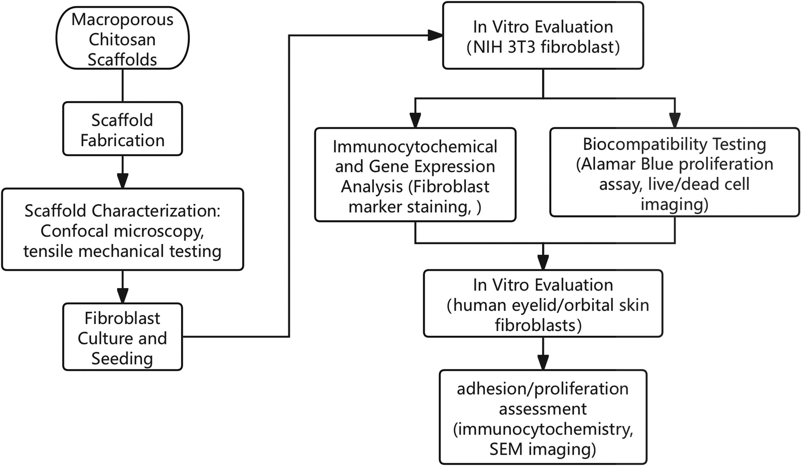
Technology Roadmap of Macroporous Chitosan Scaffolds.
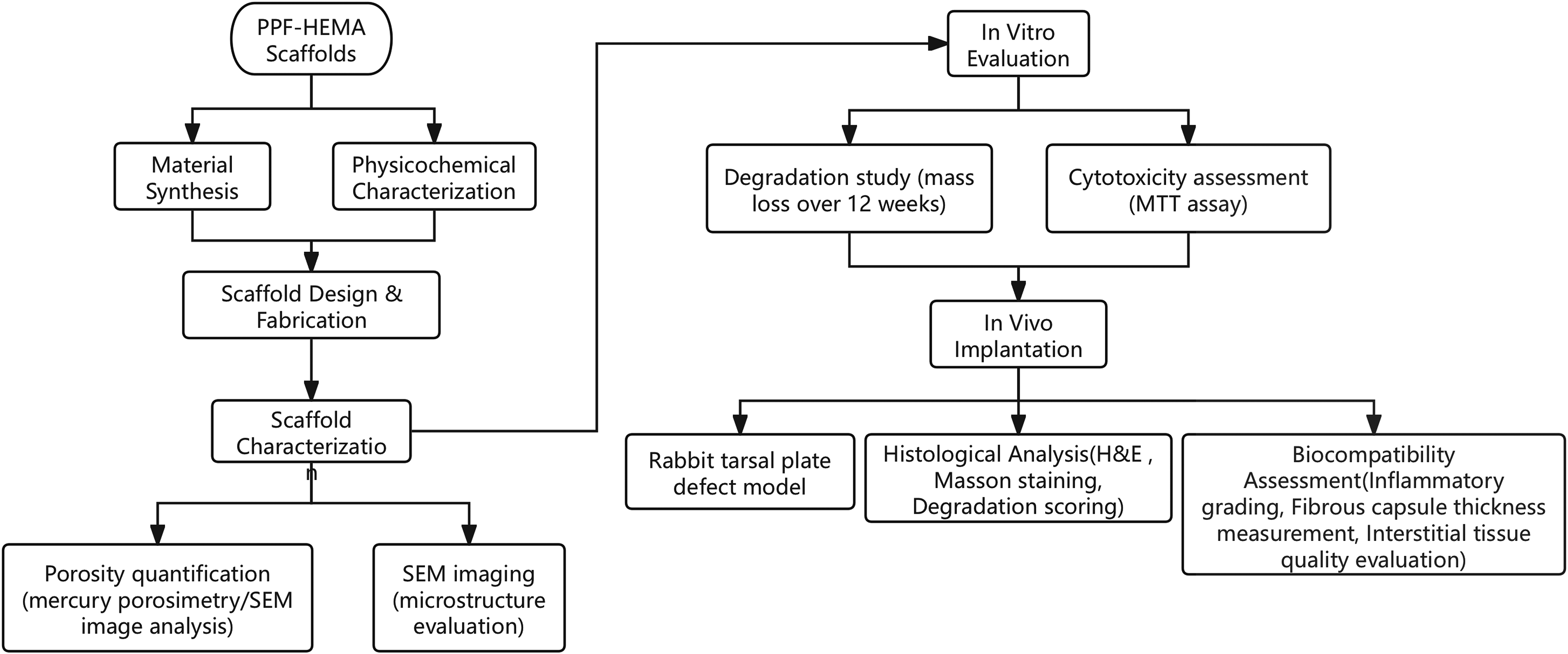
Technology Roadmap of PPF–HEMA Scaffold. PPF–HEMA, poly(propylene glycol fumarate)-2-hydroxyethyl methacrylate.
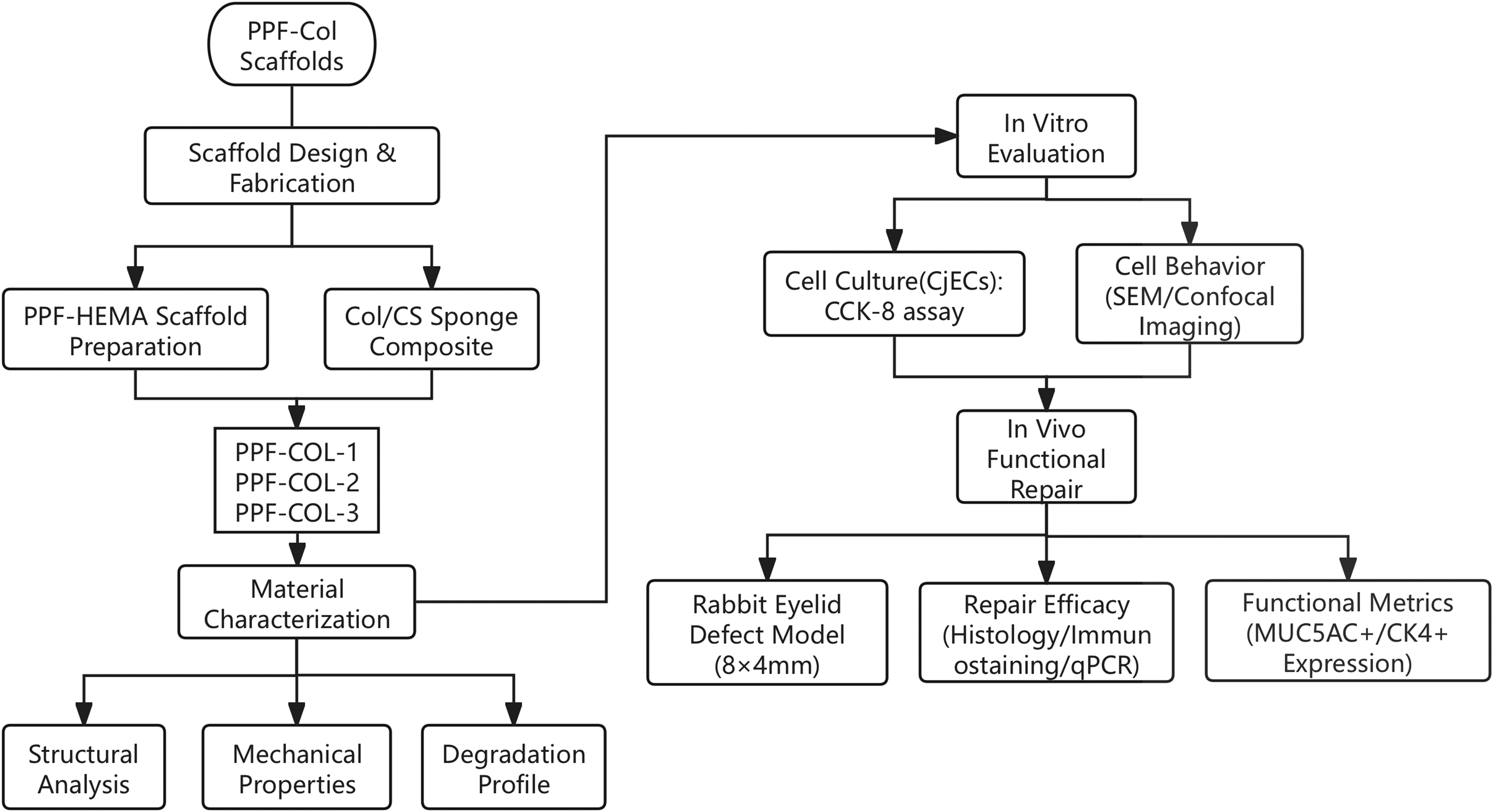
Technology Roadmap of PPF-Col Scaffold.

Technology Roadmap of DMA–PCL Scaffolds. DMA–PCL, decellularized matrix–polycaprolactone.
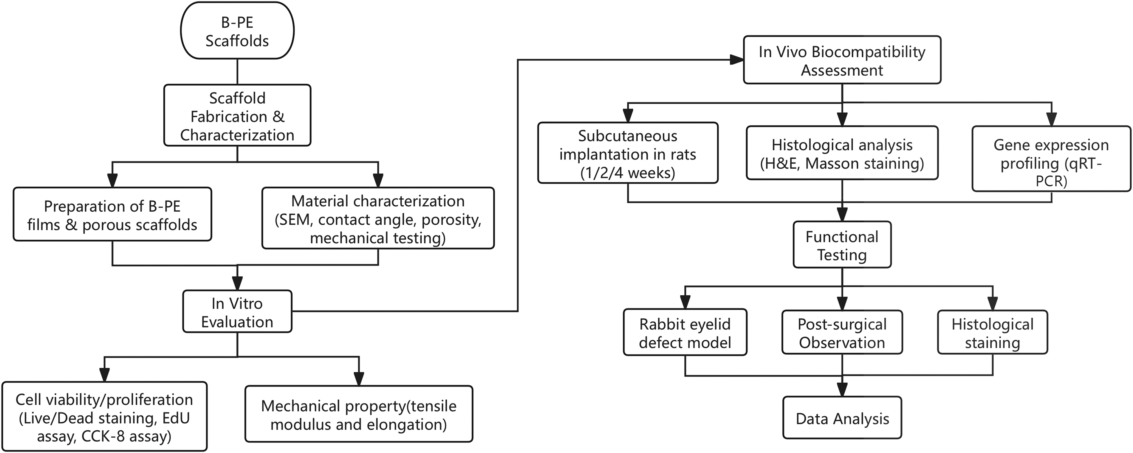
Technology Roadmap of B-PE Scaffolds. B-PE, branched polyethylene.
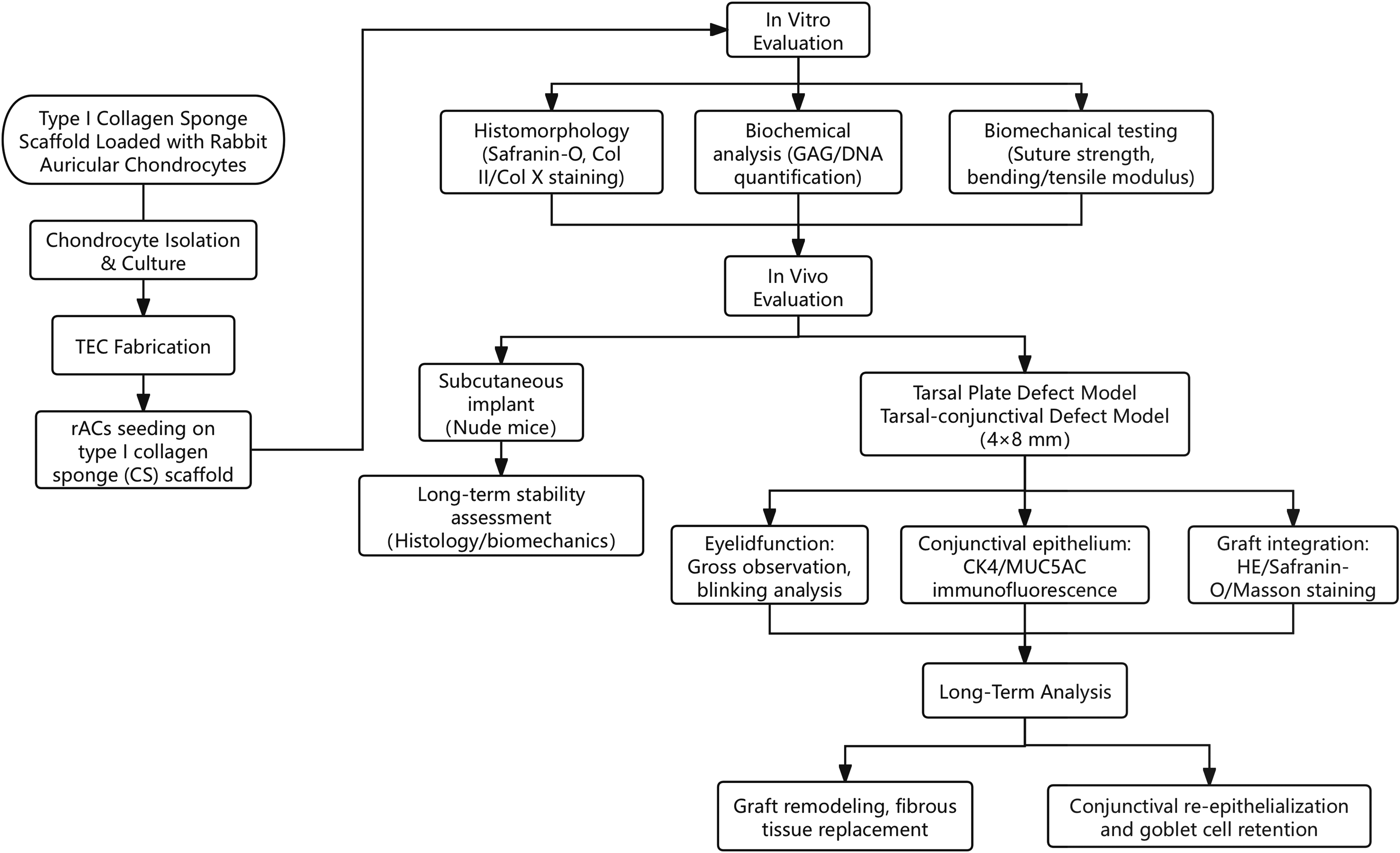
Technology Roadmap of Type I Collagen Sponge Scaffold.
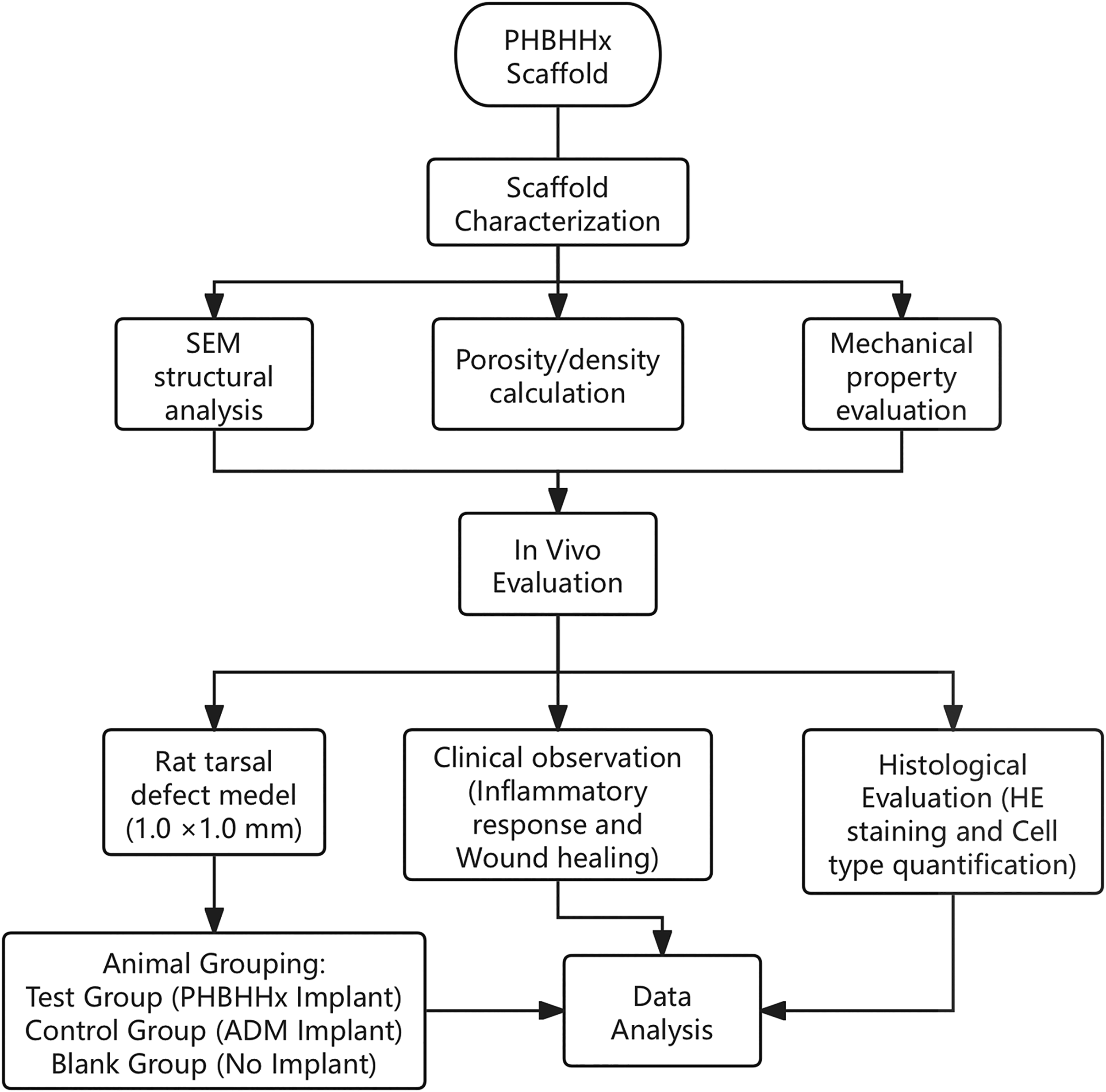
Technology Roadmap of PHBHHx Scaffolds. PHBHHx, poly(3-hydroxybutyrate-co-3-hydroxyhexanoate).

Technology Roadmap of AZM@O-PLGA Scaffolds. AZM@O-PLGA, azithromycin-loaded axially aligned pore structure.
Azithromycin-carrying and microtubule-orientated PLGA scaffolds
The technical roadmap of this study is illustrated in Figure 8.
Poly(lactic-co-glycolic acid (PLGA) is characterized by its exceptional biocompatibility and tunable degradation, which is regulated by the ratio of lactic acid to hydroxyacetic acid. This property ensures biosafety and suitability for long-term use.46–53 The material’s versatility in molecular configuration is further enhanced by functionalization and hydroxyapatite compounding, thereby promoting its application in bone tissue engineering and drug delivery.54–62
Xu et al. 63 investigated the azithromycin-loaded axially aligned pore structure (AZM@O-PLGA) scaffold for eyelid tissue repair, demonstrating its potential for application. The fabrication of the scaffold involved the implementation of temperature-controlled phase separation technology, yielding a regular vertical interconnected pore structure that emulates the microenvironment of natural tissues. In vitro experiments demonstrated the scaffold’s excellent cytocompatibility, as evidenced by the adhesion and proliferation of fibroblasts and rat lid epithelial cells along the pore direction. To ascertain the viability of the cells, a Live/Dead staining procedure was employed, which confirmed the vitality of the cells. In vivo experiments in a rabbit eyelid defect model revealed that the scaffold gradually degraded over eight weeks, integrating well with host tissues, and forming fibrous tissues similar to natural eyelid tissue. Furthermore, the azithromycin-loaded scaffold demonstrated anti-inflammatory and anti-infective properties, effectively mitigating the risk of postoperative inflammation and infection while promoting optimal tissue morphology and reducing scar formation. The degradation rate of the scaffold matched the tissue repair rate, avoiding complications from rapid or slow degradation. In summary, the AZM@O-PLGA scaffold, which exhibits structural bionicity, biocompatibility, and therapeutic effects, demonstrates significant potential as an alternative material for eyelid blepharoplasty, offering a novel approach for the treatment of eyelid defects.
The AZM@O-PLGA scaffold, which was developed for the purpose of eyelid tissue repair, was prepared using temperature-controlled phase separation technology. Scanning electron microscopy revealed a regular vertical interconnected pore structure, providing a favorable microenvironment for cell growth. In vitro studies demonstrated good cytocompatibility, with fibroblasts and rat lid epithelial cells adhering, surviving, and aligning along the scaffold’s pore direction, as confirmed by Live/Dead staining. In vivo experiments using a rabbit eyelid defect model demonstrated the scaffold’s gradual degradation over eight weeks, accompanied by its integration into host tissues and the formation of fibrous tissue resembling natural eyelid structures. The presence of azithromycin within the scaffold led to the observation of anti-inflammatory and anti-infective properties, with a resultant reduction in inflammation, the risk of infection, and scar formation. The degradation rate of the scaffold was found to be in alignment with the process of tissue repair, thereby preventing adverse effects from either too rapid or too slow degradation. This study underscores the potential of AZM@O-PLGA as a promising alternative material for eyelid blepharoplasty, offering a viable approach for addressing eyelid defects.
Discussion
A quick overview and comparison of the scaffolds can be found in Table 1.
To date, no tarsal substitute has been developed that is capable of completely replicating the structure and function of native tarsal tissue. This highlights the need for the development of novel approaches to achieve superior functional outcomes for patients.64,65 As a cornerstone of regenerative medicine, tissue engineering offers significant promise for advancing current reconstructive methods by enabling the creation of highly specialized and patient-specific tissue replacements.
In evaluating the diverse in vivo and in vitro characteristics of the scaffolds, the assessment is primarily focused on their mechanical properties and biocompatibility. In regard to mechanical properties, the modulus of elasticity of the tarsal plate, as an integral structure of the eyelid with a dual function of providing rigid support and allowing for elastic deformation, is of paramount importance and must be subjected to rigorous examination. The mean modulus of elasticity of fresh human lids is 1.73 ± 0.61 MPa, 13 and mechanical properties approximating this value are better suited to simulating the supportive and elastic roles of human lids. PPF-Col scaffolds are designed to replicate the mechanical gradient of the blepharoplane-conjunctival interface through a bilayer configuration, comprising a rigid PPF layer and a flexible collagen layer. The elastic modulus of these scaffolds can be modulated by altering the cross-linking density. However, there is a paucity of direct comparative data with human tarsal plate. Macroporous chitosan scaffolds and PHBHHx scaffolds were mechanically matched to rabbit lids by cryogel and porous structures, but the former may lose long-term support due to degradation, and the latter has not been validated for equivalence to human tissues. B-PE scaffolds, as a nondegradable material, have a modulus of elasticity comparable to that of rabbit lids with superior ductility. However, their rigidity may constrain adaptability to dynamic eyelid movements. 3D-printed DMA-PCL scaffolds are meticulously controlled for porosity (>1,000 mm) by fused deposition technology. However, they lack direct comparative data with human tissues. The technique to precisely control porosity (>90%) and mechanical strength did not provide quantitative comparisons with natural tissues. It is noteworthy that the majority of studies were confined to in vitro or short-term animal experiments, lacked long-term mechanical stability assessments, and only PPF-HEMA scaffolds explicitly mentioned elastic modulus modulation by monomer ratio (no specific value was disclosed). This underscores the pressing need for a standardized mechanical testing system in this field. Furthermore, some studies have conducted a series of physical property tests, including a suture strength test, a three-point force bending test, 41 with the aim of evaluating the physical properties of the scaffolds in greater detail. In the animal model, the removal of the grafted tissue at different time points and the subsequent performance of elastic modulus testing represent an effective means of assessing the changes in the physical properties of the stent during in vivo degradation. This method enables the assessment of the stent’s continued physical support postimplantation by quantifying the changes in its mechanical properties over time. This provides crucial data for optimizing the stent design and evaluating its potential for clinical applications.
A Comprehensive Overview of Different Scaffolds in This Review
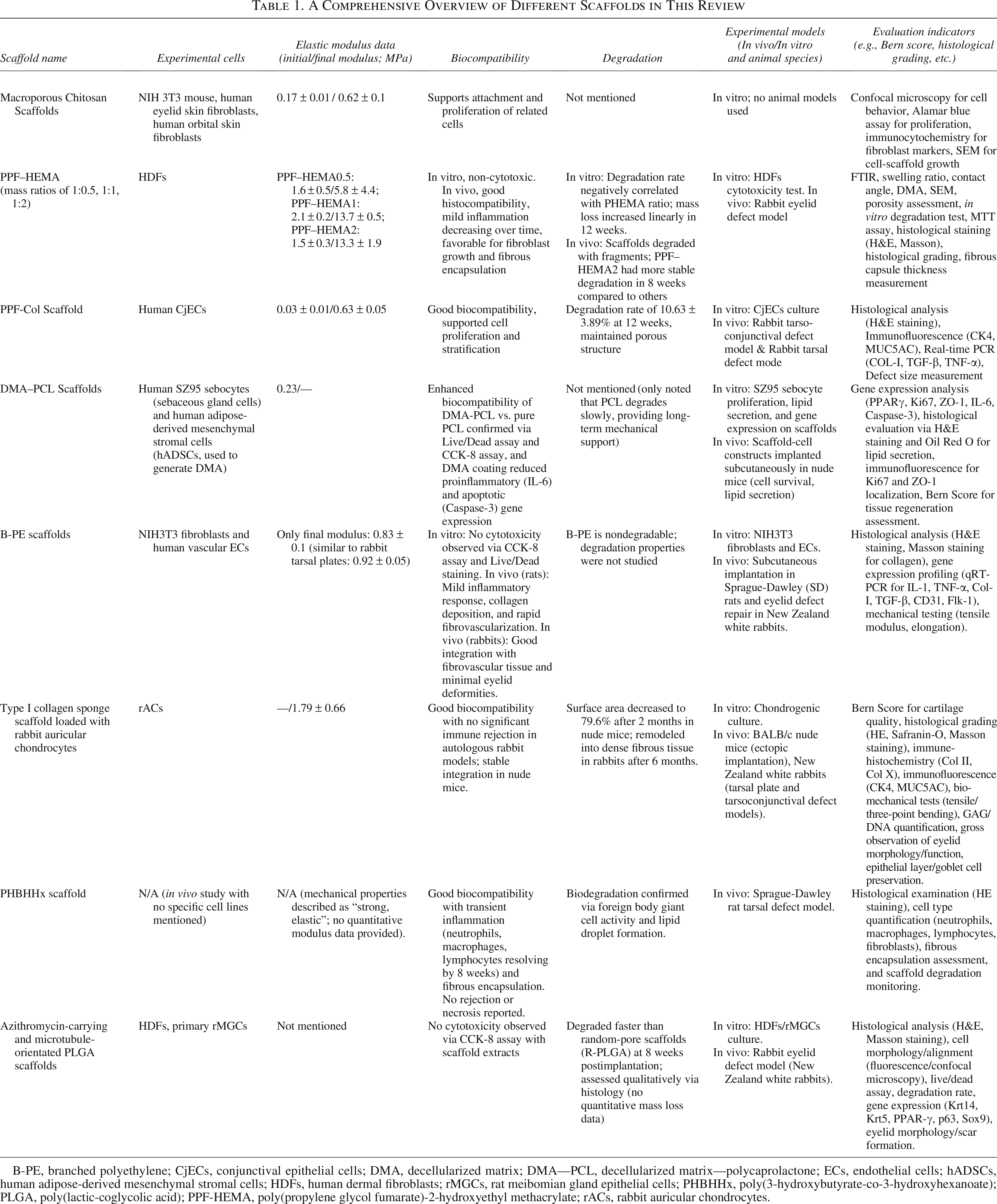
B-PE, branched polyethylene; CjECs, conjunctival epithelial cells; DMA, decellularized matrix; DMA—PCL, decellularized matrix—polycaprolactone; ECs, endothelial cells; hADSCs, human adipose-derived mesenchymal stromal cells; HDFs, human dermal fibroblasts; rMGCs, rat meibomian gland epithelial cells; PHBHHx, poly(3-hydroxybutyrate-co-3-hydroxyhexanoate); PLGA, poly(lactic-coglycolic acid); PPF-HEMA, poly(propylene glycol fumarate)-2-hydroxyethyl methacrylate; rACs, rabbit auricular chondrocytes.
The assessment of biocompatibility is conducted at the macro, cellular, and molecular levels. At the macro level, eyelid activity in animal models is monitored using photography or video to assess graft exposure and inflammation. At the cellular level, the CCK-8 assay and histopathologic methods, such as HE staining, are employed to evaluate cellular infiltration, inflammatory responses, and tissue integration. Molecular-level studies involve immunohistochemical labeling of inflammatory factors, quantitative PCR, and in vitro techniques like flow cytometry or ELISA to quantify inflammation-related cytokines. The human eyelid is not a typical example of either cartilage or fibrous connective tissue. Its main components include fibroblasts, various types of collagen, GAG, and proteoglycans. Additionally, multifunctional proteoglycans, glycoproteins, cartilage oligomeric matrix proteins, and a multitude of collagen fibers, small blood vessels, and elastic fibers are present. In the vicinity of the lid glands, a specialized ECM is observed.39,66 It is crucial to ascertain whether the introduction of a stent grafting procedure results in the formation of new collagen fibers and GAG. Macroporous chitosan scaffolds and PHBHHx scaffolds both demonstrated effective NIH 3T3 cell proliferation, yet the former exhibited reduced activity in human eyelid skin primary fibroblasts, indicating a potential species-differentiated risk. Type I Collagen scaffolds employed the Bern scoring system to assess cartilage regeneration quality, integrating it with GAG/DNA quantification to achieve objective evaluation. However, it did not analyze the effect of ocular surface microenvironment on repair. AZM@O-PLGA scaffolds innovatively introduced a drug slow-release function to inhibit the expression of inflammatory factors (TNF-α, IL-6) by locally releasing azithromycin, which elevated the biocompatibility from passive tolerance to the level of active regulation. Notably, with the exception of B-PE scaffolds, which utilized qPCR to analyze collagen deposition-related genes, the majority of studies continued to rely on conventional histological staining, a method that lacks sufficient resolution of molecular mechanisms. Additionally, while the DMA-PCL scaffold confirmed the lipid secretion function of SZ95 sebaceous gland cells, the tear film stability remained unvalidated in the eyelid defect model, underscoring the limitations of functional compatibility assessment.
Eyelid defects result in not only the loss of local tissue structure but also a significant disruption to the normal function of the ocular surface. This is particularly evident in the coating ability and stability of the tear film, which may further result in excessive tear evaporation, corneal exposure, and tear film instability. Ultimately, this can lead to exposure keratitis or even irreversible visual impairment. Consequently, when assessing the efficacy of tissue-engineered scaffolds, it is essential to consider not only their capacity for anatomical restoration but also their influence on the restoration of ocular surface function. However, the majority of current studies have concentrated on the role of scaffolds in conjunctival repair and lid replacement function, with a paucity of assessment of ocular surface-related parameters. In the extant literature, although some studies have designed specific stent structures to support conjunctival layer repair (e.g., biphasic stent design or surface modification), there is a general lack of systematic examination of key ocular surface functional parameters, such as tear film breakup time and corneal fluorescein staining score, in animal experiments. This deficiency may result in an underrecognition of potential issues with the ability of stents to support the ocular surface environment in real-world applications.
The various scaffold designs currently included in the literature represent innovative attempts at lid replacement scaffolds, each of which provides a solution to a specific problem and, to some extent, advances the development of lid replacement technology. To illustrate, PPF-Col scaffolds and PPF-HEMA scaffolds employ a heterogeneous composite strategy to enhance hydrophilicity by promoting epithelialization through the collagen layer and HEMA, respectively. However, the bilayer interface may influence eyelid stress transfer during blinking. AZM@O-PLGA scaffolds are innovatively constructed with axially aligned microtubule structure (pore size 20–50 μm), which guides the directional migration of cells. The drug-carrying system makes the scaffolds both mechanically supportive and anti-infective, providing a new paradigm for the repair of wounds at high risk of infection.56,67–69 The progressive refinement of these design options has established the foundation for the versatility and personalization of lid replacement scaffolds. Nevertheless, the optimal future lid replacement scaffold should not merely encompass the advantages of these designs but also overcome the limitations of existing scaffolds to become a comprehensive solution that combines structural replacement, functional regeneration, and long-term stability. For instance, restoring the lipid-secreting function of the meibomian glands by growing primary cultured meibomian cells on the surface of the scaffold can not only restore the integrity of the tear film but also effectively prevent postoperative complications such as dry eye. 70 This model has been first observed in DMA-PCL scaffolds, which are a type of scaffold that integrates decellularized matrices with directional printing technology to enable the first in vitro mimicry of meibomian gland lipid secretion function. However, the uneven spatial distribution of meibomian gland cells may affect functional expression. In the future, the design of meibomian gland cells and DMA-PCL scaffolds can be combined, leading to the combination of biosynthetic scaffolds and artificially cultured meibomian glands, and realizing the breakthrough of artificial tarsal plate.
In the present studies, scaffolds utilized for eyelid reconstruction were designated as “bioengineered scaffolds” or “tissue-engineered scaffolds.” This inconsistency in nomenclature may have implications for the standardization of studies and future clinical dissemination. The term “bioengineered scaffold” frequently highlights the manufacturing process and engineering design characteristics of the material, particularly innovations in structural and performance optimization. For instance, some studies have employed 3D printing to fabricate scaffolds that emulate the intricate anatomy of the eyelid. 31 However, this designation may fail to acknowledge the necessity for biological characteristics in the practical utilization of the scaffold. In contrast, the term “tissue-engineered scaffolds” is more appropriate for describing the role of these scaffolds in tissue repair and functional regeneration. The current studies commonly employ autologous cell implantation techniques to enhance the biocompatibility and functionality of scaffolds. As an illustration, in the studies conducted by Xu et al.24,39,63 the scaffolds were implanted with related cells with the objective of enhancing functional tissue regeneration. This cell-material composite design concept is more closely aligned with the fundamental principles of tissue engineering. Considering the overall trajectory of research, while these scaffolds have made notable advancements in engineering technology, their fundamental functions remain centered on tissue repair and regeneration. Designating them as “tissue engineering scaffolds” not only aligns more closely with their technical connotations but also fosters consistency across disparate studies, enhances the degree of standardization in external communication and clinical application, and ultimately facilitates a more unified understanding of their role in tissue repair and regeneration.
Future studies on eyelid replacement should prioritize the diversification of stent functions and the optimization of overall performance.1,2 In particular, the development of multifunctional stents that combine both eyelid replacement and ocular surface function restoration is a crucial area of research. This approach has the potential to achieve a comprehensive restoration of structure and function, which is a significant advancement in the field. It is also essential to establish standardized research methods, including the unification of ocular surface assessment indexes and functional evaluation systems for animal models. This will enhance the comparability and scientific validity of different studies. Furthermore, the clinical translation of lid replacement stent research represents a significant challenge. To facilitate the efficient translation of laboratory results to clinical applications, future research should prioritize the safety validation of biomaterials, optimization of production processes, and the development of clinical trial design strategies. The ideal lid replacement scaffold would provide a more reliable and comprehensive treatment option for eyelid reconstruction, ultimately improving patients’ quality of life.
Authors’ Contributions
J.L.: Conceptualization, methodology, writing—original draft, writing—review and editing, supervision, quality control, and final approval of the article. M.Z.: Literature review, writing—review and editing, and writing—original draft. M.Z.: Literature review, writing—review and editing, and critical revision. Q.W.: Writing—review and editing, project administration, and data validation. X.J.: Writing—review and editing and article formatting. Q.H.: Review and editing, quality control, and final approval of the article.
Footnotes
Author Disclosure Statement
The authors have no relevant financial or nonfinancial interests to disclose.
Funding Information
This work was supported by the Natural Science Foundation of Jiangxi Province (Grant No. 20242BAB25490) and the General Project of the Jiangxi Provincial Department of Education (Grant No.GJJ2200194).