
Abstract
Several studies suggest that the surface coating of titanium could play an important role in bone tissue engineering. In the present study, we have followed a particular biomimetic strategy where ultrasonically or electromagnetically stimulated SAOS-2 human osteoblasts proliferated and built their extracellular matrix on a titanium plasma-spray surface. In comparison with control conditions, the ultrasonic stimulation (average power, 149 mW; frequency, 1.5 MHz) and the electromagnetic stimulation (magnetic field intensity, 2 mT; frequency, 75 Hz) caused higher cell proliferation, and increased surface coating with decorin, osteocalcin, osteopontin, and type I collagen together with higher incorporation of calcium and phosphorus inside the extracellular matrix. The immunofluorescence related to the preceding bone matrix proteins showed their colocalization in the cell-rich areas. The use of the two physical stimulations aimed at obtaining the coating of the rough titanium plasma-spray surface in terms of cell colonization and deposition of extracellular matrix. The superficially cultured biomaterial could be theoretically used, in clinical applications, as an implant for bone repair.
Introduction
To obtain a stable primary stabilization and the following osteointegration or secondary stabilization of a bone implant, macroporous 1 and rough 2 surface patterns have been developed for the biomaterials. The secondary fixation is, essentially, a mechanical interlock, which requires a long immobilization period 3 and may cause bone loosening at the tissue-implant interface during the early postimplantation time.
An early osteointegration of a rough metallic surface depends mainly on the ingrowth of the bone tissue onto the material surface. Aiming at an early osteointegration in orthopedics and dentistry, Ti-alloy implants, coated with a rough Ti-alloy layer, are widely used because of their good mechanical properties and surface biocompatibility.
The material biocompatibility is of fundamental importance in determining the biological response of the host tissue. Castner and Ratner have reviewed the concept of biocompatibility and its experimental realization in the fields of biomaterials and surface science;4,5 they think the biocompatible surfaces of the biomaterials that heal as the surfaces with the characteristics of a clean, fresh wound. 4 The studies to obtain biocompatible surfaces are numerous: the nonspecific protein adsorption has been inhibited6,7 or a biomimetic strategy has been developed,8–10 for instance.
In the present work of bone tissue engineering in vitro, aiming at an accelerated and enhanced in vivo osteointegration of a rough titanium surface during the early postimplantation period, we show a particular biomimetic strategy that consists in the surface coating of titanium plasma-spray with proliferated bone cells and their extracellular matrix produced in situ; during the culture period, we have also applied an electromagnetic or an ultrasonic wave because the osteoblastic cell function can be physically modulated in terms of proliferation and differentiation.11,12
The biomaterial surfaces and the three-dimensional scaffolds for bone tissue engineering aim at healing critical-size long bone defects and maxillofacial skeleton defects due to trauma, tumor resection, and tissue degeneration.13–16 Biodegradable, osteoinductive, and osteoconductive biomaterials are ideal to follow a typical tissue engineering approach that involves the seeding and the in vitro culturing of cells in a scaffold before the implantation.
Although the biodegradation is a common requirement for biomaterials, this in vitro study was designed as a propaedeutic phase before the alternative approach of the osteointegration via the biointegration, that is, the in vivo integration of a biostable biomaterial. 17 The utilization of biostable biomaterials, with adequate mechanical properties especially in orthopedic surgery, would overcome possible problems associated with the biodegradation, such as degradation kinetics asynchronous with tissue regeneration and adverse reactions to degradation products.
Along the path leading, in future works, to the approach of the biointegration, in this propaedeutic in vitro study, we have elected the titanium plasma-spray surface. Titanium is a metal widely used in hip and knee replacements owing to its biological compatibility, particularly with bone. Titanium has been shown to be a suitable biomaterial for the culture of marrow stromal cells in an effort to create constructs for bone replacement.1,18,19 In vitro, titanium acts as scaffold for the adhesion and the osteoblastic differentiation of progenitor cells.20,21 In vivo, the material reveals itself to be osteoconductive. 1 In addition, the coating of titanium with calcium phosphates, extracellular matrix proteins, bone morphogenetic proteins, and growth factors enhances the bone formation, suggesting that the surface of titanium could play an important role in bone tissue engineering.22–26
Therefore, we have developed another type of titanium surface coating in vitro that could theoretically benefit the in vivo bone formation: we used ultrasonic or electromagnetic 11 waves, and aimed to enhance a bone cell culture over titanium plasma-spray disks, to coat their surface with bone cells and bone extracellular matrix, in other words, with a new physiological and biocompatible surface made of cell–matrix layers. Using this approach, the superficially cultured biomaterial could be theoretically used, in clinical applications, as an osteointegrable implant.
Materials and Methods
Human SAOS-2 osteoblasts from an osteosarcoma were used as cellular model: these osteoblasts were seeded onto the surface of titanium plasma-spray disks, and then cultured with the application of an ultrasound wave or an electromagnetic wave. The experimental setup permitted the study of the SAOS-2 cells as they proliferated and produced extracellular matrix under physical stimulus.
The following culture characteristics of biological interest were evaluated: the cell proliferation (by DNA extraction), the cell viability (by 3-[4,5-dimethylthiazole-2-yl]-2,5-diphenyl tetrazolium bromide [MTT] test), the cell–matrix distribution (by scanning electron microscopy [SEM] and indirect immunofluorescence staining), the extracellular matrix production (by extraction of the matrix proteins), and the distributions of calcium and phosphorus in the extracellular matrix (by energy dispersive X-ray spectroscopy [EDXS]).
Titanium plasma-spray surface
Disks (diameter, 12 mm; height, 4 mm) were cut from the titanium alloy Ti6Al4V (ISO 5832-3 standard practice). The disks were plasma-sprayed with titanium powder of the same alloy by an industrial and standardized vacuum plasma-spray procedure (Ufficio Italiano Brevetti e Marchi; Patent Number: UD2007000092 [Italian Patent pending]) (Lima-Lto S.p.A., Lima Group, Villanova di San Daniele del Friuli, Italy). According to the data sheet provided by the manufacturer (Lima-Lto S.p.A.), the resulting rough surface (Fig. 1) had the following characteristics: mean roughness depth Rz of 30 μm, thickness of plasma-sprayed layer equal to 175 ± 75 μm (evaluation according to the ASTM F1854 standard practice), static shear strength equal to 24 ± 2 MPa (ASTM F1044), static tensile strength equal to 50 ± 3 MPa (ASTM F1147), and abrasion resistance equal to 34 ± 18 mg/100 cycles (ASTM F1978).
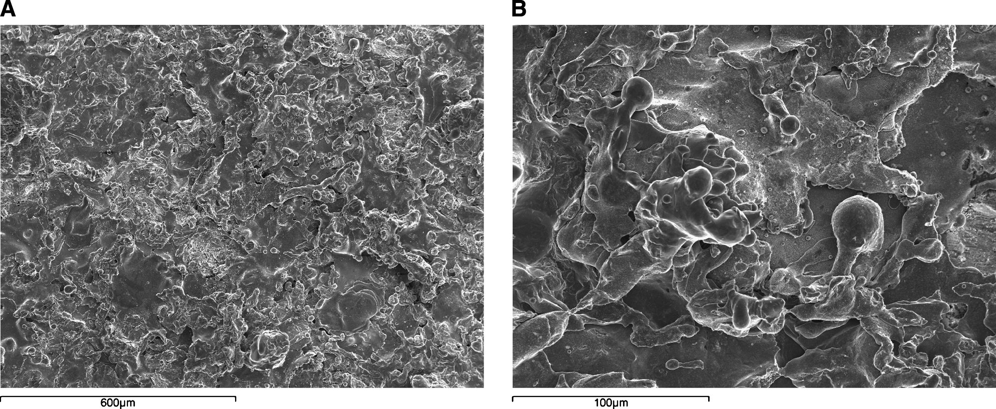
SEM images of an unseeded titanium plasma-spray disk, 100× magnification (
Cells
The human osteosarcoma cell line SAOS-2 was obtained from the American Type Culture Collection (HTB85; ATCC, Rockville, MD). The cells were cultured in McCoy's 5A modified medium with
Cell seeding
The disks were sterilized by ethylene oxide at 38°C for 8 h at 65% relative humidity. After 24 h of aeration to remove the residual ethylene oxide, the disks were placed inside the three culture systems: the static and the ultrasonic; both a standard well plate far from the electromagnetic bioreactor; and the electromagnetic, that is, a standard well plate inside the electromagnetic bioreactor. 11
A cell suspension of 4 × 10⁵ cells in 100 μL was added onto the top of each disk, and, after 0.5 h, 1 mL of culture medium was added to cover the disks. Cells were allowed to attach overnight, the static culture was continued in the standard well plate, the ultrasound stimulation was applied for the first time, and the electromagnetic bioreactor was turned on.
Ultrasound stimulation
An ultrasound stimulus was applied through the culture medium by a FAST ultrasound generator (Igea, Carpi, Italy) whose emitting surface was parallel to the seeded titanium plasma-spray surface. The mechanical wave had the following characteristics: signal frequency equal to 1.5 ± 0.03 MHz, duty cycle of 200 ± 4 μs, repetition rate equal to 1 ± 0.02 kHz, and temporal average power of 149 ± 3 mW. Low-intensity ultrasound stimulus accelerates the fracture healing in animal models 27 and in clinical studies. 12
The ultrasonic culture was placed into a standard cell culture incubator with an environment of 37°C and 5% CO2, where the electromagnetic stimulation was not detectable, and it was stimulated 20 min/day for a total of 22 days. The culture medium was changed on days 4, 7, 10, 13, 16, and 19.
Electromagnetic bioreactor
The electromagnetic bioreactor 11 consisted of a carrying structure custom machined in a tube of polymethylmethacrylate: the windowed tube carried a well plate and two solenoids, that is, Helmoltz coils, the planes of which were parallel. In this experimental setup, the magnetic field and the induced electric field were perpendicular and parallel to the disk surfaces, respectively. The surfaces of the disks were 5 cm away from each solenoid plane, and the solenoids were powered by a Biostim SPT pulse generator (Igea).
Given the position of the solenoids and the characteristics of the pulse generator, the electromagnetic stimulation had the following parameters: intensity of the magnetic field equal to 2 ± 0.2 mT, amplitude of the induced electric tension equal to 5 ± 1 mV, signal frequency of 75 ± 2 Hz, and pulse duration of about 1.3 ms. In vivo experiments demonstrated that a continuous exposure to a pulsed electromagnetic field, similar to that used in this study, stimulates the bone repair in the healing process of transcortical holes in adult horses. 28
The electromagnetic bioreactor was placed into a standard cell culture incubator with an environment of 37°C and 5% CO2. The electromagnetic culture was stimulated 24 h/day for a total of 22 days. The culture medium was changed on days 4, 7, 10, 13, 16, and 19.
Standard well plate culture
The static culture was placed into an incubator, where the electromagnetic stimulation was not detectable. The duration of the static culture was 22 days, and the culture medium was changed on days 4, 7, 10, 13, 16, and 19.
SEM analysis
At the end of the culture period, the disks were fixed with 2.5% (v/v) glutaraldehyde solution in 0.1 M Na-cacodylate buffer (pH 7.2) for 1 h at 4°C, washed with Na-cacodylate buffer, and then dehydrated at room temperature in a gradient ethanol series up to 100%. The samples were kept in 100% ethanol for 15 min, and then critical point-dried with CO2. The specimens were mounted on aluminum stubs, sputter coated with gold (degree of purity equal to 99%), and then observed with a Leica Cambridge Stereoscan microscope (Leica Microsystems, Bensheim, Germany) at 7.5 kV. The unseeded disks were observed at 100 × and 500 × magnifications, whereas the cultured disks at 200 × magnification.
EDXS was performed to qualitatively detect, with a sampling depth of 1 μm, the presence of calcium and phosphorus in the most external layer of the scanned surface. EDXS images were taken assigning spots to the detected elements.
MTT test and DNA content
To evaluate the mitochondrial activity of the seeded cells, that is, the cell viability on the titanium plasma-spray disks during the culture period, a test with MTT (Sigma-Aldrich) was performed on days 1, 10, and 22 (end of the culture period). The conditioned culture medium was replaced by a 0.5 mg/mL solution of MTT in phosphate-buffered saline (PBS) (137 mM sodium chloride [NaCl], 2.7 mM potassium chloride [KCl], 4.3 mM Na2HPO4, and 1.4 mM KH2PO4, pH 7.4), and the cell cultures were incubated for 4 h. The viable cells were able to reduce MTT into formazan crystals. After removing the MTT solution, to solubilize the formazan products, 500 μL of dimethyl sulfoxide (Sigma-Aldrich) was added, and the well plate containing the cultured disks was agitated for 20 min on a shaker. Aliquots of 200 μL were sampled, and the related absorbance values were measured at 570 nm by a microplate reader (Bio-Rad Laboratories, Hercules, CA). A standard curve of cell viability was used to express the results as percentages.
At the end of the culture period, the cells were lysed by a freeze–thaw method in sterile deionized distilled water. The released DNA content was evaluated with a fluorometric DNA quantification kit (PicoGreen; Molecular Probes, Eugene, OR). A DNA standard curve, 11 obtained from a known amount of osteoblasts, was used to express the results as cell number per disk.
Set of rabbit polyclonal antisera
L.W. Fisher (
Set of purified proteins
We have used the following purified proteins: decorin, 30 osteocalcin (immunoenzymatic assay kit, BT-480; Biomedical Technologies, Stoughton, MA), osteopontin (immunoenzymatic assay kit, 900-27; Assay Designs, Ann Arbor, MI), and type I collagen. 31
Indirect immunofluorescence staining
At the end of the culture period, the disks were fixed with 4% (w/v) paraformaldehyde solution in 0.1 M phosphate buffer (pH 7.4) for 8 h at room temperature and washed with PBS (137 mM NaCl, 2.7 mM KCl, 4.3 mM Na2HPO4, and 1.4 mM KH2PO4, pH 7.4) three times for 15 min. The disks were then blocked by incubating with PAT (PBS containing 1% [w/v] bovine serum albumin [BSA] and 0.02% [v/v] Tween 20) for 2 h at room temperature, and washed. L.W. Fisher's antitype I collagen, antidecorin, antiosteocalcin, and antiosteopontin rabbit polyclonal antisera were used as primary antibody with a dilution equal to 1:1000 in PAT. The incubation with the primary antibodies was performed overnight at 4°C, whereas the negative controls were based upon the incubation, overnight at 4°C, with PAT instead of the primary antibodies. The disks and the negative controls were washed and incubated with Alexa Fluor 488 goat anti-rabbit IgG (H + L) (Molecular Probes) with a dilution of 1:500 in PAT for 1 h at room temperature. At the end of the incubation, the disks were washed in PBS, counterstained with a solution of propidium iodide (2 μg/mL) to target the cellular nuclei, and then washed. The images were taken by blue excitation (bandpass, 450–480 nm; dichromatic mirror, DM500; barrier filter, BA515) with a fluorescence microscope (BX51; Olympus, Tokyo, Japan) equipped with a digital image capture system (Olympus) at 25× magnification. The fluorescence background of the negative controls was almost qualitatively negligible.
Extraction of the extracellular matrix proteins from the cultured disks and enzyme-linked immunosorbent assay
At the end of the culture period, to evaluate the amount of the extracellular matrix constituents over the disk surface, the disks were washed extensively with sterile PBS (137 mM NaCl, 2.7 mM KCl, 4.3 mM Na2HPO4, and 1.4 mM KH2PO4, pH 7.4) to remove the culture medium, and then incubated for 24 h at 37°C with 1 mL of sterile sample buffer (1.5 M Tris-HCl, 60% [w/v] sucrose, and 0.8% [w/v] sodium dodecyl sulfate, pH 8.0). At the end of the incubation period, the sample buffer aliquots were removed, and then the disks were centrifuged at 4000 rpm for 15 min to collect the sample buffer entrapped into the pores.
The total protein concentration in the three culture systems was evaluated by the BCA Protein Assay Kit (Pierce Biotechnology, Rockford, IL). The total protein concentration was 960 ± 135 μg/mL in the static culture, 2320 ± 261 μg/mL in the ultrasonic culture, and 2572 ± 350 μg/mL in the electromagnetic culture (p < 0.05 in the comparisons static vs. ultrasonic and static vs. electromagnetic; p > 0.05 in the comparison ultrasonic vs. electromagnetic).
After matrix extraction, the disks were incubated, once again, for 24 h at 37°C with 1 mL of sterile sample buffer, and no protein content was detected.
Calibration curves to measure decorin, osteocalcin, osteopontin, and type I collagen were performed. Microtiter wells were coated with increasing concentrations of each purified protein, from 1 ng to 2 μg, in coating buffer (50 mM Na2CO3, pH 9.5) overnight at 4°C. Some of the wells were coated with BSA as a negative control. To measure the extracellular matrix amount of each protein by an enzyme-linked immunosorbent assay (ELISA), microtiter wells were coated, overnight at 4°C, with 100 μL of the extracted extracellular matrix (20 μg/mL in coating buffer). After three washes with PBST (PBS containing 0.1% [v/v] Tween 20), the wells were blocked by incubating with 200 μL of PBS containing 2% (w/v) BSA for 2 h at 22°C. The wells were subsequently incubated for 1.5 h at 22°C with 100 μL of the L.W. Fisher's antidecorin, antiosteocalcin, antiosteopontin, and antitype I collagen rabbit polyclonal antisera (1:500 dilution in 1% BSA). After washing, the wells were incubated for 1 h at 22°C with 100 μL of horseradish peroxidase–conjugated goat antirabbit IgG (1:1000 dilution in 1% BSA).
The wells were finally incubated with 100 μL of development solution (phosphate–citrate buffer with o-phenylenediamine dihydrochloride substrate). The color reaction was stopped with 100 μL of 0.5 M H2SO4, and the absorbance values were measured at 490 nm with a microplate reader (Bio-Rad Laboratories). The amount of extracellular matrix constituents over the disk surface is expressed as fg/(cell × disk).
Statistics
The disk number was 42 in each repeated experiment (14 disks in the control culture, 14 disks in the ultrasonic culture, and 14 disks in the electromagnetic culture). The experiment was repeated four times. Results are expressed as mean ±standard deviation. To compare the results between the culture systems, one-way analysis of variance with post hoc Bonferroni test was applied, electing a significance level of 0.05.
Results
The SAOS-2 human osteoblasts were seeded onto the surface of rough titanium plasma-spray disks, and then cultured without or with a physical stimulus for 22 days. These culture methods permitted the study of the SAOS-2 cells as they modified the biomaterial surface through the proliferation and the coating with extracellular matrix. The cell–matrix distribution was compared between the three culture systems.
SEM analysis
SEM images revealed that, due to the physical stimulations, the cells proliferated and built their extracellular matrix over the available titanium surface (Fig. 2).
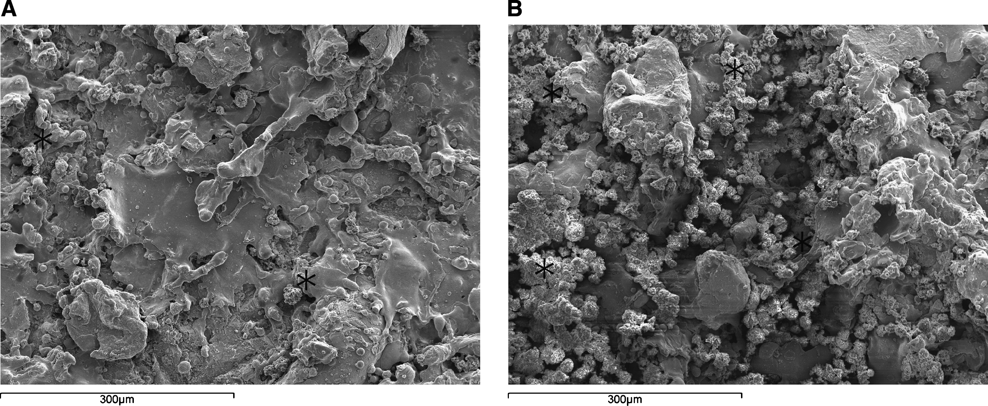
SEM images of the static (

At the end of the culture period, statically cultured cells were few and surrounded by a thin layer of extracellular matrix; therefore, wide titanium regions remained devoid of cell–matrix complexes (Fig. 2A). In contrast, the physical stimuli caused a wide-ranging coat of the rough metallic surface: several osteoblasts proliferated over the available surface, and the biomaterial roughness was tending to be hidden by cell–matrix clusters growing from the bottom (Fig. 2B, C).
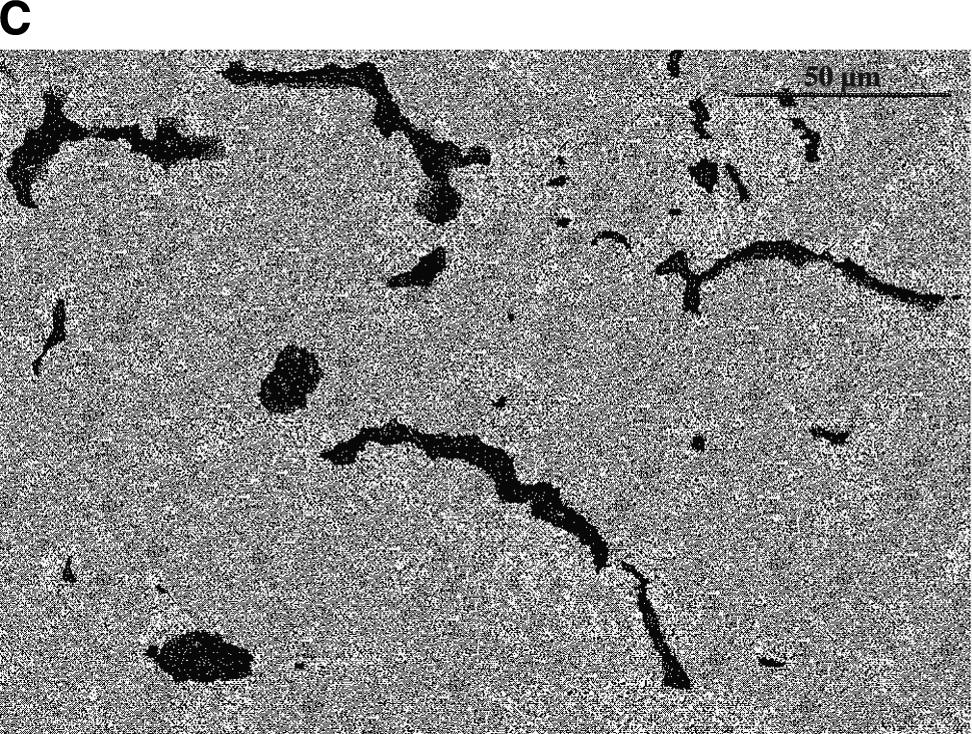
In the most external layer of the scanned surface with a sampling depth of 1 μm, EDXS images revealed traces of calcium in the control specimens (Fig. 3A), whereas calcium was abundant in physically cultured disks (Fig. 3B, C). The spectroscopy about phosphorus showed similar element distributions (data not shown). Considering the layout of calcium and phosphorus, these elements were colocalized in the biological layer onto the cultured metallic biomaterial.

EDXS images of the static (
Cell proliferation and viability
The SEM observations were confirmed by the measure of the DNA content at the end of the culture period: in the static culture the cell number per disk grew to 2.1 × 10⁶ ± 4.1 × 10⁴, in the ultrasonic culture to 5.2 × 10⁶ ± 3.6 × 10⁴, and in the electromagnetic culture to 5.6 × 10⁶ ± 3.2 × 10⁴ (p < 0.05 in the comparisons static vs. ultrasonic and static vs. electromagnetic; p > 0.05 in the comparison ultrasonic vs. electromagnetic). Because the DNA may remain entrapped in the extracellular matrix, an underestimation of culture cellularity is possible.
To evaluate the cell viability on the titanium plasma-spray disks during the culture period, an MTT test was performed. On day 1, day 10, and at the end of the culture period, the average cell viability was in the 90–95% range without statistically significant differences (p < 0.05) between the three culture systems in each day of analysis.
Extracellular matrix organization and evaluation
The immunolocalization of type I collagen showed a more intense fluorescence in the physically cultured disks, revealing the stimulation effects in terms of higher cell proliferation and more intense building of the extracellular matrix: statically cultured cells were few and surrounded by a thin discontinuous extracellular matrix (Fig. 4A), whereas the ultrasonic and electromagnetic waves permitted a better titanium coating with cell–matrix layers growing from the bottom (Fig. 4B, C). The immunolocalization of decorin, osteocalcin, and osteopontin was similar (data not shown). This observation was in agreement with the measure of the DNA content and with the SEM analysis.

Localization of the cellular nuclei (red) and immunolocalization of type I collagen (green) in the static (

To evaluate the amount of the extracellular matrix constituents over the disk surface, an ELISA of the extracted matrix was performed: at the end of the culture period, in comparison with the static culture, the physical stimulations greatly increased the coating with bone proteins (p < 0.05 in the comparisons static vs. ultrasonic and static vs. electromagnetic; p > 0.05 in the comparison ultrasonic vs. electromagnetic) (Tables 1 and 2). An underestimation of absolute protein deposition is possible because the sample buffer used for matrix extraction contained sodium dodecyl sulfate, which may interfere with protein adsorption during ELISA. These measures were in accordance with the microscope images of the extracellular matrix.
p < 0.05 in the comparisons static versus ultrasonic and static versus electromagnetic; p > 0.05 in the comparison ultrasonic versus electromagnetic. In comparison with the static or control conditions, the physical stimuli greatly enhanced the surface coating of the metallic rough biomaterial (Table 2). There was no significant difference between the ultrasonic and the electromagnetic stimulations (Table 2).
p < 0.05 in the comparisons static versus ultrasonic and static versus electromagnetic; p > 0.05 in the comparison ultrasonic versus electromagnetic.
Discussion
The aim of this study was the coating of titanium plasma-spray with extracellular matrix and osteoblasts to make the biomaterial more suitable to bone repair in vivo.
To enhance the culture of human cell line SAOS-2 over the bulk titanium plasma-spray, an ultrasound wave or an electromagnetic wave was applied to the seeded disks (the paramagnetic nature of titanium does not change the orientation of the magnetic field around and inside the metallic biomaterial); in vivo experiments demonstrated that an exposure to the preceding physical stimuli accelerates and improves the bone repair in the healing process of fractures 12 and transcortical holes. 28 In our study, the durations per day of the ultrasonic and electromagnetic treatments were different; nevertheless, both sets of wave parameters were in the useful therapeutic range as discussed by Li et al.; 32 for that reason, we hypothesize that there was no significant difference between the two physical treatments.
Further, rough titanium surface and physical stimuli were not detrimental to cell viability, and there were no significant differences between the three in vitro systems during the culture, whereas in comparison to the control conditions, the physical treatments increased the cumulative cell proliferation around 2.5-fold. This result was consistent with the rise in prostaglandin secretion33,34 and in cell proliferation in response to an ultrasonic or electromagnetic wave as reported by Li et al.. 32
A temporal and functional pattern of the gene expression characterizes the osteoblast maturation process, which can be divided into the proliferation, differentiation, and mineralization stages, 35 and the effects of ultrasonic and electromagnetic waves depend on the maturation stages of the osteoblasts.36,37 The increase in bone formation depends on the increase in extracellular matrix synthesis, 38 and the type I collagen synthesis is upregulated at the proliferation stage, when the osteoblasts are not confluent, and downregulated at the subsequent stages.35,39 Studies reported a significant increase in extracellular matrix synthesis when the osteoblast-like cells were subjected to electromagnetic 40 or ultrasonic 37 treatments.
In this study, the mature SAOS-2 osteoblasts were in the proliferation stage on the surface of titanium plasma-spray disks. According to the preceding studies, the physical stimuli caused a significant increase in extracellular matrix synthesis: in comparison with control culture, the surface deposition of type I collagen was approximately 9–11 times as great. Type I collagen is the most important and abundant structural protein of bone matrix, and decorin is an important proteoglycan considered a key regulator for the assembly and the function of many extracellular matrix proteins. Decorin plays a major role in the lateral growth of collagen fibrils; in comparison, the incorporation of decorin inside the matrix was approximately eight to nine times greater than with control conditions. The immunolocalization of type I collagen and decorin showed their colocalization in the cell-rich areas.
Other fundamental matrix constituents are osteopontin and osteocalcin. Osteopontin is a glycosylated bone phosphoprotein secreted during the early stages of the osteogenesis before the onset of the mineralization, it binds calcium, it is probably involved in the regulation of hydroxyapatite crystal growth, and, through specific interaction with the vitronectin receptor, it promotes the attachment of the cells to the bone matrix, whereas osteocalcin is secreted after the onset of the mineralization, and it binds to bone minerals. The surface deposition of osteopontin and osteocalcin was around 9–9.7 times as great as with control culture. The immunolocalization of these matrix proteins showed their colocalization in the cell-rich areas, like type I collagen and decorin. Considering the role of osteopontin and osteocalcin in the matrix mineralization process, we observed, qualitatively, a concordant higher incorporation of calcium and phosphorus inside the extracellular matrix due to the physical stimuli.
The preceding results could be explained with a signaling model. The ultrasonic and electromagnetic treatments raise the net Ca2+ flux in the osteoblast cytosol and the release of the intracellular Ca2+.32,37,41 According to Pavalko's diffusion-controlled/solid-state signaling model, the increase of the cytosolic Ca2+ concentration is the starting point of signaling pathways, which cause the secretion of prostaglandins enhancing the osteoblast proliferation, and which target specific bone matrix genes. 42
In this study, elaborating an idea of Castner and Ratner,4,5 we physically enhanced the coating of titanium plasma-spray with osteoblasts and with extracellular matrix: we followed a particular biomimetic strategy where the seeded cells built a new biocompatible surface over the biomaterial, making it very useful for the biointegration. 17 The idea of Castner and Ratner and a discussion about the concept of biocompatibility follow. When a biomaterial is implanted in a biological environment, a nonspecific and nonphysiologic layer of adsorbed proteins mediates the interaction of the surrounding host cells with the material surface. 43 The body interprets this protein layer as a foreign invader that must be walled off in an avascular and tough collagen bag.5,44 Therefore, the biomedical surfaces must be developed so that the host tissue can recognize them as self. Castner and Ratner think the biocompatible surfaces of the biomaterials that heal as the surfaces with the characters of a clean, fresh wound: 4 these self-surfaces could obtain a physiological inflammatory reaction around the biomaterials leading to normal healing, leading to physiological osteointegration in bone tissue engineering. In the present study, we followed a particular biomimetic strategy: we obtained a surface coating of the biomaterial, over which the seeded and physically stimulated osteoblasts built a new biocompatible surface made of cell–matrix layers, that is, a physiological surface with the characters of a clean, fresh wound.4,45
The choice of the cell line was justified. An ability to induce the formation of new bone at a specific site would represent a significant advance in bone repair and tissue engineering. This property seems to belong to SAOS-2 cells. These osteoblasts uniquely contain an osteoinductive activity, whereas other human osteosarcoma cells, such as U-2 OS, cannot replicate that bone-inducing ability. 46 Devitalized SAOS-2 cells, extracts, and secretions induced the formation of new bone when implanted subcutaneously in nu/nu mice.47,48 These osteoblasts can be grown, virtually indefinitely, to produce large quantities of osteoinductive factors, such as bone sialoprotein and osteonectin. 46
The use of a cell line showed the potential of the ultrasonic and electromagnetic stimulations; nevertheless, by appropriately tuning the parameters of the two waves and the culture time, a better result could be obtained with autologous bone marrow stromal cells instead of SAOS-2 osteoblasts for total immunocompatibility with the patient.
In this study, we have demonstrated that the titanium plasma-spray can be suitable to matrix coating under physical stimulation, making the biomaterial very useful for the biointegration, that is, the in vivo integration of a biostable biomaterial. 11 We have reported that, under physical stimulation, the coating with matrix constituents was greatly enhanced. We could suppose that the seeded osteoblasts adhered to the titanium plasma-spray through serum-adsorbed proteins,49,50 and that they then adhered to secreted matrix proteins, especially type I collagen. The stimuli made more adhesion proteins available; in other words, they permitted more efficient development of the biomaterial–matrix–cell system.
In conclusion, we theorize that the obtained cultured self-surface could be used fresh, that is, rich in autologous cells and matrix, or after sterilization with ethylene oxide, that is, rich only in autologous matrix. In future work, we intend to use our constructs, which are rich in autologous matrix, as a simple, storable, tissue engineering product for the bone repair. 45
Footnotes
Acknowledgments
The authors thank Prof. L.W. Fisher, Dr. S. Setti, Dr. R. Cadossi, Dr. A. Facchini, Mr. D. Picenoni, and Mr. A. Mortara for support. The FAST ultrasound generator and the Biostim SPT pulse generator were gifts from Igea (Carpi, Italy). This work was supported by Fondazione Cariplo Grants (2004.1424/10.8485 and 2006.0581/10.8485) to F.B.; by PRIN Grant (2006) from Italian Ministry of Education, University and Research, to L.V.; and by FIRB Grant (RBIP06FH7J) from Italian Ministry of Education, University and Research, to M.G.C. De A.
Disclosure Statement
All the authors state that no competing financial interests exist.