
Abstract
In the present study, we examined the possible utility of a three-dimensional culture system using a thermo-reversible gelation polymer to isolate and expand neural stem cells (NSCs). The polymer is a synthetic biologically inert polymer and gelates at temperatures higher than the gel–sol transition point (∼20°C). When fetal mouse brain cells were inoculated into the gel, spherical colonies were formed (∼1% in primary culture and ∼9% in passage cultures). The spheroid-forming cells were positive for expression of the NSC markers nestin and Musashi. Under conditions facilitating spontaneous neural differentiation, the spheroid-forming cells expressed genes characteristic to astrocytes, oligodendrocytes, and neurons. The cells could be successively propagated at least to 80 poly-D-lysines over a period of 20 weeks in the gel culture with a growth rate higher than that observed in suspension culture. The spheroids formed by fetal mouse brain cells in the gel were shown to be of clonal origin. These results indicate that the spheroid culture system is a convenient and powerful tool for isolation and clonal expansion of NSCs in vitro.
Introduction
NSCs are defined as cells that have the capacity of self-renewal and multipotency to differentiate into different neuronal lineages, including neurons, astrocytes, and oligodendrocytes. A number of different methods for isolating NSCs have been developed. NSCs can be isolated by sorting using specific markers such as CD243,4 and CD133. 5 This approach is relatively specific and enables recovery of cells with defined characteristics, but it needs sophisticated equipment and techniques, it causes substantial damage to cells, and collection of a large amount of cells is difficult. Weiss et al. 6 found that NSCs were enriched and propagated in floating cell aggregates (neurospheres) on a nonadhesive substrate. Neurosphere culture was later used for propagation and characterization of stem cells or stem-like cells of various origins. 7
Although neurosphere culture is convenient and reliable for isolation and propagation of NSCs, the resulting neurospheres are heterogeneous, being composed of various stem and/or progenitor cells with varying potentials and differentiated cells of various cell lineages.8,9 This is because when neuronal cell preparations containing NSCs are inoculated in suspension, they readily aggregate to form small neurospheres composed of heterogeneous cell populations, and the growing neurospheres adhere and aggregate even during the propagation stage. This lack of clonality hampers characterization of initially isolated individual cells and quantitation of NSCs in a given cell preparation. As a method for clonal propagation of NSCs, cultivation of single NSCs in 96-well culture plates has been developed.8,10,11
An alternative approach for single-cell culture is to cultivate cells in a gel that prevents cellular movement and aggregation.12–14 Recently, we found that a thermo-reversible gelation polymer (TGP) was useful for clonal propagation of stem cells from embryonic mouse skin. 15 In the present study, we examined the possible utility of TGP for clonal propagation of NSCs. Single NSCs derived from a mouse embryonic brain grew well in a three-dimensional culture using TGP, maintaining multipotency for differentiation up to 80 poly-D-lysine (PDL) over a period of 20 weeks.
Materials and Methods
Animals
Female C57BL/6 mice with confirmed pregnancy were purchased from Japan SLC (Hamamatsu, Japan). Green fluorescent protein (GFP)–transgenic mice were provided by Dr. Masaru Okabe (Osaka University) and maintained in the Animal Section, Advanced Science Research Center, Okayama University. All animals were handled in conformity to the institutional code for animal experiments.
Primary culture
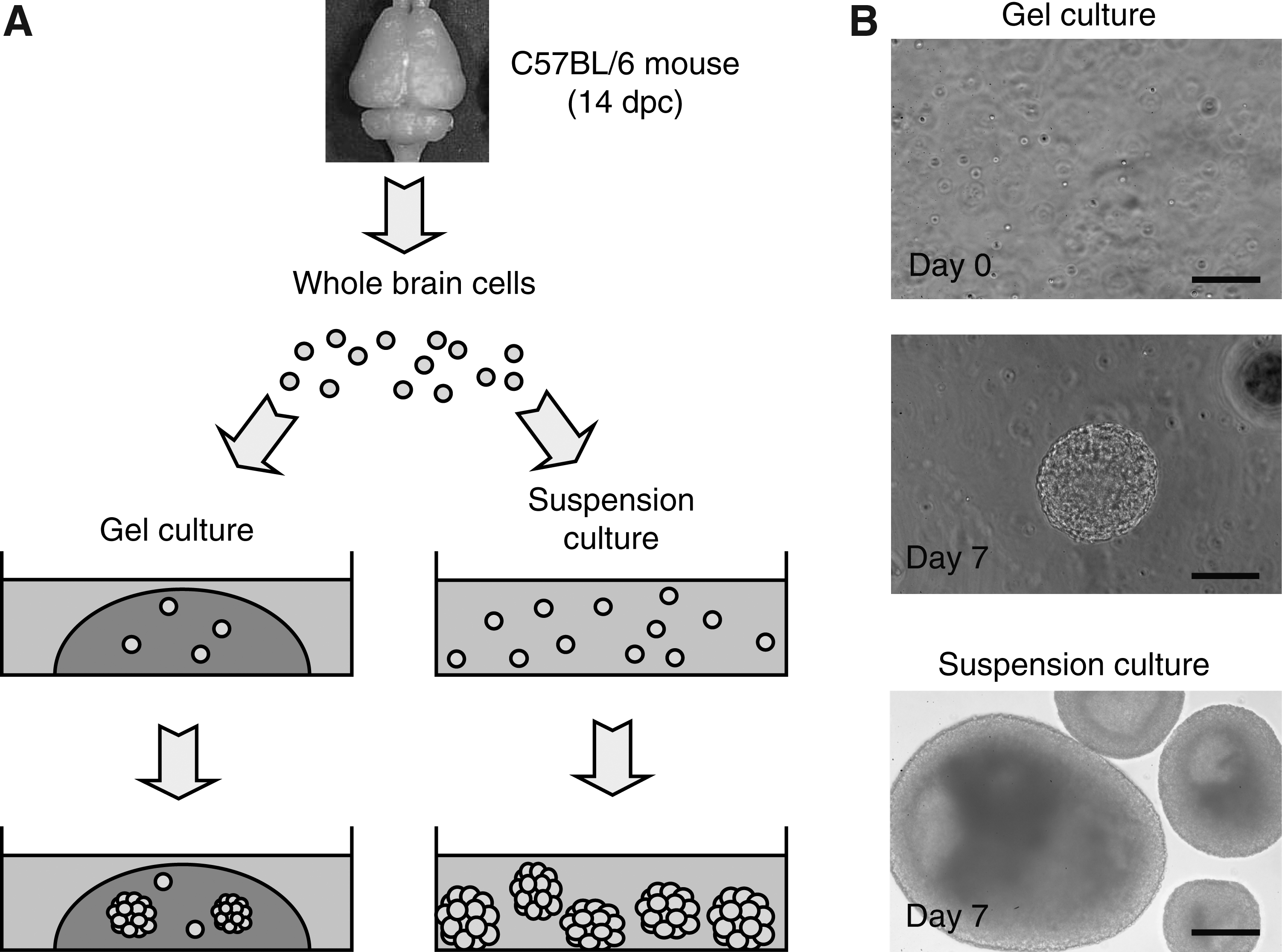
Whole brains were resected from fetal mice at 14 days postcoitus (dpc) and suspended in a cold 1:1 mixture of Dulbecco's modified Eagle's medium and Ham's F-12 (Sigma-Aldrich, St. Louis, MO). Brain cells were mechanically dissociated by gentle pipetting and passed through a cell strainer (40 μm; BD Falcon, Bedford, MA).
For gel culture, we used a TGP (Mebiol Gel; Mebiol, Tokyo, Japan). The TGP is a purely synthesized biocompatible polymer that consists of conjugates of polyethyleneglycol and poly-N-isopropylacrylamid (1:2). 16 Molecular weight of the TGP is higher than 100,000. It is a biologically inert and is in a fluid state at temperatures lower than the gel–sol transition point (∼20°C). The transition is completely reversible with no hysteresis. To prepare the gel, powder TGP was reconstituted with Neurobasal medium (Invitrogen, Carlsbad, CA) using the amount indicated by the manufacturer, gently shaken overnight at 4°C, and then kept undisturbed at 4°C for at least 3 days before use. For primary gel culture, the brain cells were mixed with the TGP solution to obtain a final concentration of 1 × 10⁶ cells/mL (the volume of cell suspension should be less than 10% of the TGP solution) and seeded in 24-well plates at 0.25 mL per well. After gelation by incubating at 37°C for 5 min, 1 mL of Neurobasal medium containing 2% B-27 supplement (Invitrogen), 20 ng/mL bFGF (Sigma-Aldrich), and 20 ng/mL EGF (Sigma-Aldrich) was added to each well. For primary suspension culture, 2.5 × 10⁶ cells in 5 mL of the same medium were inoculated into a 60-mm low-cell-binding dish (Nunc, Roskilde, Denmark).
Passage of cells
In the TGP gel culture, shperoids were collected by brief centrifugation after liquefying the gel by cooling on ice. The cells were dissociated by gentle pipetting in a cell dissociation solution, Accumax (Innovative Cell Technologies, San Diego, CA), washed with cold medium, and seeded again in the TGP gel at a concentration of 2 × 10⁴ cells/mL. For suspension culture, cells were dissociated as described above, and 2.5 × 10⁶ cells in 5 mL medium were inoculated in a 60-mm low-cell-binding dish.
Incorporation of bromodeoxyuride
Cells in the TGP gel culture were incubated with 40 mM bromodeoxyuridine (BrdU) for 1 h and fixed with 10% formalin overnight at 4°C. Paraffin sections were hydrated, briefly boiled in a 10 mM citrate buffer, and immunostained for BrdU with a monoclonal antibody against BrdU (DAKO, Glostrup, Denmark).
Neural differentiation
Spheroids formed by fetal mouse brain cells in the TGP gel were transferred onto a PDL-coated cover glass in Neurobasal medium containing 10% fetal bovine serum, 20 ng/mL bFGF, and 20 ng/mL EGF.
Immunocytochemistry
For immunostaining, cells cultured on a cover glass were fixed with cold methanol at −20°C for 20 min. Spheroids were manually squashed on a slide glass and fixed with 4% parafolmaldehyde for 50 min at room temperature. The specimens were washed three times with 0.1% Triton 100 in PBS (PBST), incubated in 5% goat serum (20% for the detection of O4) in PBST for blocking, and then incubated with primary antibodies at 4°C overnight. Mouse anti-MAP2 and mouse anti-S100β antibodies were purchased from Sigma, (St. Louis, MO) and mouse anti-β-III tubulin, mouse anti-GFAP, mouse (Sigma), mouse anti-O4, mouse anti-Nestin, and rabbit anti-Musashi antibodies were provided by Chemicon (Temecula, CA). After rinsing three times (5 min each time) with PBST supplemented with 1% goat serum, the specimens were treated with an antimouse IgG or antirabbit IgG antibody (Chemicon) for 1 h at room temperature. Fluorescent signals were observed using a fluorescence microscope (Olympus IX71; Olympus, Tokyo, Japan) or a confocal fluorescence scanning microscope (LSM510; Zeiss, Jena, Germany) after counterstaining of cell nuclei with DAPI or PI.
Results
Spheroid formation by fetal mouse brain cells in the TGP gel
A single-cell suspension of whole brain cells was prepared from C57BL/6 mice at 14 dpc and inoculated into the TGP gel (Fig. 1A). The cells grew and formed spherical colonies. The size of the spheroids was smaller than that of neurospheres formed by a conventional suspension culture when observed at the seventh culture day (Fig. 1B). The frequency of spheroid formation by fetal brain cells of 14 dpc was 1.0% in primary culture in the TGP gel (Table 1). The frequency increased to ∼9% at the first passage and remained stable during passage cultures up to four passages. In a conventional suspension culture, the frequencies of neurosphere formation were 0.91% in primary culture and 1.8% in secondary culture. When spheroids formed in primary TGP gel culture were transferred into suspension culture, the frequency of neurosphere formation was 6%. These results indicate that the TGP gel used in the present study supports formation of mouse fetal brain cells into spheroids.
Formation of neurospheres by fetal mouse brain cells. (
To determine the frequency of spheroid formation in the thermo-reversible gelation polymer (TGP) gel, 2 × 10⁴ cells/mL of brain cells were inoculated for primary culture and 5 × 10³ cells/mL were inoculated in the first passage and thereafter. The number of spheroids formed was determined after 7 days. Neurosphere formation in a suspension culture was determined under similar conditions except for inoculation of 1.5 cells/mm2 for primary culture and 1 cell/mm2 for secondary culture. The number of spheroids was divided by the number of cells inoculated.
Expression of NSC markers in spheroid-forming cells in the TGP gel
To characterize the spheroid-forming cells in the TGP gel, the spheroids were immunostained for nestin and Musashi, representative marker genes of NSCs. As shown in Figure 2, almost all cells in the spheroids were homogeneously positive for expression of nestin and Musashi. Only limited number of cells in spheroids formed in the gel after cultivating for 5 days were positive for astroglial markers GFAP and S100β, an oligodendroglial marker, O4, and neuronal markers MAP-2 and β-III-tubulin (Fig. 3). A similar staining profile was observed in spheroids formed in the suspension culture. These results indicate that most of cells comprising spheroids in the TGP gel have a property characteristic to NSCs like cells in spheroids formed by the suspension culture.

Expression of NSC markers in fetal mouse brain cells cultured in the TGP gel for 7 days. Sections of the spheres were stained for nestin and Musashi. Ab (−), a negative control without application of the first antibody. PI, propidium iodide. Scale bars: 100 μm. Color images available online at

Expression of a NSC marker, nestin, and neuronal differentiation markers in spheroids formed in the gel culture or in the suspension culture. Specific signals are in green, and nuclei are stained with PI in red. Scale bars: 50 μm. Color images available online at
Neural differentiation of spheroid-forming cells in the TGP gel
To examine differentiation potential of spheroid-forming cells in the TGP gel in vitro, the spheroids formed in primary culture were transferred onto a PDL-coated cover glass in differentiation medium and cultivated for 7 days to promote spontaneous differentiation. Immunocytochemistry revealed that the cells were positive for marker proteins for astrocytes (GFAP and S100β), for oligodendrocytes (O4), and for neurons (MAP-2 and β-III tubulin) (Fig. 4). These results indicate that the spheroid-forming cells in the TGP gel showed multipotency for differentiation into different neuronal lineages.

Differentiation of NSCs isolated by the TGP gel culture. Neurospheres formed by primary culture of fetal mouse brain cells in the TGP gel were dissociated and cultured further on a PDL-coated cover glass with Neurobasal medium containing 10% FBS in the absence of EGF and bFGF for 7 days. Cells positive for GFAP, S100β, O4, MAP-2, and β-III tubulin were observed. Ab (−), a negative control without application of the first antibody. Nuclei were counterstained with propidium iodide. Scale bars: 20 μm. Color images available online at
Proliferation capacity of spheroid-forming cells in the TGP gel
To monitor the mode of proliferation of fetal brain cells in the TGP gel, we examined incorporation of BrdU into nuclei. Incubation with BrdU for 1 h resulted in positivity in BrdU incorporation in the majority of cells (Fig. 5), indicating that the cells grew rapidly and homogeneously regardless of positions within spheroids. Long-term culture revealed that growth rate of the spheroid-forming cells in the TGP gel was higher than that observed in suspension culture (Fig. 6). The cells grew stably with a population doubling time of 42 h up to 80 PDLs in the TGP gel and 64 h up to ∼50 PDLs in suspension over a period of 20 weeks.
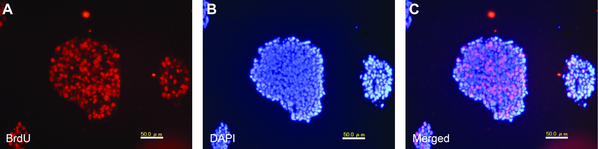
Incorporation of BrdU into fetal mouse brain cells in neurospheres formed in the TSG gel. The cells were cultured in the gel for 7 days and then incubated with 40 mM BrdU for 1 h. Incorporation of BrdU was detected using an anti-BrdU antibody (

Growth curve of fetal mouse brain cells cultured either in the TGP gel or in suspension. After isolation of sphere-forming cells by a primary culture of fetal mouse brain cells in the TGP gel, the cells were dissociated and further cultivated either in the TGP gel or in suspension for a period of 20 passages. Inoculation size for each passage was 2 × 10⁴ cells/mL in the TGP gel culture and 5 × 10⁵ cells/mL in the suspension culture. The number of cells was determined at every passage. DT, doubling time.
Clonality of spheroid-forming cells in the TGP gel
Conceptually, spheroids were formed through clonal expansion of single cells inoculated into the TGP gel. To confirm this, we daily followed single cells, starting shortly after inoculation. As shown in Figure 7, single cells rapidly grew to form spheroids in a clonal manner. When spheroid-forming cells derived from GFP-trangenic mice and those from GFP-negative C57BL/6 mice were inoculated in a 1:1 mixture into the TGP gel, both types of cells segregated to form either purely GFP-positive or GFP-negative spheroids (Fig. 8A). In constract, inoculation of the mixed cell population into suspension culture resulted in formation of spheroids composed of both GFP-positive and GFP-negative cells (Fig. 8B). These results demonstrate that spheroids formed in the TGP gel culture were formed through clonal expansion of single brain cells.

Clonal expansion of fetal mouse brain cells in the TGP gel. After primary culture in the TGP gel, sphere-forming cells were dissociated, seeded in the gel at a low density (1000 cells/mL gel), and observed daily. Pictures of a representative cell are shown. Scale bars: 200 μm. Color images available online at
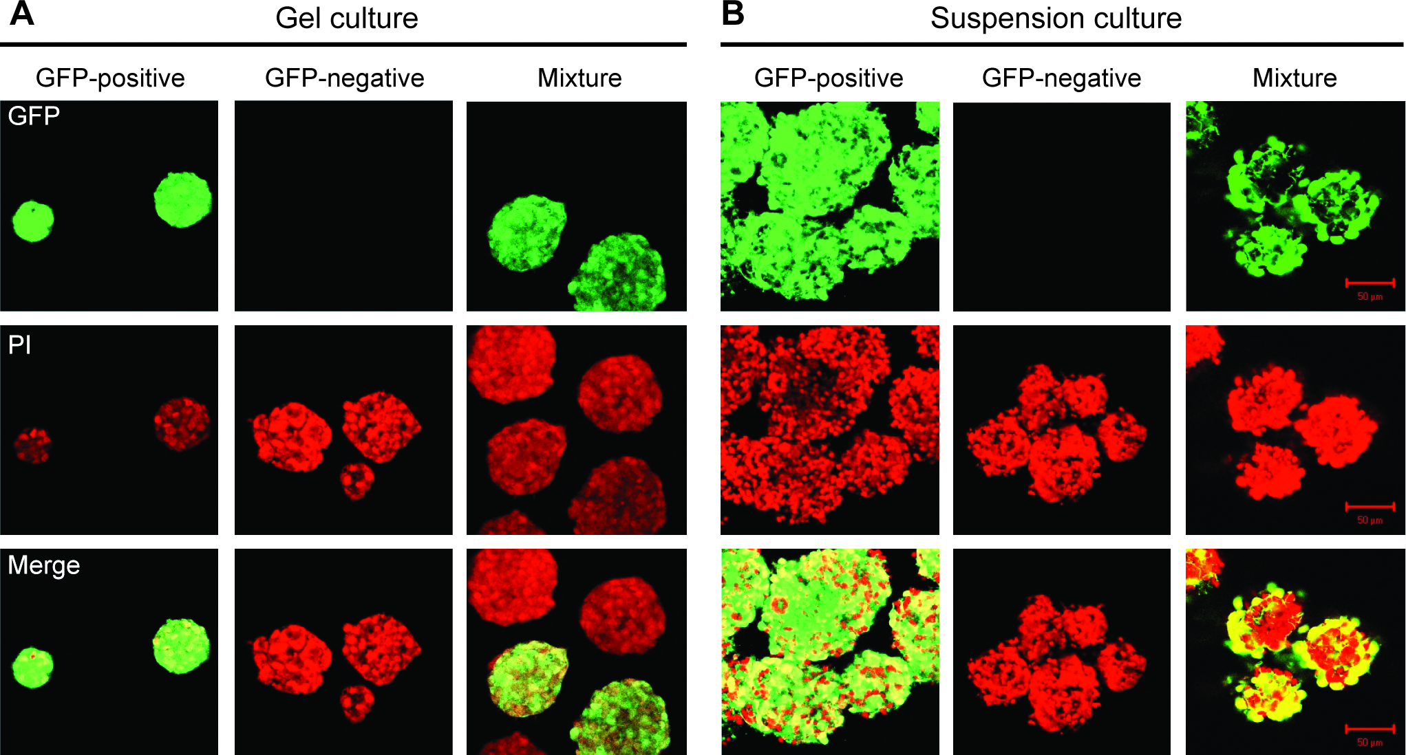
Homogeneity of neurospheres formed in the TGP gel. (
Discussion
In the present study, we examined the possible utility of a TGP for isolation and expansion of NSCs in vitro. NSCs are a tissue-specific subtype of self-renewing and multipotent cells that can give rise to all neural populations. 17 The spheroid-forming cells in the TGP gel were positive for the NSC markers nestin and Musashi (Fig. 2). When cultured under conditions facilitating spontaneous neural differentiation, the spheroid-forming cells derived from fetal mouse brains showed a capacity to differentiate into astrocytes, oligodendrocytes, and neurons (Fig. 4). The spheroid-forming cells could be propagated to at least 80 PDLs over a period of 20 weeks, maintaining a stable proliferation capacity with a doubling time of 42 h (Fig. 6). Spheroid-forming cells in the TGP gel can thus be regarded to be similar to NSCs that are isolated as neurospheres by conventional suspension culture.
When neural cells are inoculated into a suspension culture, the cells readily aggregate and eventually form neurospheres that are derived from more than one sphere-forming cell and even include cells lacking the capacity for sphere formation by themselves. 18 Cell aggregation is a common event during neurosphere formation as revealed by a chimeric neurosphere formation assay7,19 or by tracking cell fate. 18 Neurospheres formed by suspension culture were confirmed to be heterogeneous by molecular phenotyping. 9 The results of our analysis using GFP-positive and GFP-negative cells accorded with those observations (Fig. 8B). Thus, the formation of neurospheres in suspension culture is a complicated process involving cell proliferation and aggregation. 19 On the other hand, the TGP gel culture enables neural cells to undergo clonal expansion to form single-cell-derived homogeneous spheroids (Figs. 7 and 8A). Clonal propagation of stem cells from primary culture has profound significance because it is the only way to determine the range of differentiation potential and self-renewing capacity of individual cells. For this purpose, a multi-well culture of single NSCs has been developed.8,10,11 The present gel culture using a TGP, however, is clearly simpler and more convenient and reliable.
Several extracellular matrix proteins, including collagen and Matrigel, have been used for clonal expansion of cells in three-dimensional culture. Materials from biological source, however, cannot be absolutely free from contamination of unknown substances, including pathogens. The TGP used in the present study is a purely synthesized biocompatible polymer that consists of conjugates of polyethyleneglycol and poly-N-isopropylacrylamid. 15 The TGP is biologically inert. Hishikawa et al. reported that collagen gel by itself altered the gene expression profile of mesenchymal stem cells but that TGP did not. 20 When we cultured SH-SY5Y cells, a human neuroblastoma cell line that can be induced to differentiate into neuronal cells, 21 the cells showed an elongated shape and formed many processes in a collagen gel or in Matrigel. In the TGP gel, however, the colonies that formed were completely round, and no interaction between cells and the matrix was indicated (data not shown). This inertness of the TGP is probably the reason why NSCs could be stably propagated for a long period (80 PDLs, 20 weeks) in the TGP gel culture (Fig. 6; Table 1).
Cells that compose tissues in vivo can only grow in an anchorage-dependent manner in vitro. Thus, colony formation in three-dimensional culture matrices is a selection system for cells with a capacity of anchorage-independent growth such as cancer cells and cells that do not form solid tissues in vivo. Stem cells possess normal cell properties, form solid tissues in vivo, and generally grow with attachment to culture substrates in a monolayer culture. However, many stem cells have been demonstrated to grow in suspension, including NSCs5,6 and mammary gland cells. 22 Stem cells from fetal mouse skin and human cornea were isolated as spheroid-forming cells in TGP gel.13,16 Embryonic stem cells and induced pluripotent stem cells were also shown to form spheroids in TGP gel (data not shown). The spheroid-forming induced pluripotent stem cells stably expressed a stem cell marker, Nanog. These results indicate that the TGP is a potent tool for isolation and propagation of stem cells of a broad variety of origins.
In conclusion, the spheroid culture system using TGP is useful for isolation and clonal expansion of NSCs in vitro.
Footnotes
Acknowledgments
We thank Dr. H. Yoshioka for providing the TGP Mebiol Gel. This work was supported by a grant from the Ministry of Education, Culture, Sports, Science, and Technology of Japan (20659062).
Disclosure Statement
For this study, the TGP was provided by Mebiol Inc. The commercial value, however, was very limited and the company was not involved in the study at all and not in a position to affect the conclusion.