
Abstract
Objective:
Osteoarthritis (OA) is a disease that limits the mobility of patients and is of considerable economical importance. Up to now, despite the increasing number of patients with OA, treatments to manage the disease remain symptomatic, designed to control pain, and improve function and quality of life limiting adverse events. With the aim to explore a new approach to treat OA patients suffering from early degenerative lesions of hyaline cartilage, we transplanted in an experimental animal model of OA a hyaluronan-based scaffold (Hyaff®11) seeded with mesenchymal stem cells (MSCs) obtained from bone marrow and expanded in culture.
Design:
Rabbit knee joints were bilaterally subjected to anterior cruciate ligament transection to surgically induce OA. After 8 weeks, the time necessary to the development of cartilage surface damage, animals were treated with MSCs seeded onto Hyaff-11 scaffold in the left condyle and unseeded Hyaff-11 in the controlateral knee. Untreated rabbits were used as controls. All the animals were sacrificed at 3 and 6 months after surgery. Histological, histomorphometric, and immunohistological evaluations were performed.
Results:
OA changes developed in all animals subjected to anterior cruciate ligament transection. The predominant macroscopically observed OA changes were mild (lateral femoral condyle) or moderate (medial femoral condyle) ulcerations. Statistically significant differences in the quality of the regenerated tissue were found between the implants with scaffolds carrying MSCs compared to the scaffold alone or controls in particular at 6 months.
Conclusions:
From the observations, it is possible to demonstrate that Hyaff-11, a hyaluronan-based scaffold, has potential for MSC implantation and that may have application for the treatment of early OA in humans.
Introduction
Autologous chondrocyte implantation (ACI) is a widely used therapeutic strategy for the repair of damaged cartilage.7,8 The data obtained at long-term follow-up showed good clinical results together with the formation of new tissues with some hyaline and/or fibrous features, and a longer period is required for a complete regeneration.9,10 The indications for ACI are symptomatic full-thickness chondral injuries or osteochondritis dissecans,11,12 and the treatment requires the harvest of healthy cartilage tissue from which chondrocytes are liberated and expanded in vitro. 13 An improvement of this technique could be the use of different cell populations, easily obtained without invasive procedures, representing an interesting alternative in the treatment of cartilage defects.
Mesenchymal stem cells (MSCs) are multipotent cells that are present in adult bone marrow, can replicate as undifferentiated cells, and have the potential to differentiate to lineages of mesenchymal tissues, including cartilage, bone, and fat. 14 MSCs therefore present themselves as a promising cell source for the regeneration of cartilage, as they possess chondrogenic differentiation potential, are easily obtainable in high numbers, and are expandable in vitro without loosing their differentiation potential. 15 Due to these characteristics they are used in preclinical studies in vitro 16 and in vivo both in animals17,18 and in humans19,20 with the aim to regenerate cartilage tissue in combination with different suitable scaffolds. Hyalograft-C construct has been successfully used in ACI in clinical application as reported by Marcacci et al. 21 It is made of hyaluronan, a natural component of extracellular matrix, and it appears to be able to enhance the re-acquisition of the chondrogenic phenotype, 22 to downregulate the expression and production of many molecules involved in cartilage degeneration,23,24 and to encourage the differentiation of MSCs to chondrogenic phenotype. 25
To explore a new approach to treat OA patients suffering for early degenerative lesions of hyaline cartilage, we propose to transplant in an experimental animal model of OA a scaffold seeded with MSCs, obtained from bone marrow and expanded in culture. To this end, rabbit knee joints were subjected to anterior cruciate ligament transection (ACLT) to surgically induce OA. The therapeutic outcome of ACLT reconstruction by means of this strategy was evaluated in terms of histomorphometry, histology, and immunohistochemistry looking for the expression of some typical extracellular matrix proteins and metalloproteinases that have been shown to be particularly involved in cartilage degradation in OA rabbit ACLT model. 26
Materials and Methods
Scaffold
The scaffold, kindly provided by FIDIA Advanced Biopolymers (Abano Terme, Italy), was sterilized by γ-irradiation. The scaffold was made of Hyaff-11, a polymer derived from the total esterification of sodium hyaluronate (80–200 kDa) with the benzyl alcohol on the free carboxyl groups of glucuronic acid along the polymeric chain. 27 Hyaluronic acid ester is prepared by treating a quaternary ammonium salt of hyaluronic acid with an esterifying agent in a suitable aprotic solvent at a controlled temperature. The configuration used in this study was a pad of nonwoven mesh composed of a random array of polymer fibers having a diameter of 10 μm. In aqueous solution, the material hydrates, which consequently results in swelling of the fibers, and it undergoes complete degradation by spontaneous hydrolysis of the ester bonds in 2 months. 28 Studies in vivo in rat on the biodegradation of Hyaff-11 have shown that the material disappears after about 4 months after implantation, 29 and in rabbit osteochondral defects, in less than 3 months. 30
Animal experimental design
European and Italian laws on animal experimentation were strictly followed throughout the study, and the animal experimental protocol was approved by the Rizzoli Orthopaedic Institute Technical Scientific and Ethical Committee and by the other public authorities as requested by Italian Law according to EC rules (Law by Decree, 27 January 1992, no. 116).
Twelve-month-old 62 New Zealand adult male rabbits (4.0 ± 0.5 kg body weight) were used and housed singly in stainless steel cages. They were allowed ad libitum commercial rabbit pellets (Mucedola; Settimo Milanese, Milan, Italy) and water during the entire pre- and postoperative stabling period and were permitted free cage activity without joint immobilization. The animals were maintained at 20–22°C, with a relative humidity of 40–60% and a photoperiod of 12/12 h, light and dark. All surgical procedures were performed under general anesthesia and with the use of sterile techniques. Anesthetic premedication was obtained with 35mg/kg body weight ketamine (Ketavet; Farmaceutici Gellini, Aprilia, Italy) and 5 mg/kg body weight xylazine (Rompun; Bayer Italia s.p.a., Milan, Italy) administered by intramuscular injection. Anesthesia was maintained by means of a mixture of 2% forane (Fluothane; Zeneca, Macclesfield, UK) and oxygen/nitrous oxide (1/0.4 L/min) delivered by an automatic ventilator.
For ACLT procedure, a 2 cm skin and capsular incision was made, and right and left ACL were exposed through a medial parapatellar cut. To achieve optimal observation of the ACL, the patellar bone was displayed laterally, and the knee was placed in full flexion. To avoid spontaneous reattachment, the ACLT was associated to the removal of a small fragment of tissue between the two ligament stumps. The incision was sutured in a routine fashion.
An antibiotic (Flumequine; ATI s.r.l., Ozzano Emilia, Italy) and analgesic (Ketoprofene; Rhone-Poulenc-Rorer, Milan, Italy) therapy was administered immediately after each surgery and for 2 days thereafter. Postoperatively, the animals were permitted cage activity without immobilization.
The 62 animals were distributed among different groups:
Sham operation group (12 animals): A sham control arthrotomy without ACLT was performed in both knees. Four animals were sacrificed at 8 weeks, four at 3 months, and the other four at 6 months. OA group (18 animals): Articular cartilage of both distal femoral epiphyses was submitted to ACLT to induce a rabbit OA model,31,32 and left untreated. Six animals were sacrificed at 8 weeks, six at 3 months, and the other six at 6 months. Treated groups (32 animals): after 8 weeks from ACLT, performed as reported above, articular cartilage of both femoral epiphyses was treated, respectively: Left femoral condyle: Hyaff-11 seeded with autologous MSCs (MSC-HA group). Right femoral condyle: Hyaff-11 alone (HA group). Sixteen animals were euthanized at 3 months, and the others at 6 months.
All the animals (sham operation group, OA group, and treated group) were sacrificed under general anesthesia by an intravenous lethal injection of Tanax (Hoechst Roussel Vet GmbH, Wiesbaden, Germany). Both femurs were explanted and cleaned from soft tissues, and the samples were used to perform histomorphometric, histological, and immunohistochemical evaluations.
A schematic representation of the experimental design is reported in Figure 1.

Schematic representation of the experimental model.
Isolation, expansion, and differentiation of MSCs
Ten days before the transplantation procedure, 4 mL of bone marrow was aspirated under general anesthesia from the iliac crest of each rabbit of the experimental group. Bone marrow–derived MSCs were isolated using Ficoll-Hypaque density gradient (d = 1.077 g/mL) from Pharmacia Biotech (Uppsala, Sweden). Cells were washed twice and resuspended in growth medium α-MEM (Sigma, St. Louis, MO) supplemented with 15% FBS (Gibco–Invitrogen, Carlsbad, CA), 0.1 M glutamine, penicillin–streptomycin 10,000 units/mL and 10,000 μg/mL, respectively (Gibco, Carlsbad, CA), gentamycin 50mg/mL (Biological Industries, Kibbutz Beth Haemek, Israel), and once a week L-ascorbic acid 50 μg/mL (Sigma). Then, the cells were counted and plated at a concentration of 2 × 10⁶ cells/T150 flask.
After 7 days, the culture medium was renewed, removing thus the nonadherent cells and selecting the MSCs given their proved capacity of attaching to the plastic of culture flasks. 33 Once the cells were confluent, they were expanded in vitro and replated at 28,000 cells/cm2. The cells were cultured under conventional monolayer culture conditions at 37°C in a humidified atmosphere of 5% CO2. The medium was changed every 3–4 days. After the third passage, 2 × 10⁶ MSCs were resuspended in 250 μL of chondrogenic medium (αMEM containing TGF-β1 [R&D System, Minneapolis, MN] at 10 ng/mL) and seeded onto 1.5 × 1.5 cm Hyaff-11 nonwoven meshes in 35 mm Petri dishes.
The scaffolds were returned to the incubator for 3–4 h, observed under the microscope to grossly verify cell adhesion to polymer surfaces, added with 3 mL of chondrogenic medium, and used within 2–3 h. Unseeded Hyaff-11 meshes were also prepared.
Autologous grafting procedure
About 8 weeks after creating the OA model that exhibited chondral lesions, the 32 animals of the treated groups underwent the second operation of autologous grafting, under general anesthesia. The knee joint was again opened, the patella was luxated laterally to expose the femoral condyles, and two pads of Hyaff-11 nonwoven tissue (with and without cells) were gently inserted over the surfaces without the need for any fixation method because of the intrinsic adhesive properties of the hyaluronan scaffold. 21
The implants roughly covered all the articular surface, as it was impossible to identify a single lesion's spot and to be reasonably sure that they completely cover the condyle. These implants were put on grossly obvious lesions. The patella was repositioned, and the capsule was repaired with 4-0 vicryl broken sutures by checking the grafting position. Finally, the subcutaneous tissues and skin were closed with a broken 5-0 nylon suture. Sixteen animals were sacrificed at 3 months, and 16 animals at 6 months.
Histological and immunohistochemical analyses
Histological and immunohistochemical analyses were performed on the half of total number of rabbits used and specifically on the middle region of medial and lateral femoral condyles from untreated and treated animals.
Specimens were fixed in 10% neutral buffered formalin for 48 h, and decalcified with a 4% HCl, 5% formic acid decalcificant solution for about 14 days. After decalcification, the femoral condyles were cut into four pieces from medial to lateral condyle along the sagittal plane, and all pieces were embedded in paraffin (Fig. 2). Serial sections of 5–6 μm thickness (microtome HM 340E; Carl Zeiss, Oberkochen, Germany) in a transversal plane were cut. Some sections were stained with Safranin-O or Fast Green to assess proteoglycans and collagen in the matrix. Other sections were immunostained with antibodies against type II and I collagen, metalloproteinase-1 (MMP-1), and MMP-3. After deparaffination, in the sections used for type I and II collagen analyses, an epitope unmasking with 1 mg/mL pronase (Sigma P-5147) in phosphate buffered saline (PBS) at 37°C for 15 min was performed. A blocking step with peroxidase blocking solution for both MMP-1 and MMP-3 was also performed.
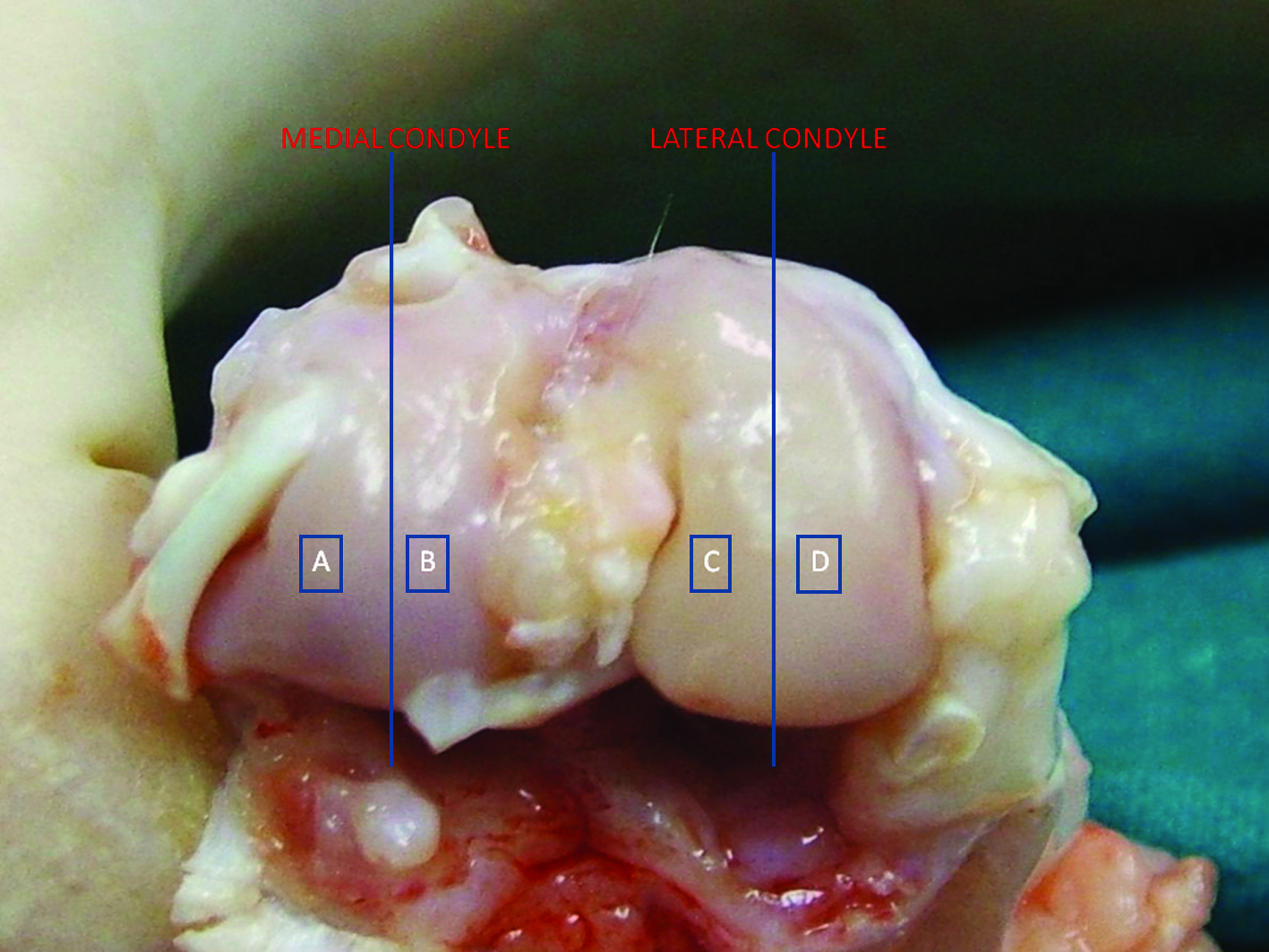
Schematic representation of the rabbit femoral condyle indicating the divided regions used for histology. Color images available online at
After washes, samples for type II and I collagen were incubated at room temperature for 30 min in 1× PBS containing 10% of normal goat serum (Dako, Carpinteria, CA) to prevent nonspecific bindings. The slides were then incubated with primary antibodies, respectively, anti-rabbit type II collagen (mouse monoclonal antibody IgG1; Hybridoma Bank II, Department of Biological Sciences, University of Iowa) diluted 1:200, anti-rabbit type I collagen (mouse monoclonal antibody IgG1; Sigma), anti-human/rabbit MMP-1 (mouse monoclonal antibody IgG2; Chemicon, Temecula, CA) diluted 1:50, and anti-rabbit MMP-3 (mouse monoclonal antibody IgG1; Chemicon) diluted 1:50 in 0.04 M trizma base saline (TBS) pH 7.6 containing 1% bovine serum albumin for 1 h at room temperature. Slides were washed three times with 0.04 M TBS pH 7.6, and those for type II and I collagen were incubated with goat anti-mouse biotin labeled (Kirkegaard & Perry Laboratories, cat. no. 16-18-07) and diluted 1:200 in 0.04 M TBS pH 7.6 containing 1% bovine serum albumin at room temperature for 30 min; the other samples were incubated with anti-mouse poly HRP for 30 min. After washing, an incubation for 1 h at room temperature with a streptavidin–alkaline phosphatase conjugate (Boehringer Mannheim GmbH, Mannheim, Germany) was performed for type I and II collagens. Type II and I collagen proteins were detected with new fucsin kit for alkaline phosphatase substrate (Kit New Fucsin Substrate System; Dako); the others were detected with HRP Kit, biotin free (Anti-Mouse Poly HRP IHC detection Kit, Chemicon). Negative controls were performed by omitting the primary antibody. Slides were counterstained with hematoxylin and mounted on glycerol gel. Histological sections were observed with a Nikon Eclipse 90i.
Histomorphometric analysis
Histomorphometric analysis was performed on the other half of total number of rabbits used and specifically on both medial and lateral femoral condyles from untreated and treated animals. Specimens were fixed in 4% formalin, cut into four pieces, and embedded in methacrylate. Histomorphometry was performed with image analysis Kontron KS 300 software (Kontron Electronics, Eching bei Munchen, Germany). A total of three central slices for each condyle were analyzed, and read blindly by two expertise researchers. The semi-quantitative histological grading criteria of Kraus' modified Mankin score was used.34,35 The histological score represents the sum of five parameters: articular cartilage structure, Toluidine blue staining, cellularity, osteophyte presence, and tidemark integrity. This modified Mankin score has a possible maximum score of 21 points and a minimum of 0 that translates to a completely normal cartilage.
Further, a quantitative evaluation of cartilage thickness (CT), cartilage surface fibrillation index (FI), and subchondral bone plate thickness (SBT) was carried out. Briefly, CT was expressed in micrometer as the mean of 10 measurements perpendicular to the articular surface; cartilage surface FI was calculated according to the method developed by Pastoureau et al. 36 by dividing the length of the cartilage surface border by the length of a standardized measured area × 100 (expressed in percentage); SBT was measured in micrometer and represented the mean of 10 measurements from the cartilage–bone interface to the top of the epiphyseal marrow space, as reported by Huebner et al. 37
Statistical analysis
Statistical analysis was performed using the SPSS v.12.1 software (SPSS, Chicago, IL). Data are reported as mean ±standard error of the mean (SEM). Because normal distribution and homogeneity of variance was not verified by Levene's test, the nonparametric Kruskal-Wallis followed by Mann-Whitney U-test methods were used to analyze data among groups within each experimental time. Individual trend was also analyzed for OA group by performing Friedman following by Kendall W tests (data not shown).
Results
Macroscopic features of cartilage
Varying degrees of OA features developed in all animals subjected to ACLT. Degenerative changes were seen typically on the medial condyle rather than on the lateral condyle. The severity of the disease depended upon the time from ACLT to death. At three different times evaluated (8 weeks, 3 months, and 6 months), all the gross characteristics of OA were seen, including fibrillation, erosion, and osteophyte formation.
A cartilage tissue regeneration was evident in the MSC-HA–treated group particularly at 6 months. This process was shown also in the HA-treated group even if at less extent.
Histological and immunohistochemical evaluations
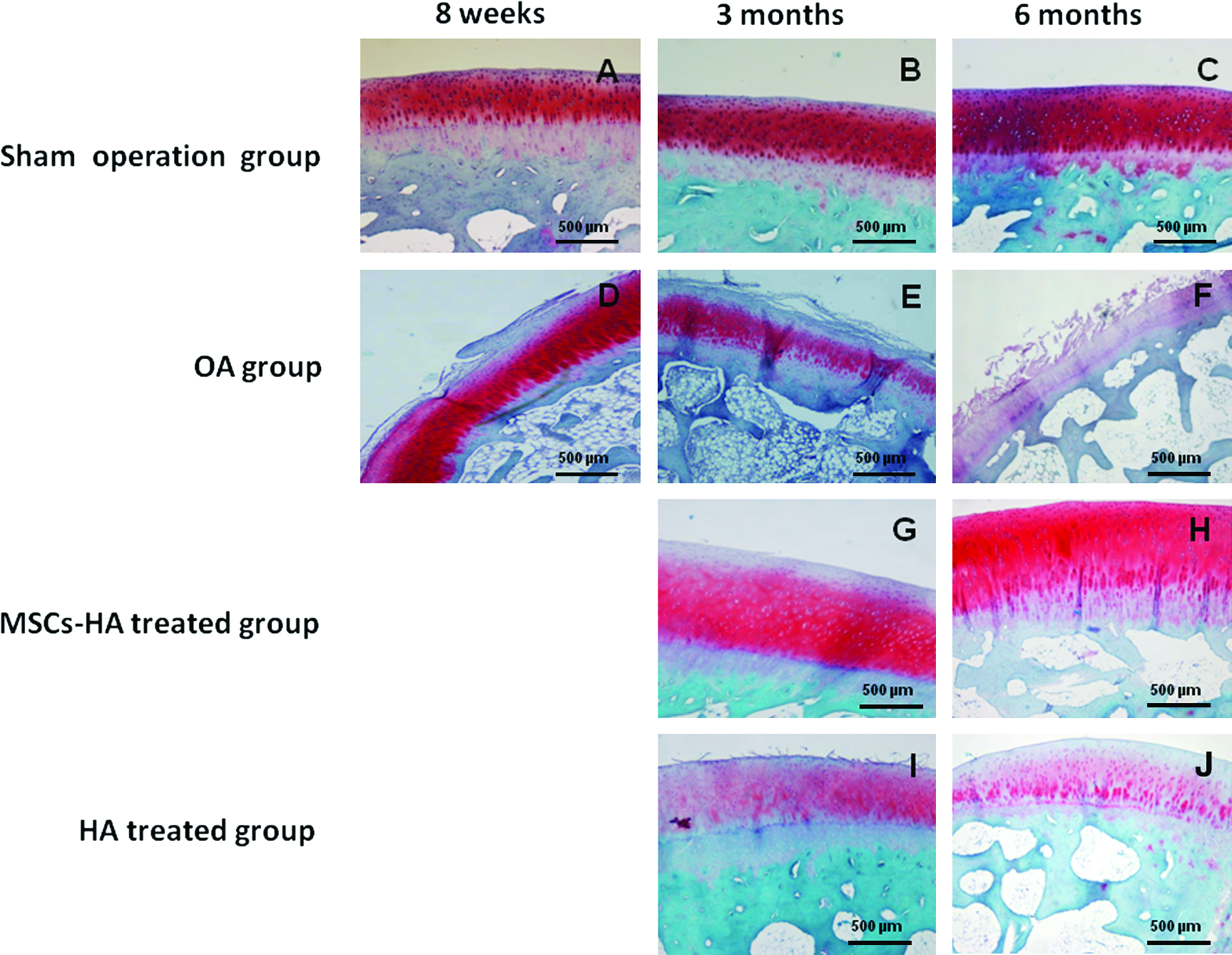
Cartilage tissues from sham-operated group presented the typical hyaline features with smooth cartilage surface, regular cellular organization, and normal proteoglycan content after 8 weeks (Fig. 3A), 3 months (Fig. 3B), and 6 months (Fig. 3C). Paraffin-embedded sections of articular cartilages harvested after 8 weeks (Fig. 3D), 3 months (Fig. 3E), and 6 months (Fig. 3F) from ACLT stained with Safranin-O clearly showed the progression of degenerative OA changes during the times evaluated particularly in the medial condyle. In particular, superficial fibrillation, proteoglycan depletion, cracks' extension, and articular cartilage reduction were observed.
(
The treatment with MSC-HA led to the formation of a new cartilage tissue with hyaline-like features: smooth surface, an increase in cellular component, and a higher presence of proteoglycan component particularly at 6 months (Fig. 3H) than at 3 months (Fig. 3G).
Some degrees of tissue remodeling were noticed also in HA group (Fig. 3I, J).
In sham-operated group, immunohistochemical analysis showed a positivity for type II collagen at extracellular matrix level at 8 weeks (Fig. 4A), 3 months (Fig. 4B), and 6 months (Fig. 4C). In OA group, type II collagen was positive at 8 weeks (Fig. 4D), but became negative at 3 (Fig. 4E) and 6 (Fig. 4F) months. MSC-HA group showed collagen type II positivity at extracellular level at 3 months (Fig. 4G) and also at cellular level at 6 months (Fig. 4H). HA group, instead, showed only a mild positivity confined to extracellular matrix at 6 months (Fig. 4J).

(
Collagen type I analysis was negative in the sham group at 8 weeks (Fig. 5A), 3 months (Fig. 5B), and 6 months (Fig. 5C). Collagen type I staining was instead positive in the control group after ACLT at 8 weeks (Fig. 5D), 3 months (Fig. 5E), and 6 months (Fig. 5F), particularly at the cellular level. In the MSC-HA–treated group collagen type I became negative at both experimental times evaluated (Fig. 5G, H). HA-treated group showed a mild positivity, which is higher at 3 months (Fig. 5I) than at 6 months (Fig. 5J).
(
In sham-operated group, MMP-1 was negative in all the samples evaluated (Fig. 6A–C). In OA group, MMP-1 immunostaining showed a diffuse dark-brown positivity in chondrocytes of the superficial layer at 8 weeks (Fig. 6D), 3 months (Fig. 6E), and 6 months (Fig. 6F). MSC-HA–treated group did not show the presence of positive cells at both experimental times evaluated (Fig. 6G, H), while the positivity remained evident in the group treated with HA alone both at 3 (Fig. 6I) and 6 (Fig. 6J) months. As seen for MMP-1, sham-operated group was negative to MMP-3 at all experimental times (Fig. 7A–C), while OA group showed positive cells localized at the superficial layer at 8 weeks (Fig. 7D), 3 months (Fig. 7E), and 6 months (Fig. 7F). Both the treated groups were instead negative at 3 (Fig. 7G, I) and 6 (Fig. 7H, J) months.

(

(
Histomorphometric analyses
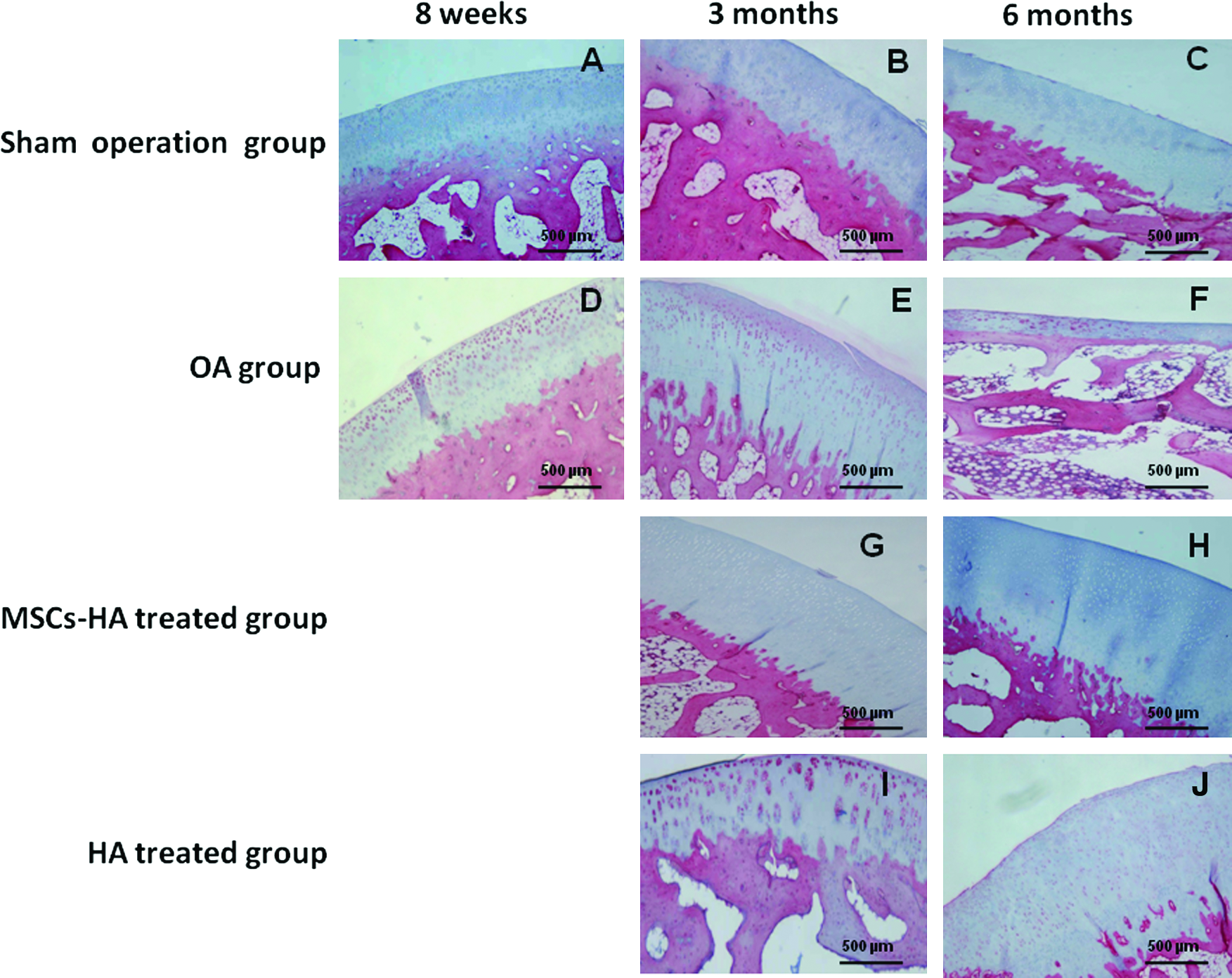
The histomorphometric parameters performed in OA group after 8 weeks, 3 months, and 6 months confirmed the development of articular degenerative processes, resulting to be significantly different from those of treated groups for the Kraus' modified Mankin score, which takes into account several parameters as reported in Materials and Methods section (Fig. 8A). Articular cartilage structure in the sham-operated group was normal, smooth, and uninterrupted (data not shown), while in the OA group was irregular with superficial clefts that increased and extended into the middle zone at 3 and 6 months.

(
MSC-HA group showed mild surface irregularity at both 3 and 6 months. HA group showed instead a worsening condition at both times compared to the MSC-HA–treated one.
Toluidine blue showed uniform staining throughout the articular cartilage in the sham group (data not shown), while in the OA group, we observed a loss of proteoglycan staining in the superficial and middle zones, which increased with time. MSC-HA group showed only a mild loss of proteoglycans in the superficial zone, while HA group showed the same findings as OA group at both experimental times evaluated.
Concerning cellularity, a normal distribution was observed in the sham group (data not shown), while a diffuse hypercellularity and cloning formation was observed in the OA group. A mild hypercellularity was observed in both treated groups at the two experimental times. Osteophyte presence was not observed in the sham group (data not shown), and in the OA group medium-sized osteophytes were seen. MSC-HA group showed the presence of few small osteophytes, while HA group showed medium-sized osteophytes. Tidemark was intact in all the groups.
Mean CT of the medial femoral condyle was 380 μm in the sham group. This value decreased significantly in OA group during the experimental times evaluated, whereas it increased in the MSC-HA group at 3 and 6 months. HA group showed instead a significant decrease of CT value compared to MSC-HA group at 6 months (Fig. 8B).
FI was lower in the OA group than in the MSC-HA and HA groups at all experimental times without significant differences (Fig. 8C).
Mean SBT showed a significant increase in OA group at 3 and 6 months compared to treated groups. MSC-HA group showed an SBT value significantly lower than that of OA and HA groups at both experimental times analyzed (Fig. 8D).
Discussion
OA is a degenerative joint disease generally characterized by progressive cartilage degeneration, subchondral bone changes, osteophyte formation, and low-grade synovitis. 38 Increases in mechanical stress and changes in biochemical factors within affected joints are thought to be primarily responsible for progression of the disease. 39 Although many details of OA pathogenesis in humans have remained elusive, it is clear that this is a complex multifactorial disease process involving cartilage catabolism and anabolism, as well as changes in other tissues in the joints. 40 Currently, there is no disease-modifying treatment for OA. While available treatments for early OA are based on symptomatic relief, irreversible joint disability in advanced OA usually requires surgical intervention to relieve pain and improve joint function. For the last two decades, animal models have played a critical role in improving understanding of the early events occurring during the disease progression because available human joint tissues usually represent advanced OA.41,42 Many models have been characterized and used to study the histological and biochemical changes occurring during OA.43,44 In rabbits, it is reported that 3–8 weeks after ACLT, articular cartilage exhibits degenerative changes.31,32,45 Our study shows that 8 weeks after ACLT is a proper time to preclinically evaluate disease-modifying treatments of OA. We demonstrated that OA changes, including cartilage fibrillation and erosion, periarticular osteophytosis, and proteoglycan depletion, develop sequentially and progressively with time in a rabbit model of OA based on surgically induced joint instability. The first degenerative changes in the surgically induced OA model were found in the superficial zone of cartilage and showed focal fibrillation of the articular surface, increase of chondrocyte proliferation, and matrix disorganization increasing over time.
The use of autologous cell implantation in repairing cartilage lesions is promising and associated with several potential long-term benefits. 12 However, because of the high rate of progression to OA among patients as a result of acute or chronic injury, the possibility to apply this technology to reduce at least the progression of the disease is of great interest.
Recently, the use of MSCs, isolated from bone marrow, is becoming a valid alternative to the use of adult chondrocyte cells,19,20,46 avoiding the harvest of a health cartilage biopsy and the expansion of the cells in vitro. After appropriate induction, MSCs are able to differentiate into several mesodermal tissues, including bone, cartilage, and muscle.15,47
It is possible to speculate that the beneficial effects of these cells might reflect, in part, some trophic and protective activities they exert on injured cells and tissues. As reported by Caplan 46 MSCs, themselves, secrete a broad spectrum of bioactive macromolecules contributing in the injury response by providing a broad array of paracrine factors that serve to structure regenerative microenvironments in tissue damages.
These processes are ultimately favored by an opportune behavior as the growth onto three-dimensional structures that provide the right signals. 48
Hyaluronan-based scaffold has been reported to downregulate the expression of some catabolic factors 23 and to favor the differentiation process of MSCs onto cartilage-differentiated cells. 25 Our data showed that OA defects, created by ACTL in a rabbit model, were repaired with hyaline-like cartilage after implantation of MSC-HA. The repair process with MSC-HA was superior to HA implantation alone and to the untreated tissues, as evidenced by the Kraus'modified Mankin score, which takes into consideration some cartilage structure parameters that are particularly affected during OA development.
The morphological, histological, and immunohistochemical data confirm in general the formation of a new cartilage with some hyaline-like features in the MSC-HA group even if some degree of tissue remodeling was observable also in the group treated with HA alone.
Both the treatments seem to downregulate some molecules involved in catabolic processes as MMP-1 and MMP-3 as observed by other authors of our group, 24 suggesting an important active role also in vivo of the cell-seeded scaffolds on degradative pathways occurring in OA disease. This is not surprising because, as previously reported, hyaluronan-based biomaterials are able to supply the ideal environment to recapitulate embryonal events.49,50
In conclusion, despite the limitations of the present study, that rely on the not well-defined MSC characterization (due to their number of days in culture and their exposure to FBS and ascorbic acid) and the lack of an MSC control group, our preliminary results seem to demonstrate that the use of these cells in combination with Hyaff-11 scaffold reduces the development of early/mild OA lesions in a rabbit model and opens up the possibility of future therapy based on tissue engineering.
Footnotes
Acknowledgments
Thanks to Elettra Pignotti for statistical evaluation. Thanks to Patrizia Rappini and Graziella Salmi for assistance in the preparation of the manuscript, and to Luciano Pizzi for technical assistance.
This study was supported by Fondazione Monte dei Paschi di Siena.
Disclosure Statement
No competing financial interests exist.