
Abstract
The description of a microencapsulation procedure for Wharton's jelly mesenchymal stem cells (WJMSCs) is reported. The applied method is based on the generation of monodisperse droplets by a vibrational nozzle. An ionic alginate encapsulation procedure was utilized for the microbeads hardening. Different experimental parameters were analyzed, including frequency and amplitude of vibration, polymer pumping rate, and distance between the nozzle and the gelling bath. The produced barium–alginate microbeads were characterized by excellent morphological characteristics as well as a very narrow size distribution. The microencapsulation procedure did not alter the morphology and viability of the encapsulated WJMSCs. In addition, the current paper reports the functional properties in terms of secretive profiles of both free and encapsulated WJMSCs. The analyzed factors were members of the family of interleukins, chemokines, growth factors, and soluble forms of adhesion molecules. These experiments showed that despite encapsulation, most of the proteins analyzed were secreted both by the free and encapsulated cells, even if in a different extent. In conclusion, the described encapsulation procedure represents a promising strategy to utilize WJMSCs for possible in vivo applications in tissue engineering and biomedicine.
Introduction
Many studies suggest that MSCs may differentiate toward cells of different lineages, including chondrocytes, adipocytes, osteocytes, miocytes, neurons, and tenocytes.3–10 Because of their peculiar characteristics, MSCs are considered to be very important for several applications in the field of regenerative medicine, including the development of cell-based therapies and tissue repair procedures.2,11–13
Different adult human tissues have been considered as MSC sources, including bone marrow, trabecular bone, adipose tissue, peripheral blood, synovium, skeletal muscle, dental pulp, and periodontal ligament.4,11–16 Although bone marrow still represents the main and most investigated source of adult MSCs, the isolation and use of these cells still present some drawbacks. For instance, the number of MSCs found in bone marrow decrease progressively starting at age 17, and the harvesting techniques are invasive, often causing severe infections, bleeding, and chronic pain for donors.17,18
Looking for alternative MSC sources, fetal tissues,19,20 extraembryonic tissues such as placenta, and amniotic fluid,21,22 umbilical cord blood,23,24 and stroma25,26 have recently been considered. Umbilical cord, due to the unique morphological properties, represents an interesting alternate source for MSCs, especially if compared to umbilical cord blood.27,28 Umbilical cord, usually weighing 40 g and spanning between 60 and 65 cm in length, contains a special primitive connective tissue called Wharton's jelly. 29 This jelly acts as a protective tissue for vessels and contains into its stromal compartment cells with specific mesenchymal characteristics, called Wharton's jelly MSCs (WJMSCs). 30 Therefore, Wharton's jelly can be an ideal, unique, easily accessible, and uncontroversial source for early MSCs due to the simple collection procedure once umbilical cord is routinely discharged at parturition. It has recently been described that WJMSCs are able to support long-term maintenance of hematopoietic stem cells. 31 Therefore, it is reasonable that WJMSCs, like MSCs collected from other tissues, 32 can release many soluble molecules, including interleukins, chemokines, and many growth factors. Unfortunately, to the best of our knowledge, in literature there is limited data available about the qualitative and quantitative characterization of factors secreted by WJMSCs. The ability to self-renew and to differentiate into several cell types makes the use of MSCs particularly attractive for the development of innovative therapeutic strategies aimed at repairing and replacing damaged tissues and makes them a very promising cell source for tissue engineering applications.
Nevertheless, several factors still hurdle the extensive clinical use of cell-based therapy. The protection of implanted cells from the host's immune response is of primary importance. To solve this problem, one of the most promising approaches is represented by cell's encapsulation within semipermeable capsular systems.33,34 Encapsulation protocols have also been proposed to maintain the cell phenotype longer than in monolayer, 35 to prolong the cell viability and therefore to sustain cell function. 36
With respect to encapsulation/immobilization procedure for MSCs, Langer and coworkers have reported a methodology to enhance the vascular differentiation of human embryonic stem cells by encapsulating the cells in a bioactive scaffold based on a photopolymerizable dextran hydrogels comprising either insoluble or soluble endothelial growth factors. 37 Moreover, a protocol aimed at maintaining human embryonic stem cell in culture, for a period of up to 260 days, without exposure to animal cells has recently been published. This procedure is based on the encapsulation of cells in 1.1% (w/v) calcium alginate hydrogels. 38
The use of collagen-based hybrid fibers for tissue engineering was also presented, based on the synthesis of a cell-encapsulated fibrous scaffold by interfacial polyelectrolyte complexation; human MSCs encapsulated in the fibers showed a higher proliferation rate than those seeded on the scaffold. 39 Finally, alginate beads were also tested to study the chondrogenesis of human MSCs. 40
With the aim of proposing WJMSCs for tissue engineering application, the current paper describes (a) the isolation of human WJMSCs, (b) the production of barium (Ba)–alginate microbeads by a vibrating nozzle approach for WJMSCs encapsulation, and finally (c) in vitro characterization of WJMSCs encapsulated into alginate microbeads, including viability, proliferation, and secretive profile.
Materials and Methods
WJMSCs: isolation procedure and culture conditions
Human umbilical cords (all from natural deliveries) were collected after mothers' consent and approval of the Ethics Committee of the University of Ferrara and S. Anna Hospital. Harvesting procedures of Wharton's jelly from umbilical cord were conducted in full accordance with the Declaration of Helsinki as adopted by the 18th World Medical Assembly in 1964 and successively revised in Edinburgh (2000) and the Good Clinical Practice guidelines.
Cords were processed within 4 h, and stored at 4°C in sterile saline until use. Typically, the cord was rinsed several times with sterile phosphate-buffered saline (PBS) before processing and was cut into pieces (2–4 cm in length). Blood and clots were drained from vessels with PBS to avoid any contamination. Single pieces were dissected, first separating the epithelium of each section along its length, to expose the underlying Wharton's jelly. Later, cord vessels (the two arteries and the vein) were pulled away without opening them. The soft gel tissue was then finely chopped.
The same tissue (2–3 mm2 pieces) was placed directly into a 75-cm2 flask for culture expansion in 10% fetal calf serum (Euroclone S.p.A., Milan, Italy) Dulbecco's modified Eagle's medium low-glucose medium supplemented with antibiotics (penicillin 100 mg/mL and streptomycin 10 mg/mL) at 37°C in a humidified atmosphere of 5% CO2. After 5–7 days, the culture medium was removed and thereafter changed twice a week. At a ∼70–80% confluence, cells were scraped off by 0.05% trypsin/ethylene diamine tetraacetic acid (EDTA) (Gibco, Grand Island, NE) (2 min), washed, counted by hemocytometric analysis, assayed for viability, and thereafter used for further in vitro experiments or for encapsulation procedures.
Flow cytometric analysis
The WJMSCs were analyzed for expression of MSC surface marker molecules, by direct immunofluorescent staining, as reported in the literature.41,42 Briefly, cell pellets were resuspended in PBS and incubated with fluorescein isothiocyanate (FITC)– or phycoerythrin (PE)–conjugated mouse anti-human antibodies CD45-PE, CD34-FITC, CD90-FITC, CD105-PE, CD44-FITC, and CD29-PE (DakoCytomation; Dako, Glostrup, Denmark) for 15 min at 4°C. Monoclonal antibodies with no specificity were used as negative control.
Antibody-treated cells were then washed with PBS and spinned down. Cell pellets were resuspended in 400 μL of PBS and analyzed by FACS Scan (Becton Dickinson, Franklin Lakes, NJ). For each sample, 20,000 events were acquired and analyzed using the CellQuest software (Becton Dickinson European HQ, Erembodegem Aalst, Belgium).
Encapsulation of WJMSCs
Monodisperse alginate beads containing WJMSCs were prepared using an encapsulation device that is based on a vibrating nozzle (Encapsulator Research Inotech, Dottikon, Switzerland) according to the experimental procedure previously described.33,34 The encapsulator is composed by a 2-L glass reaction vessel with stainless steel top and bottom plates. The top plate contains a feed-line connected to a syringe and a vibrating nozzle. A nozzle with an internal diameter of 300 μm was used. The flow of alginate to the nozzle is achieved by a precision syringe pump. The production of WJMSC-filled alginate microcapsules was optimized by changing the following experimental parameters: vibrational frequency (freq), vibrational amplitude (amp), alginate pumping rate (pump), and distance between the nozzle and the surface of the gelling bath (height) (see Table 1). Before encapsulation, WJMSCs were suspended in a 1.5% (w/v) aqueous solution of highly purified sodium alginate (Stern Italia, Milano, Italy) at a concentration of 8–12 × 106 cells/mL. The generated microdroplets were dropped into an isotonic barium chloride solution (1.2%; w/v); after gelation (3 min), the microbeads were washed twice with saline and cultured in 10% fetal calf serum (Euroclone S.p.A.) Dulbecco's modified Eagle's medium low-glucose medium supplemented with antibiotics (penicillin 100 mg/mL and streptomycin 10 mg/mL) at 37°C in an humidified atmosphere of 5% CO2.
Dimensional and morphological characterization of microbeads
The morphology of Ba–alginate microbeads was evaluated by optical stereomicroscopy (Nikon SMZ 1500 stereo microscope, Tokyo, Japan). Microbead size and size distribution were determined by photomicrograph analyses (Eclipsenet version 1.16.5; Laboratory Imaging s.r.o. for Nikon Italia, Firenze, Italy). Microbead samples, immediately after preparation and at intervals after storage under different conditions, were deposited onto a microscope slide and examined microscopically. A sample of 100–300 beads was examined, and the mean size was determined.
Experimental design and statistical analysis
To study the effect and the influence of different experimental parameters (i.e., frequency, pump, and height) on the dimensional and morphological characteristics of alginate microbeads, a randomized central composite face-centered design consisting of 17 runs was used. The experimental design and the evaluation of the experiments were performed by the PC software MODDE 8.0 (Umetrics AB, Umea, Sweden), followed by multiple linear regression algorithms.
Viability analysis of WJMSCs
Before and after encapsulation, the viability of the cells was analyzed both by colorimetric assay with [3-(4,5-dimethylthiazol-2-yl)-2,5-diphenyltetrazolium bromide] MTT (thiazolyl blue) and by double staining with propidium iodide (PI) and Calcein-AM according to the manufacturer's instructions. The MTT assay, based on the conversion of the yellow tetrazolium salt MTT to purple formazan crystals by metabolically active cells, provides a quantitative determination of viable cells. 43 After 3, 6, and 9 days of culture 200 μL of MTT was added to each well of beads, and the plate was incubated at 37°C. After 2 h the MTT crystals were solubilized with 50% dimethyl formamide; after 24 h spectrophotometric absorbance of each sample was measured at 570 nm. For the PI and Calcein-AM analysis, cells were visualized under a fluorescence microscope (Nikon, Optiphot-2; Nikon Corporation, Tokyo, Japan) using the filter block for fluorescein. Dead cells stained red, while viable ones appeared green.
Induction and evaluation of osteogenic differentiation
For the analysis of osteogenic differentiation, both free WJMSCs (growing in monolayer) and microbead-entrapped cells were incubated in human MSC osteogenic differentiation medium (Lonza, Walkersville, MD) for 21 days.
The extracellular matrix composition was analyzed by Fourier transform infrared spectroscopy. Cell layers were collected in ammoniated water (50 mM ammonium bicarbonate, pH 8.0), lyophilized, and analyzed by FT-IR Spectrometer Perkin Elmer, Spectrum 100 (Perkin Elmer, Salem, MA). Absorption spectra were collected from 4000 to 600 cm−1. After 21 days of incubation in standard and osteogenic conditions, microbeads were assayed for alkaline phosphatase (ALP) activity. Microbeads were incubated at 37°C in a 50 mM EDTA solution (pH 7.0) for 2 min to dissolve the Ba–alginate microbeads and obtain the free cells. The cells were lysed with 300 μL of 0.2% Triton X-100. ALP activity was assayed by measuring, after 30 min of incubation at 37°C, the conversion p-nitrophenylphosphate to p-nitrophenol, using an enzyme-linked immunosorbent assay reader at 405 nm wavelength. ALP activity was normalized to total cellular proteins, determined by the Bradford protein assay protocol and expressed as U/mg of protein. One unit was defined as the amount of enzyme that hydrolyzes 1 μmol/min of p-nitrophenylphosphate. For Alizarin Red S staining, free WJMSCs (growing in monolayer) and microbead-entrapped cells were fixed and then stained with 40 mM Alizarin Red S solution (pH 4.2) at room temperature for 10 min. Samples were then rinsed five times with distilled water and washed three times in PBS on an orbital shaker at 40 rpm for 5 min each to reduce nonspecific binding. The stained matrices were microphotographed by an optical microscope (Nikon, Optiphot-2; Nikon Corporation, Tokyo, Japan). For real-time polymerase chain reaction (PCR) analysis, Ba–alginate microbeads were dissolved as previously reported, and total RNA was isolated from the WJMSC-free cells using Total RNA Isolation system (Promega, Madison, WI). Two micrograms of total RNA was reverse transcribed with the Improm-II RT System (Promega). mRNA of target genes was quantified by real-time PCR using the ABI Prism 7700 system and TaqMan probes 5′AACCCAGAAGGCACAGACAGAAGCT3′ for runt related gene 2 (RUNX-2) (Applied Biosystems, San Francisco, CA). PCR was carried out in a final volume of 25 μL. After a 10 min preincubation at 95°C (denaturation) and 1 min at 60°C (annealing/elongation), the mRNA levels were corrected for glyceraldehyde 3-phosphate dehydrogenase mRNA levels (reference gene) and normalized to a calibrator sample (control cells).
Determination of WJMSC secretory pattern by Bio-Plex analysis
After 72 h of in vitro cell culture, the medium from WJMSCs growing as monolayer and embedded in Ba–alginate microbeads was collected and analyzed for a set of selected proteins. To compare and normalize the data of adherent and encapsulated WJMSCs, the microbeads were washed twice with PBS before the analyses and incubated at 37°C, for 15 min, with 500°μL of 50 mM EDTA (Sigma, St. Louis, MO) in PBS buffer. EDTA was then neutralized with additional 10 mL PBS, and cells were collected by centrifuging at 400 g for 15 min. Pellets obtained from WJMSCs growing as monolayer and embedded in Ba–alginate microbeads were lysed with 50 μL buffer (NaCl 150 mM; Tris-HCl pH 7.4 20 mM; EDTA 1 mM; ethylene glycol tetraacetic acid (EGTA) 1 mM; Triton X-100 1%; Na3VO4 0.1 mM; phenylmethanesulphonyl fluoride (PMSF) 1 mM; proteinase inhibitory cocktail (PIC) 0.1% v/v). About 10 μL of each sample was tested with the Bradford method to determine proteins content. 44 Concentrations of interferon-alpha2 (IFN-α2), interleukin-1alpha (IL-1α), IL-2 receptor α, IL-3, IL-12 (p40), IL-16, IL-18, cutaneous T-cell-attracting chemokine (CTACK), growth regulated oncogene-α (GRO-α), hepatocyte growth factor (HGF), intercellular adhesion molecule-1 (ICAM-1), leukemia inhibitory factor (LIF), monocyte chemotactic protein-3 (MCP3), macrophage colony stimulating factor (M-CSF), macrophage migration inhibitory factor (MIF), monokine induced by IFN-Gamma (MIG), beta-nerve growth factor, stem cell factor, stem cell growth factor-beta (SCGF-β), stromal cell-derived factor 1α (SDF-1α), tumor necrosis factor-beta, tumor-necrosis-factor related apoptosis inducing ligand (TRAIL), and vascular cell adhesion molecule-1 (VCAM-1) were simultaneously evaluated using a commercially available multiplex bead–based sandwich immunoassay kit (Human 23-plex; Bio-Rad Laboratories, Milano, Italy). Bio-Plex analysis was performed following the manufacturer's instructions. About 23 distinct sets of fluorescently dyed beads loaded with capture monoclonal antibodies, specific for each cytokine, were used. Secretion and standard samples (50 μL/well) were incubated with 50 μL of premixed bead sets in a prewet 96-well microtiter plate. After incubation and washing, 25 μL of fluorescent detection antibody mixture was added and left to react for 30 min under gentle shaking; samples were then washed and resuspended in the assay buffer. Standard calibration curves for each protein were used, ranging from 2 to 32000 pg/mL; the minimum detectable dose was 2 pg/mL. The formation of the different immunocomplexes was measured by the Bio-Plex Protein Array System (Bio-Rad Laboratories). A 50 μL volume sample was withdrawn from each well, and the fluorescent signal of a minimum of 100 beads per region (chemokine/cytokine) was measured. To compare directly secretive content of adherent and encapsulated WJMSCs, all values were normalized with respect to the total protein amount.
Data analysis and statistics
Statistical analysis was performed by one-way analysis of variance followed by the Student's t-test. A p-value <0.05 was considered statistically significant. Cytokine analysis was performed with Bio-Plex Manager software version 3.0 (Bio-Rad Laboratories). Standard levels between 70% and 130% of the expected values were used. In general, at least five standards were accepted and used to establish standard curves following a five-parameter logistic regression model. Sample concentrations were immediately interpolated from the standard curves. Values were expressed as pg/mL +standard deviation.
Results and Discussion
Isolation and characterization of WJMSCs
As a source of human MSCs, solely Wharton's jelly was used instead of the whole cord (easier and faster to treat); this choice was made with the aim of isolating a relatively homogeneous cell population, possibly avoiding any epithelial cell contamination. For the isolation of WJMSCs, a slightly modified version of a previously published procedure was used. 26 Can and Karahuseyinoglu have indeed recently reported in a review article (dealing with the umbilical cord stroma as source for stem cells) that a collagenase treatment is critically important, since there is always a risk of over digestion of the cellular external lamina, causing the prevention of cell adhesion to the culture substrate after isolation. 26 In this respect, collagenase, trypsin, and hyaluronidase treatments were omitted, in agreement with other investigators,45–47 observing that skipping the enzymatic digestion treatment favors cell adhesion and viability.
When in vitro cultured, the primary cells, isolated from Wharton's jelly, displayed an MSC-like phenotype. After 3 days of culture, the WJMSCs formed adherent colonies, reaching confluence after 10–14 days. High percentage of cells appeared viable, as demonstrated by Calcein-AM stain (Fig. 1A), and displayed a spindle shape resembling fibroblasts (Fig. 1B, C). The immunophenotypical profile of WJMSCs was determined by flow cytometric analysis (Fig. 1D–I). There are, as of yet, no known MSC-specific markers. Nevertheless, according to previous studies,28,48,49 cell surface markers such as CD90 (Thy-1), CD29 (β-1 integrin), CD44 (hyaluronan receptor), and CD105 (SH2, endoglin) were analyzed to attribute an MSC-like immunophenotype. Flow cytometry showed that, since first passage, the WJMSCs expressed these markers (Fig. 1D–G). The hematopoietic marker CD45 and the hematopoietic/endothelial marker CD34 were not detectable in these cells (Fig. 1H, I), indicating that WJMSCs were not contaminated with cells of hematopoietic or endothelial origin.

Phenotype characterization of Wharton's jelly mesenchymal stem cell (WJMSC) monolayers. (
Rigorous identification of MSCs requires demonstration of their capability to differentiate along specific mesenchymal lineages when induced to do so. In this context, we are interested in demonstrating the ability of WJMSCs to differentiate into osteoblasts. The osteogenic differentiation of expanded WJMSCs was induced by the addition of dexamethasone, β-glycerophosphate, and ascorbate, and was assessed after 21 days by monitoring the production of mineralized extracellular matrix, as a marker for terminally differentiated MSCs into osteoblasts. 30 As shown in Figure 1J and K, we detected the presence of calcium by Alizarin red staining, and the presence of apatite by the Fourier transform infrared spectroscopic assessment of mineral content from the induced WJMSC.
Encapsulation of WJMSCs in alginate microbeads
WJMSCs were embedded into alginate microbeads by an encapsulation device based on a vibrating nozzle (see scheme in Fig. 2). The encapsulation procedure was relatively simple and consisted of a limited number of steps. To achieve complete biocompatibility, essential for mammalian cells, the encapsulation procedure was conducted at room temperature under physiologic pH and tonicity using a pyrogen-free alginate solution. The resulting Ba–alginate microbeads were elastic and transparent, facilitating the microscopic observation of the WJMSCs' viability and morphology during the in vitro studies. The hardening of alginate solution was accomplished by an ionic gelation procedure based on barium chloride. The use of barium ions (instead of more often used calcium ions) resulted in the formation of mechanically stable microbeads with an extremely high biocompatibility, preserving the in vitro and in vivo viability of the embedded cells. 34
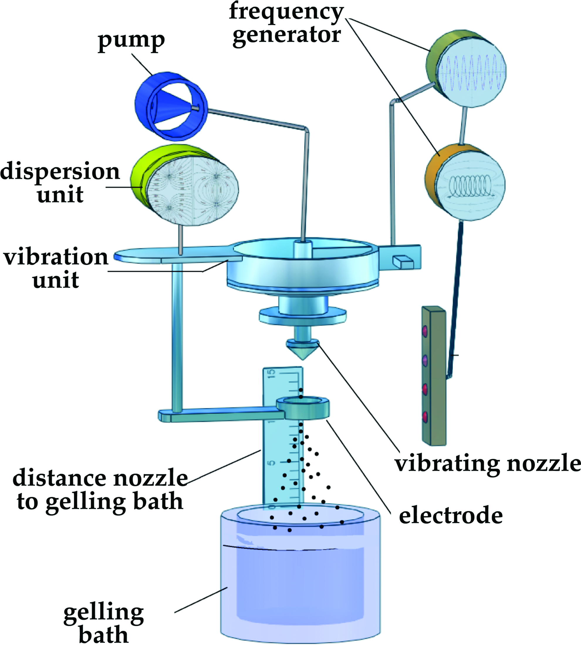
Schematic representation of the encapsulation device based on a vibrating nozzle. Color images available online at
A classical intuitive approach (Changing One Separate Factor a Time) was used to define the experimental setup, including the selection of the crucial experimental parameters. This approach was chosen to evaluate which factor(s) could mostly affect the morphology of the produced alginate microbeads in terms of general geometry, surface characteristics, and dimensions. Following the above procedure, only one experimental parameter was varied at a time (see Table 1), while all the others remained constant. Experiments were performed by changing the following parameters: frequency of vibration (freq), amplitude of vibration (amp), polymer pumping rate (pump), and distance between the nozzle and the gelling bath (height). The frequency was calculated from knowledge of the alginate solution viscosity for the nozzle diameter used, to obtain the desired microbeads size and morphology (mean diameter was between 550 and 650 μm with a spherical shape). The vibrating frequency of the nozzle varied from 100.0 to 140.0 Hz, while the rate of polymer pumping was investigated in the range 7.5–9.5 (mL/min) (see Table 2). The investigation of these parameters revealed that increasing both factors led to a decrease in particle size (data not shown), but if the vibrating frequency was set too high (>200.0 Hz), a progressive number of coalescences (formation of clusters of microbeads partially fused together) were detectable in the microbead population (data not shown). The amplitude of the vibration applied to the nozzle (ranged from 1.0 to 6.0 mm) had only a slight effect on the microbead characteristics such as size, sphericity, and presence of coalescences. On the contrary, the distance between the nozzle and the gelling bath (range, 100.0–140.0 mm) had a great effect on the microbead morphology. In fact, a distance of 100.0 or 120.0 mm caused the formation of microbeads with an elliptic shape (formation of tails). Considering the results of this set of experiments (summarized in Table 2), it can be concluded that among the four different experimental variables, only the amplitude of the vibration has minor effect on the microbead formation, while all the others exert significant morphological and dimensional changes in the alginate beads. The microbeads with the best morphology (Fig. 3A, B) were produced by the following setup: a frequency of 150 Hz, an amplitude of 1.0 mm, a pump flow rate of 8.5 mL/min, and an height of 140.0 mm.

Optical (
The morphological characteristics of microcapsules were assessed on at least 200 microcapsules/batch, considering the presence of (a) tails, (b) coalescences, and (c) the surface characteristics. All the reported microbeads were prepared with an alginate concentration of 1.5% (w/v). Data represent the average of three independent determinations on different microbead batches.
Effect of alginate concentration on microcapsule characteristics
Once the instrumental setup was achieved, we focused our attention on another important experimental parameter: the concentration of alginate solution. Different percentages were tested, ranging from 0.5% to 3.5% (w/v), and the effect on the production of microbeads was considered as well. At the first observation, it should be mentioned that using the automated vibrating nozzle instrument, we succeeded in obtaining microbeads using alginate concentration up to 2.0% (w/v). On the contrary, using higher concentrations of alginate (>2.0%, w/v), we found that the resulting solution was too viscous to be efficiently converted in microdroplets. Samples of microbeads produced with an alginate concentration of 0.5%, 1.0%, 1.5%, and 2.0% were analyzed for morphology, size, and size distribution. Table 3 and Figure 3 report the results of the above experiments showing that microbeads produced with an alginate concentration between 1.0% and 2.0% (w/v) were spherical and characterized by a smooth surface. Lowering the alginate concentration down to 0.5% (w/v) caused partial breaking of beads, resulting in particles with an irregular shape. This behavior was attributed to the mechanical stress caused by the landing in the hardening barium chloride solution. Moreover, at the same concentration, some coalescences were detectable. We obtained alginate microbeads with a smaller mean diameter and homogenous dimensional distribution using a polymer concentration of 1.5% (w/v). When the alginate concentration was increased up to 2% (w/v), the size distribution became broader and the mean diameter increased from 630 μm (in the case of an alginate concentration of 1.5%) to 645 μm (see dimensional distribution plots reported in Fig. 3E, F). On the other hand, alginate microbeads prepared with lower polymer concentrations (0.5% and 1.0%, w/v) formed some coalescences (see Table 3), although the size distribution remained acceptably narrow.
Data represent the average of three independent batch determinations.
Calculated on the dry weight of the alginate powder.
SD, standard deviation.
Encapsulation of WJMSCs in alginate microbeads: A design of the experimental approach
After performing the Changing One Separate Factor a Time study, a “design of the experiments” optimization and a screening of the experimental parameters were also conducted. The design of the experiments reduces the number of experiments and provides statistical information about the effects of different variables and their possible interactions.
The parameters frequency, pump, and height were chosen as variables and tested at three levels. Therefore, in our case we selected 14 experiments by a randomized central composite face-centered design that requires fewer trials together with three center points to have an estimation of the experimental error (Table 4).
Data represent the average of three independent determinations.
By examining the results the main observation was that a change in height value from a low to a high level (80–160 mm) results in an increase of tail formation as well as of coalescences. On the other hand, the frequency and pump parameters influence their high and low levels causing the increase of the both responses (see Table 4).
Three-dimensional graphs of the investigated factors are given in Figure 4 showing the influence of factors on tails (panels A–C) and coalescences formation (panels D–F).

Design of the experimental analysis for the production of alginate microbeads containing WJMSCs. Response surface plots of the responses: “tails” (
After investigating the factor influence, the validity and the significance of the model were estimated by analysis of variance (ANOVA). All the data obtained fit well the model determining a good reproducibility of the studied model. We get a large regression coefficient R 2 that is a necessary condition for a validity model with a significant power of prediction of the model Q 2 (see Table 5).
n is the number of experiments; R2 is the percentage of the variation of the response explained by the model; R2Adj. is the fraction of the variation of the response explained by the model adjusted for the degree of freedom; Q2 is the percentage of the variation of the response predicted by the model; SDY shows the variation due to the replicated observations adjusted for degrees of freedom and in the same units as Y; RSD shows the variation of the response not explained by the model, adjusted for degrees of freedom and in the same units as Y. This is the residuals standard deviation.
Viability, proliferation, and osteoblastic differentiation of encapsulated WJMSCs
A crucial issue that should be always investigated in the case of embedding/seeding protocols for cell scaffolding concerns the effect of the encapsulation procedure on the growth and the activity of cells. For this purpose, the viability of WJMSCs encapsulated in alginate beads was determined by the live/dead test. Beads were incubated with Calcein-AM (a marker of living cells; fluorescent signal was monitored using 485 nm excitation wavelength and 530 nm emission wavelength) and with PI (a marker of cell death; excitation, 535 nm; emission, >610 nm), as described in the Materials and Methods section. The observation of the fluorescent images recorded immediately after the encapsulation procedure at the typical excitation wavelengths (Fig. 3C, D) indicated that the cells were highly viable (>95%). To strengthen these data, the cell viability was determined after different lengths of culture time (up to 9 days) using two alternative procedures, namely, the double staining with a Calcein-AM cell viability assay kit (Fig. 5A–E) and the MTT test (Fig. 5F). About 100 alginate beads were incubated with thiazolyl blue, and the presence of formazan salts, marker of the viable cells, was reported in the graph as percentage of viable cells respect to day 0 (rated to 100%). During each day of measurement, we observed a steady but insignificant (p > 0.05) decrease in formazan absorption during the first 10 days.
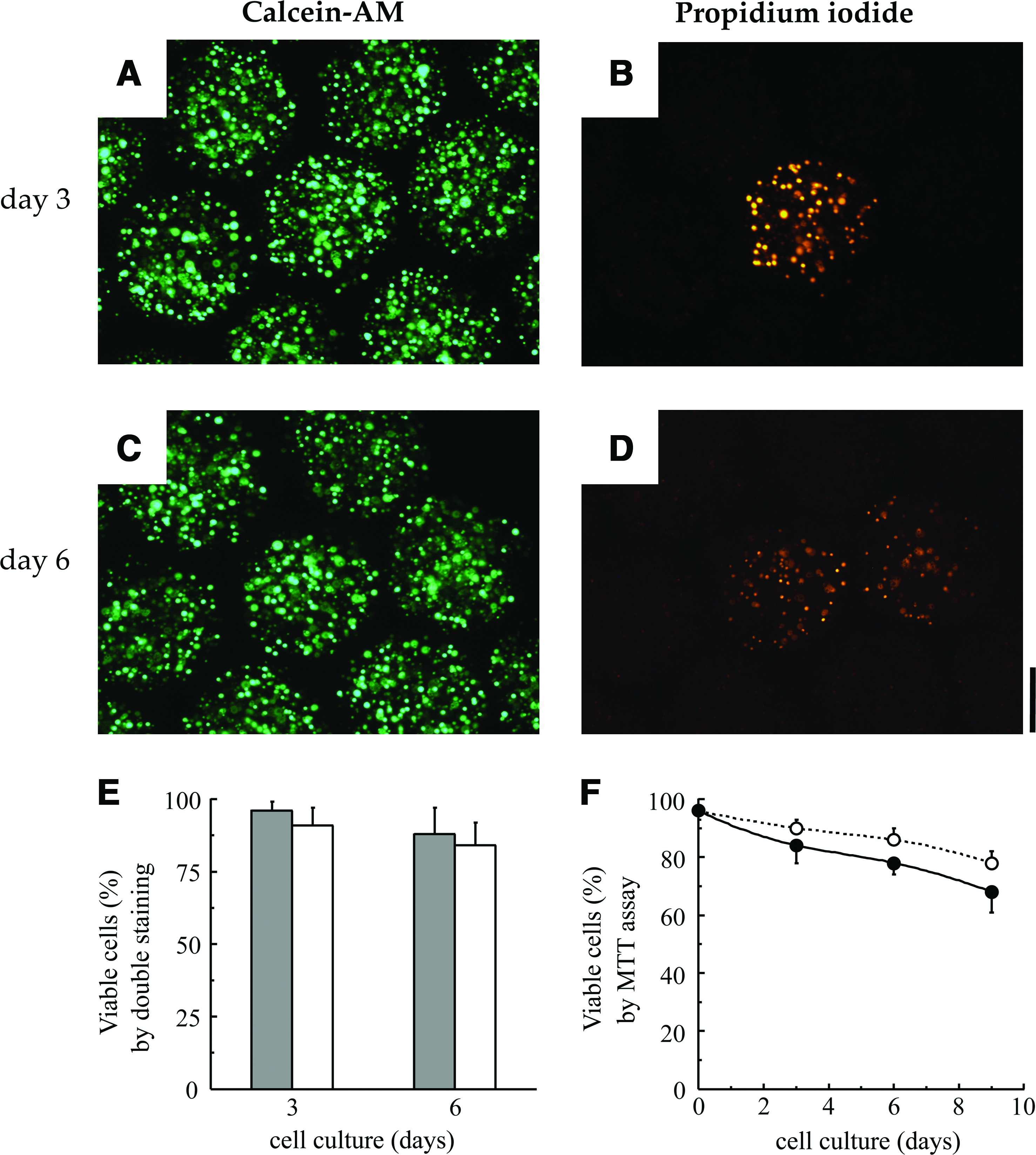
Viability of WJMSC encapsulated in alginate microbeads, as determined by double staining assay with Calcein-AM and propidium bromide (
The ability of the WJMSCs entrapped in alginate to differentiate in osteoblasts was assessed at day 21 of osteogenic induction (WJMSCs/alg/ost) by a number of classical criteria (Fig. 6) and compared to WJMSCs entrapped in alginate in the absence of osteogenic medium (WJMSCs/alg). The WJMSCs/alg/ost cells showed an appreciable increase of ALP activity, an early marker for osteoblast differentiation (Fig. 6A). The WJMSCs/alg/ost cells showed also an increase of the expression of a bone-specific gene such as runt related gene 2 (Runx2) analyzed by quantitative real-time PCR (Fig. 6B). Similarly, mineralization was present in WJMSCs/alg/ost cells (Fig. 6C). All together, these results suggest that the cells were undergoing osteogenic differentiation despite encapsulation.
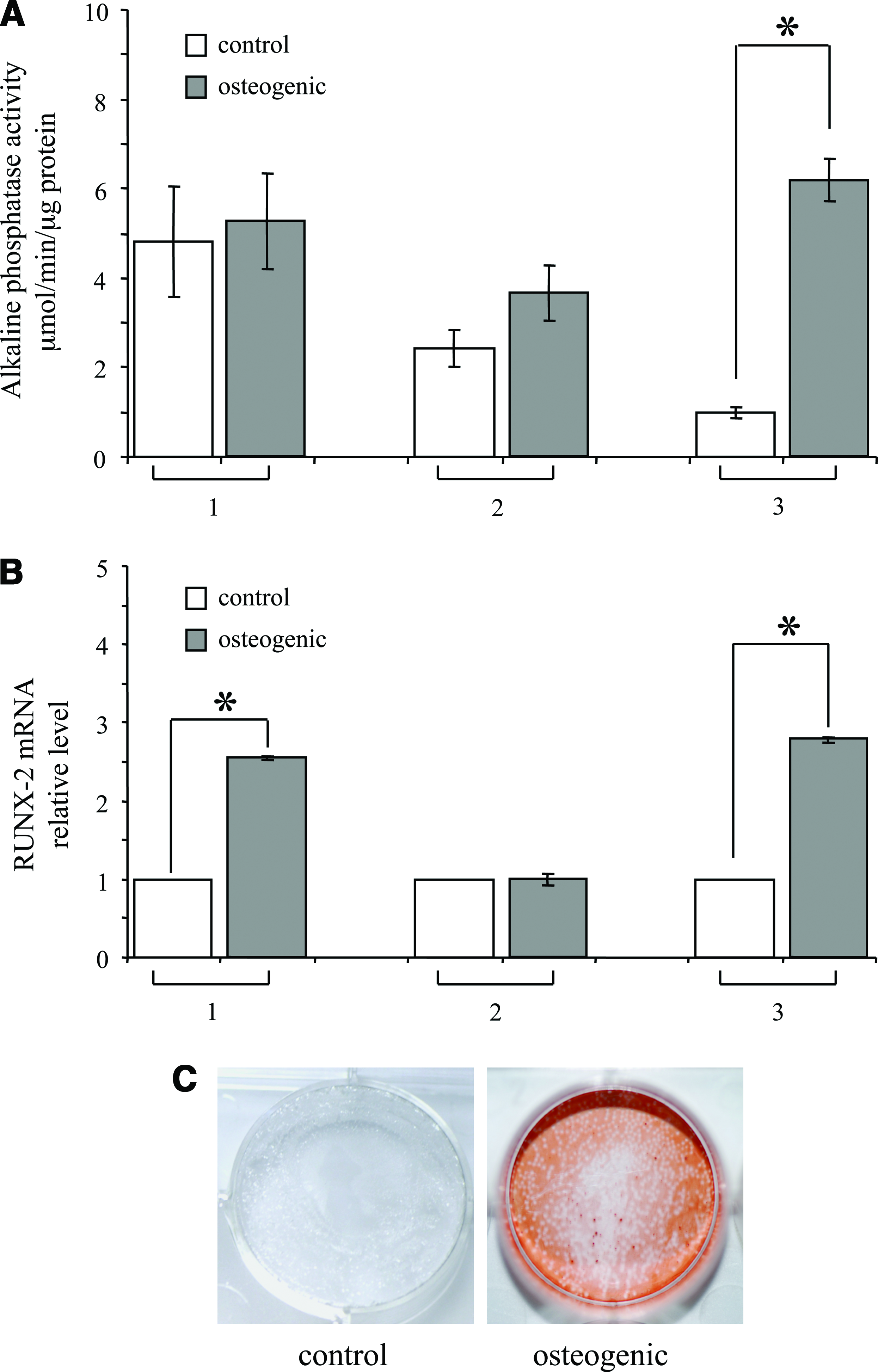
Osteogenic differentiation of WJMSCs encapsulated in alginate microbeads. WJMSCs from different isolations (samples #1, #2, and #3) were cultured in standard (open bars) and osteogenic conditions (gray bars). After 21 days, alkaline phosphatase activity (
Secretive profile: WJMSCs in alginate beads versus adherent WJMSCs
The secretive profile of both adherent (free) and alginate-encapsulated WJMSCs was analyzed by multiplex bead-based sandwich immunoassay (shortly, Bio-Plex). As reported in Table 6, the panel of analyzed factors includes members of the family of interleukins, chemokines, growth factors, and soluble forms of adhesion molecules. It should be underlined that all the proteins analyzed have a molecular weight lower than the molecular weight cut off of the microbead membrane. 34 Most of the proteins analyzed are secreted both by the free and encapsulated cells, even if to a different extent. The levels of IFN-α, IL-2Rα, TRAIL, M-CSF, IL-12, ICAM-1, SCGF-β, CTACK, VCAM, and SDF-1α, were higher in the supernatant from free cells. In particular, IFN-α, IL-2Rα, TRAIL, M-CSF, IL-12, ICAM-1, and SCGF-β were significantly higher in the supernatant from free cells (p < 0.05). On the contrary, MCP3, GRO-α, MIF, and HGF were more abundantly present in the supernatant from the encapsulated cells. In particular, MCP3 and GRO-α were significantly higher in the supernatant from free cells (p < 0.05). Finally, some factors (IL-1α, IL-18, beta-nerve growth factor, stem cell factor, tumor necrosis factor-beta, LIF, and MIG) were not detectable or detected at very low levels (IL-3 and IL-16). Without forcing the speculations on these preliminary data, nevertheless, some considerations may be done. The decrease of SCGF-β production by encapsulated WJMSCs may be correlated to their cytostaticity. In fact, SCGF-β is a cytokine of the C-type lectin family acting in hematopoietic stem/progenitor cells to support their proliferation. 50 In addition, an appreciable decrease in the production of factors strictly associated with the immune response including IFN-α (−63%), IL-12 (−60%)51,52 was observed in encapsulated cells. IL-3 and IL-16 became undetectable after encapsulation. This should be interpreted as a benefit in consideration of an effective role of alginate in the protection of the cells from the host's immune response. In this respect, it should be highlighted that the optimized microbeads have shown an excellent biocompatibility and immunoprotection capabilities, as demonstrated by in vivo studies conducted up to 8 months of transplantation in the peritoneal cavity of non-obese diabetic (NOD) mice. The microbeads hardened with barium chloride were freely floating in the peritoneal cavity and morphologically intact, with the majority of the microbeads free of fibrotic tissue overgrowth. In addition, at 8 months of transplantation, the encapsulated cells were extraordinarily viable. 34
Data represent the average of four independent experiments conducted in triplicate.
WJMSCs, Wharton's jelly mesenchymal stem cells; IL-1α, interleukin-1alpha; n.d., not detectable; β-NGF, beta-nerve growth factor; SCF, stem cell factor; TNF-β, tumor necrosis factor-beta; IFN-α, interferon-alpha; SCGF-β, stem cell growth factor-beta; HGF, hepatocyte growth factor; MIG, monokine induced by IFN-Gamma; LIF, leukemia inhibitory factor; CTACK, cutaneous T-cell-attracting chemokine; VCAM-1, vascular cell adhesion molecule-1; TRAIL, tumor-necrosis-factor related apoptosis inducing ligand; M-CSF, macrophage colony stimulating factor; SDF-1α, stromal cell-derived factor 1α; ICAM-1, intercellular adhesion molecule-1; GRO-α, growth regulated oncogene-alpha; MCP3, monocyte chemotactic protein-3; MIF, macrophage migration inhibitory factor.
At the same time, the encapsulation may lead to modifications of WJMSCs, inducing a secretion increase of specific proteins such as GRO-α, MCP3, and HGF. At present, we do not know the correlation between the functionality of encapsulated WJMSCs and the over-production of two important chemokines (GRO-α and MCP3) involved in MSC chemotaxis53,54 and a pleyotropic cytokine of mesenchymal origin (HGF) promoting migration and survival of MSCs. 55 Nevertheless, for example, it is interesting to underline that HGF is one of the factors with therapeutic potential in regenerative medicine greatly studied in the last years.55,56 In addition, many researchers are looking for the best approach to maintain the therapeutic level of HGF at the repair site for endogenous stem cell recruitment. 57
Concluding Remarks
In this paper, we present the procedure for the encapsulation of WJMSCs in alginate-based microbeads, and its effects on cell viability and secretive profile analyzed by multiplex Bio-Plex technology. This is, to the best of our knowledge, the first report showing the characterization of human WJMSCs in view of their potential use in cell therapy and tissue repair. It is evident from the literature that multipotential MSCs possess a wide therapeutic applicability. Because of their fetal origin,28,46 WJMSCs have been shown to share properties of both bone marrow MSCs and embryonic stem cells. Up to now, the ability of MSCs to secrete cytokines was determined only by mRNA profile, enzyme-linked immunosorbent assay, or microarray analysis, and the experiments were performed using animal MSCs or human MSCs obtained from other sources, such as adipose tissue, cord blood, and bone marrow.32,58–61 Interestingly, our data on the secretion of proteins determined by Bio-Plex suggest that encapsulated WJMSCs maintain a secretive activity. Therefore, it is possible to conclude that alginate does not prevent cell functionality; on the contrary, in some cases it may promote it. Further studies are required to design and select the appropriate scaffold for specific application of MSCs to the tissue of interest. Nevertheless, our data support the idea that alginate-encapsulated WJMSCs may have an important role for tissue engineering strategies, and may be useful for improving a regenerative medicine approach for tissue repair based on MSCs. This latter point appears particularly important to the potential application of WJMSCs in clinic; in fact, issues about the safety of therapies based on free Wharton's jelly–derived MSCs should be considered, including the possibility to produce immunogenic responses or to induce tumor formation.
Footnotes
Acknowledgments
This research was supported by grants from Programma di Ricerca Regione Emilia Romagna-Universita' 2007–2009 (to R.P.). EU Project COCHISE and the Fondazione Cassa di Risparmio di Padova e Rovigo also provided funding (to R.G.). E.L. is a recipient of a fellowship from the Fondazione Cassa di Risparmio di Cento. The authors thank Dr. John Skelton, English expert at the University for Foreigners of Perugia, for his kind help in the revision of the linguistic aspects of the article.
Disclosure Statement
No competing financial interests exist.