
Abstract
Two-dimensional micro-computed tomography (micro-CT) slices can be reconstructed into three-dimensional (3D) models that demonstrate capillary beds. This study focused on the acquisition of data necessary to create scaffolding that directly mimics the unique structural patterns of a microvascular tree system. The Microfil™ vascular contrasting method was compared to the Baston's methylmethacrylate corrosion casting (BMCC) method to determine which provided the most accurate and high-resolution results for 3D micro-CT reconstruction derived from the two-dimensional micro-CT slices of the capillary beds. It was determined that the BMCC, a method traditionally used in the scanning electron microscopic analysis of the microvasculature, was the best method for representing capillary lumina for micro-CT scanning. The removal of tissues from the BMCC cast resulted in samples that eliminated background material, thus increasing the X-ray contrast levels of the CT images. This provided for a more complete and more distinguishable high-resolution image of the represented capillary lumina. Images created with this BMCC method were reconstructed in a stereolithography file format as 3D mesh structure for later importing into computer-aided design (CAD) software. The resulting Bio-CAD, then, can be used to guide the more accurate fabrication of the microvascular scaffolding and then serve as the framework for tissue engineering of microvascular structures. Results from this study clearly indicated that the BMCC method is superior to the Microfil method for accurate and complete high-resolution imaging of capillary beds.
Introduction
The generation of biological tissues is a natural phenomenon that requires the cells that comprise the tissues to orchestrate its engineering. Much of this process is due to interactions between cells and their surrounding environments 8 that have evolved from millions of years of adaptive evolution leading from single cellular to multicellular organisms. The key structure in vivo that allowed evolvement of multicellularity from small unicellular aquatic organisms to large animals with complex tissue structures was the evolution of a vascular system. Clearly, the reconstruction of complex tissue structures in vitro would open a large window to significant therapeutic applications. This bioengineering potential, however, will not be reached without first reconstructing the vascular tree in vitro9–11 from complete and accurate models of the capillary bed.
Micro-computed tomography (micro-CT) is commonly used to generate three-dimensional (3D) tissue structures, although there has been limited success in using this method for the reconstruction of complete and accurate capillary beds.12–17 While micro-CT has the resolving potential to create 3D tissue structures,18,19 limitations arise due to the nature of contrast agents used in this technique 12 : they allow for the demonstration of large vessels adequately but do not result in high-resolution imaging of small vessels, such as those found in capillary beds.20–22
A technique to overcome the difficulties with micro-CT contrast agents hampering complete observation of capillary bed systems is the utilization of corrosion cast methodology such as Batson's methylmethacrylate corrosion casting (BMCC) (Polysciences, Warrington, PA) because of its proven success in creating durable vascular casts.23,24 Viscosity issues were long ago addressed with this technique allowing for complete perfusions of capillary beds.25–27
The goal of this study was to demonstrate our ability to acquire two-dimensional micro-CT image slices that could be reconstructed into models that clearly mimic capillary beds. To accomplish this, this study compared the BMCC casting methodology to the Microfil™ (Flow Tech, Inc., Carver, MA) contrasting agent methodology to determine which resulted in the most accurate and high-resolution imaging of capillary beds.
Materials and Methods
High-resolution micro-CT was used to completely resolve capillary bed systems. Specimens were prepared with both Microfil microvascular method and modified Batson's #17 method.
The ethics approval for procedures carried out on animals used in this study was obtained from University of South Florida's Institutional Animal Care and Use Committee, established in accordance with the U.S. National Instituted of Health's Guidelines for humane care and the University of Ghent's Faculty of Veterinary Medicine's Ethics Committee for Animal Experiments, Ghent, Belgium.
Euthanization of animals
The rabbit was a 1-year-old female New Zealand White rabbit weighing 2.2 kg and was euthanized by intravenous injection of 1 mL T61® (embutramide 200 mg, mebenzoniumiodide 50 mg, tetracaine hydrochloride 5 mg, and dimethylformamide et aqua dest. q.s. ad 1 mL; Intervet, Mechelen, Belgium) into the marginal ear vein. The mouse was an 8-week-old male Swiss mouse weighing 27 g and was killed by cervical dislocation.
Vascular perfusion
A ventral incision was made exposing the animal's thoracic cavity. An appropriately gauged cannula was inserted into a large artery supplying blood to the tissue. Using a three-way valve, heparinized normal saline solution was perfused through the tissues' vasculature to prevent blood clotting. Perfusion was done at a pressure of 100 mmHg until blood was cleared from the tissue to be vascularly casted. If venous perfusion was necessary veins would be perfused at a pressure of 20–40 mmHg to prevent rupture of the vessel's wall. The valve was switched to perfuse a freshly prepared Batson's #17 solution,28,29 modified with the addition of methyl methacrylate, into the animal's tissues (Polysciences catalog # 07349). Batson's resin was allowed to polymerize within the animal's tissue. Curing of the polymer took 2–3 h and was done with specimen in an ice bath. This slowed polymerization and minimized distortion of the cast during this exothermic reaction. Tissue was then immersed into a 25% potassium hydroxide solution and allowed to stand for 24–48 h as necessary. The solution corroded the tissues away from the polymerized plastic within the vascular tree lumens. The ethics approval of procedures carried out on animals at the University of South Florida, used in this study, were granted by its Institutional Animal Care and Use Committee, established in accordance with the U.S. National Instituted of Health's Guidelines for humane care and use of animals. Procedures carried out on animals at the University of Ghent were approved by the Faculty of Veterinary Medicine's Ethics Committee for Animal Experiments.
Modified Batson's formula
A freshly prepared Batson's #17/methyl methacrylate working solution consisted of 50 mL of Base Monomer, 7.5 mL of solution B Batson's #17 catalyst, 10 drops Promoter solution C, 20 mL of methyl methacrylate monomer (MMA; BDH Chemicals, Poole, United Kingdom), and 5 mg of one of the color dyes for observation.
Microfil®, contrasting method
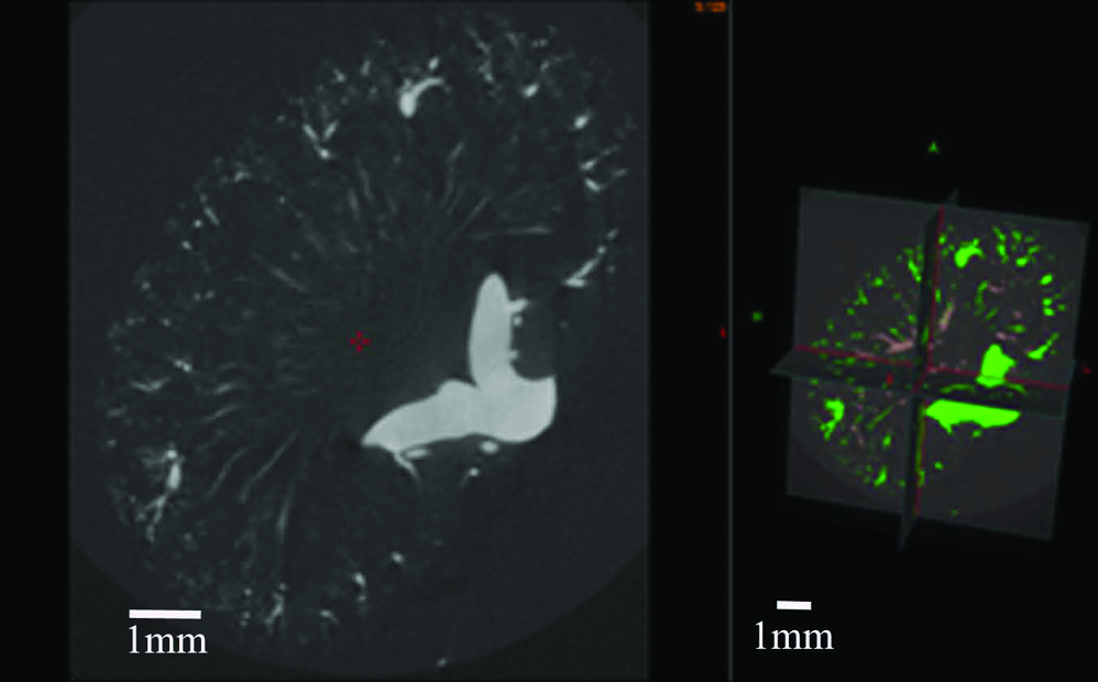
The vascular clearing procedure mentioned above was used followed by perfusion with Microfil's microvascular formula, 30 used as a contrast agent 31 for micro-CT (see Table 1). The tissue was cleared using glycerol 32 to make the Microfil visible to the eye facilitating dissecting the surrounding tissue under the stereomicroscope. Small sections were taken from the cortical region of the kidney and scanned with high-resolution micro-CT (Tables 2 and 3) to capture 3D images of glomeruli.
Micro-CT scanning of specimens
The Skyscan 1172 high-resolution micro-CT and the 1076 IN-VIVO micro-CT scanners were both used to gather the necessary image data to reconstruct vascular tree structures for scaffold design. The instruments were produced by Skyscan (Kontich, Belgium). Skycan 1076 is designed for the in vivo scanning of whole animals and has an image field of 68 mm. The 1076 was used on rabbit lung and kidney for complete scans of their larger vasculatures. For microvasculature representations, the Skyscan 1172 was used for its ability to move both the sample stage and the X-ray camera. This allows us magnification adjustments where we can optimize the spatial resolution and image quality. Smaller specimens were prepared from both the Batson's and the Microfil microvascular preparations and scanned on the 1172. The specimens used to obtain the images created in this paper are listed in Tables 2 and 3 along with the operating and equipment specifications utilized.
Stereolithography model preparation
Three-dimensional models were created in stereolithography file format using Skyscan's computed tomography analyzer (CTan). The resulting models were viewed in 3D using Skyscan's computed tomography volume (CTvol).
Assembly of computer hardware for image processing
After slice set reconstruction of the micro-CT scans, data sets were used to construct 3D models of the imaged vascular structures using a HP xw4300 workstation with Windows XP Professional. An Intel Pentium 4 640 Supporting Hyper thread technology 3.0 GHz/2 800 FSB was used with 3 GB Duo channel DDR2 640 memory.
Scanning electron microscopy preparation
Vascular cast needed very little additional processing for scanning electron microscopy (SEM). Gold/palladium sputter coating was used to provide an electron interactive surface for surface structure observation. Imaging and photography of vascular castings was performed using the Hitachi S570.
Results
Using CTan analysis and CTvol observation software, stereolithographic images were created. The resulting volumetric images were constructed from various tissue types (Tables 2 and 3) using both micro-CT–scanned corrosion casts of vascular networks (Figs. 4, 6, 7, and 9) and micro-CT–scanned contrast agent–filled vascular networks (Figs. 1–3).

(
(
A 3D projection of mouse kidney vasculature created from micro-CT image data (image pixel size, 40 μm).

(
Resolution of capillary structures
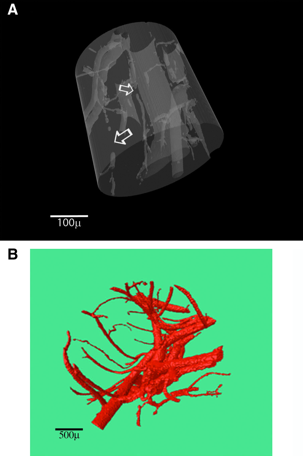
Tissue scanned with Microfil as a contrasting agent, even with small tissue samples (Fig. 1A, B), demonstrated very few, if any, capillary structures, even at high-resolution micro-CT scanning (Tables 2 and 3). Region of interest (ROI) models constructed from high-resolution micro-CT of Microfil-prepared samples demonstrate the incomplete filling of capillary system (see arrow points in Fig. 2A). Micro-CT–scanned Microfil specimen, after 3D reconstruction, can be adapted to show a continuous microvascular structure (Fig. 2B) with image sweep variation of the despeckling image processing plug-in for Skyscan's CT-Analyser version 1.8.1.3.
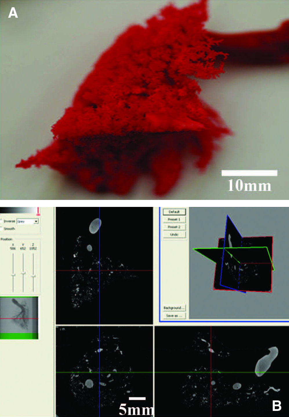
The mouse tissue was perfused with Batson's 17 solution. The blue pigment mixed in the Batson's casting resin allowed us, using stereolight microscopy, to visually analyze the successful vascular perfusion of the casted tissue (Fig. 5). This method also allowed us to be selective in our further processing of tissue for capillary bed imaging. This strategy is not apparent in past investigations.

Stereomicrograph of lung from a mouse perfused with Batson's methylmethacrylate based casting resin. Color images available online at
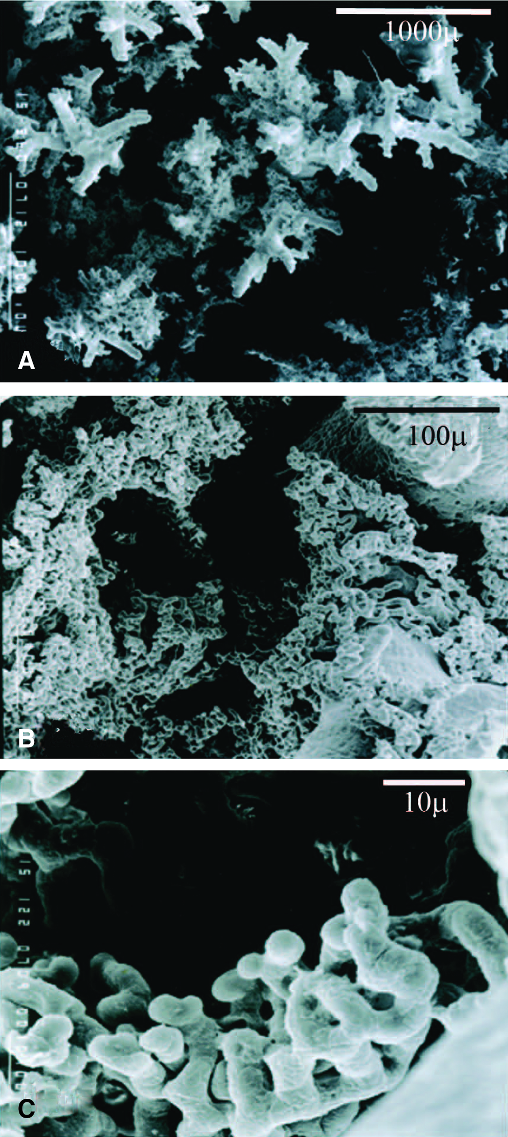
The rabbit vascular casts were good for whole-organ representations. Figure 6A is a casting of a whole rabbit lung taken from the animal casting. Figure 6B is a 3D view of a micro-CT image data set acquired from this casting using the Skyscan 1176. To capture high-resolution images of capillary beds, a small portion was taken from whole lung vascular casting (Fig. 6A) for high resolution with micro-CT (Fig. 7C). Micro-CT data captured from the lung vascular cast were used to construct the 3D model presented in Figure 7B. Figure 8A–C show, at increasing magnifications, an SEM analysis of the vascular cast shown in Figure 7A of rabbit lung. In Figure 8B, the capillary network surrounding an alveolar air sac becomes clearly visible. Figure 8C shows the complexity of these networks and their association with the neighboring alveolar air space. These networks along with their associations were not easily demonstrated with micro-CT. The prescreening of specimens with SEM allows us to narrow our searching of the casts for areas representative of the capillary bed structure desired.
(

(
(
Modeling the complete capillary bed structure
The best reconstruction results for capillary structures by micro-CT were obtained with the modified Batson's procedure described in this report. Micro-CT of the rabbit skin's vasculature showed a cast with Batson's that completely filled most of the vascular tree system of the dermal and subdermal regions. Figure 9B shows, using SEM, the fine capillary detail present in the dermal vascular corrosion cast from Figure 9A. Figure 9C demonstrates a 3D model created from the micro-CT images from a portion of this vascular corrosion cast. The 3D model contains these capillary structure as well as the structures of larger blood vessels (Fig. 9C). This 3D model clearly represents the arteriole and venous blood supply system with continuous microvasculature and capillary bed structures demonstrated at the SEM level.

(
Discussion
The 3D observation of a vascular tree's complete capillary bed is the most important step in our CAD process. Micro-CT was used to capture image data directly from BMCC of vascular tree system. Previous micro-CT investigations that illustrate complete16,33–35 3D reconstruction of capillary bed structures demonstrate that there are major obstacle against their complete observation (Table 1). Contrasting agents do not produce sufficient contrast between the agent in the capillary lumen and the surrounding tissues.21,36–39 Viscous polymers and gels consistently fill capillary beds. 31 Low-viscosity, iodine-based in vivo contrast agents are metabolized quickly, creating unstable images that fade rapidly from within the capillary lumina.40–42 Reconstructing images of the whole vascular tree system at once gives rise to further problems.43,44 Intensity thresholding inhibits reconstruction from clearly contrasting lumen-filled capillaries from their surrounding tissues, and at the same time demonstrates the larger vasculature. When making comparisons between micro-CT scans of a mouse kidney's vasculature contrasted with Microfil and a BMCC vascular cast of a rabbit kidney, we found that the ability to demonstrate the smaller vessels was restricted more in Microfil. Incomplete perfusion, likely due to the endocytosis-induced vacuole formation (Fig. 1B), reported caused by metal additive for contrasting X-ray image45,46 used in Microfil. Also, it was not possible to find an intensity threshold that eliminated back ground noise from the tissues surrounding blood vessels and demonstrate the complete microvascular and the larger diameter blood vessels. This is likely because large vessels have a greater amount of X-ray absorption than the capillaries with inconsistent amounts of X-rays absorbing contrast agent within their structure. The small variations in the contrast levels of capillaries from background tissues make capillaries indistinguishable from surrounding tissues without introducing large amounts of background noise. In eliminating background noise, much of the contrast agent's image of the capillary's lumen is lost. Imaging of Microfil samples necessarily implies the sacrifice of some of the microvascular details, even at high-resolution CT scanning levels.47,48
The use of heavy metals to increase the density of contrasting agents (see Table 1) causes a toxic response by endothelial cells. The endocytosis of heavy metal–based contrasting agent by the endothelial cell encompassing the blood vessel lumina (see Fig. 1B) 45 creates vacuoles that causes cytoplasmic swelling and can lead to significant narrowing of microvascular lumina. The subsequent narrowing slows or blocks completely contrast agent's profusion, resulting in incomplete filling of the capillary beds20,30,43,49–52 and its inaccurate micro-CT imaging.
For complete observation of capillary bed systems, both the capillary bed and larger vessels on the opposite side of the contrasting agent's entrance into the capillary bed system need to filled. To overcome the obstacles with micro-CT contrast agents hampering complete observation of capillary bed systems, we used BMCC corrosion casts (see Figs. 8 and 9). The corrosion away of tissue surrounding the vascular cast also allows us to examine samples with SEM before micro-CT scanning. With this added step, it can be determined whether the perfusion of the casting material has entered the necessary regions of the capillary bed structures and whether the necessary image data can be obtained from the sample chosen.
The use of the BMCC method without adding barium sulfate or other heavy metal prevents vacuole formation in endothelial cells of the microvascular walls allowing capillary lumina to stay open during the perfusion with BMCC. Lowering the polymerization rate during perfusion can increase the BMCC casting medium's ability to flow through the capillary beds. Precise control of polymerization time will allow for complete filling of the blood stream with the BMCC casting medium before its viscosity begins to increase to a level limiting its flow through the microvasculature. If care is not taken in these areas, polymerization can occur in the vascular system before perfusion of the contrasting agent has completely filled the blood vasculature.
Specimen's size and thickness is an issue addressed with the eroding away of tissues, characteristic of the BMCC technique. With the removal of surrounding tissues, the X-ray tube can be set at lower acceleration voltages, allowing the smallest diameter of the capillaries luminal casts, 5–7 μm, the ability to block the X-rays' path to exposure, making their structures resolvable. This precludes the need to add X-ray opaque materials, which can interfere with the perfusion, to the Batson's polymer.
The results here show that corrosion casting with BMCC method creates representations of continuous vascular tree structures that are micro-CT detectable without the use of contrasting metals, such as barium, which have been shown to block microvascular and capillary lumina.45,52 The eroding away of tissues, characteristic of the BMCC technique, gives significant reduction in background noise (see Figs. 4A, 6B, 7C, and 9D), and this technique provides clean structures from which 3D models can then be created as mesh structures in a stereolithography file format. These files promise to be an excellent resource for further Bio-CAD refinements of vascular tree–mimicking scaffolds.
Conclusion
Corrosion casting can be used to overcome the viscosity and contrasting issues that prevent capillary imaging with micro-CT. Perfusion issues have long ago been worked out using corrosion casting techniques. The eroding away of all tissues characteristic of corrosion casting rids micro-CT images of the background noise problems seen during the intensity thresholding of micro-CT images with tissue still present. The results are images that can then be created and stored as mesh structures in a stereolithography file format, compatible with software-guided fabrication. Whereas investigators are now beginning to attempt to use our imaging strategy for the reverse engineering tissue scaffolds for large defects in soft tissues, 53 without the necessary vascular supply system native to tissue structure, attempts at engineering functional tissue structure from these types of scaffold are feudal.
Footnotes
Acknowledgments
We would like to acknowledge Drs. Pieter Cornillie and Paul Simoens from the Department of Morphology at the Faculty of Veterinary Medicine, Ghent University, Ghent, Belgium, for their assistance with the modified Batson's #17 corrosion casting method used in this report. Also, many thanks to John Sled, Ph.D., at the Mouse Imaging Centre of Toronto Canada for his help with mouse kidney samples; Tim Sledz at Micro photonics USA for his assistance with micro-CT data; the technical staff of the Laboratory of Cell Biology & Histology and the Micro-CT Scan Research Group of the university of Antwerp for their help and guidance with histological and micro-CT specimen preparations; Dr. Phil Salmon at the Skyscan Company, Belgium, for his help with micro-CT scanning; and Drs. Bert Masschaele and Manuel Dierick of Ghent University Department of Subatomic and Radiation Physics for their technical advice with micro-CT image processing. This research has been supported by the NSF grant awards IGERT DGE #0221681 and S-STEM DUE #0807023, and the Alfred P. Sloan Foundation Minority Ph.D. program at USF.
Disclosure Statement
No competing financial interests exist.