
Abstract
To investigate the propriety of decellularized porcine corneas as a source of lamellar corneal xenografts, we treated porcine corneas with (1) freezing, (2) three freezing–thawing, (3) hypertonic saline, (4) hyperosmolar glycerol, (5) trypsin/sodium dodecyl sulfate/Dispase, and (6) DNase/RNase. After processing, we examined the cells and collagen structures of the decellularized corneas using hematoxylin–eosin staining, terminal deoxynucleotidyl transferase-mediated nick end labeling (TUNEL) assay, and transmission electron microscopy. Cell viability was also assessed via organ culture. In addition, the outcomes of porcine anterior lamellar corneal xenografting were evaluated in rabbits. Graft integration and corneal thickness were assessed using anterior optical coherence tomography, and the corneas were histologically examined sequentially after transplantation. We found that porcine corneas treated with hypertonic saline-based decellularization had little immunogenicity with intact collagen structures. The porcine corneal xenografts decellularized with the hypertonic saline-based method were well integrated into the adjacent host tissues and remained clear in rabbit eyes for more than 6 months.
Introduction
Therefore, it is important to seek an alternative to donor corneal allografts. Corneal replacements have been widely studied. Synthetic replacement materials include keratoprostheses and natural, corneal equivalent biomaterials, which are tissue engineered using cultured cells and extracellular matrix (ECM). However, at present, there are no clinically usable substitutes because of problems related to biocompatibility and mechanical or optical properties.2,3 Consequently, xenogeneic corneas are worthy of investigation as a human replacement. Xenografts are more readily available and are convenient for clinical application, except with regard to their xenoimmunogenicity.
The porcine cornea is a promising source for human corneal substitutes; the porcine cornea has similar refractive properties and size compared to the human cornea.4–6 Moreover, the routine use of porcine organs for transplantation is regarded as ethically acceptable. Further, genetically modified pigs, such as α1,3-galactosyltransferase knockout pigs and hDAF transgenic pigs, have been widely studied and produced for possible clinical application. 7 However, corneal full-thickness xenografts elicit severe immune rejection.8–10 In addition, our previous reports have shown that lamellar corneal xenografts without the corneal endothelium still undergo rejection, even though only the anterior portions of the diseased cornea are replaced.11,12 This indicates that corneal stromal cells (keratocytes) can cause immune rejection in xenotransplants, a phenomenon that is not common in allotransplants.13,14 Therefore, we hypothesized that porcine corneal stroma that has been deprived of cells could be used as donor tissue for partial-thickness lamellar keratoplasty (replacing just the anterior portions of the diseased cornea) in humans who have corneal stromal opacities and healthy endothelium. Moreover, in recent years, the paradigm of corneal transplantation has shifted to one in which only the diseased area is replaced. 15
Many acellular biological materials have been used clinically to repair defects in various organs, and various methods of tissue decellularization have been studied. However, the ability of these materials to decellularize the corneal stroma has not been evaluated. Further, efficient cornea processing methods have not been established to minimize the transplant immunogenicity and pathogenicity, while retaining the transparency. Accordingly, the purpose of this study was (1) to compare various current techniques and develop novel techniques to reduce the immunogenicity of porcine corneal stroma, which remains biologically and optically compatible with human application, and (2) to investigate the propriety of porcine cornea as a source for lamellar corneal xenografts in a rabbit model.
Materials and Methods
The experimental protocol was approved by the Institutional Review Board and Institutional Animal Care and Use Committee of Seoul National University Hospital.
Decellularization of porcine corneas
Porcine corneas were obtained from adult inbred miniature pigs (>12 months old) immediately after death. The epithelium was scraped off after treatment with 100% ethanol, and the corneas were thoroughly washed in phosphate buffered saline (PBS) with antibiotics for three periods of 30 min with agitation. The 250-μm-thick anterior lamella was prepared from the pig cornea using an 8.0-mm-diameter trephine (Kai industries, Seki City, Japan). The corneas were randomized into seven groups, with the following treatments applied (n = 5 in each group): (1) Fresh corneas without any treatment (control group). (2) Corneas placed in a freezer at −20°C for 1 week. One week later, the corneas were taken out of the freezer and allowed to warm to room temperature (freezing group). (3) Corneas placed in a 50 mL tube, frozen with liquid N2 for 15 min, and taken out to thaw rapidly at 37°C. This procedure of rapid freezing and thawing was repeated three times (three freeze–thaw group). (4) Corneas transferred into 1.5 mol sodium chloride solution at 37°C for 24 h, then placed in a solution of 0.05% trypsin/0.02% ethylenediaminetetraacetic acid (EDTA; Sigma-Aldrich, St. Louis, MO) at 37°C for 48 h, and washed with PBS three times (hypertonic NaCl group). (5) Corneas placed in 98% glycerol solution at 4°C for 21 days, then washed with PBS, and placed in a solution of 0.05% trypsin/0.02% EDTA (Sigma-Aldrich) at 37°C for 48 h, followed by PBS washing three times (hyperosmolar glycerol group). (6) Corneas soaked in a 0.25% trypsin solution at 4°C for 24 h, followed by treatment with a 0.1% sodium dodecyl sulfate (SDS) solution at room temperature for 6 h, followed by treatment with 560 U/L of a Dispase solution at 4°C for 12 h and treatment with a 0.1% SDS solution at room temperature for 6 h before being washed three times with PBS (trypsin/Dispase/SDS group). (7) Corneas immersed in 0.1 M NaOH solution at 25°C for 2 h, then placed in a container of 40 U/mL DNase and RNase at 37°C for 1 h on a shaker, followed by thorough washing with PBS (DNase/RNase group). The corneas in each group were stored at 4°C until use.
Ex vivo evaluation of decellularized corneal stroma
Hematoxylin–eosin staining
The porcine cornea from each group was sectioned and stained with hematoxylin–eosin (H&E). The H&E-stained slides were observed under a light microscope (Olympus Optical, Tokyo, Japan).
TUNEL staining
To determine the mechanism of decellularization in porcine corneas, we performed terminal deoxynucleotidyl transferase–mediated nick end labeling (TUNEL) staining using the ApopTag® Plus Fluorescein in situ apoptosis detection kit (Chemicon International, Billerica, MA) according to the manufacturer's protocol. Apoptotic cells were observed under a fluorescent microscope (BX-61; Olympus, Tokyo, Japan).
Transmission electron microscopy
To evaluate the ultrastructural damage to corneal cells and collagen, the cornea from each group was fixed with 2.5% glutaraldehyde PBS (pH 7.2) at 4°C overnight and postfixed in 1% osmium tetroxide–PBS for 1 h. Samples were then washed and dehydrated through serial dilutions of ethanol. Samples were mounted onto stubs, sputter-coated with gold by Polaron SC-500 (VG Microtech, Sussex, United Kingdom), and finally examined with transmission electron microscopy (JEM-1400; Jeol, Tokyo, Japan).
Organ culture
To validate the viability of corneal stromal cells in each group, the lenticles were chopped and placed in a medium for keratocyte culture including DMEM/F-12, 10% FBS, and 1% penicillin–streptomycin (Lonza, Basel, Switzerland). Keratocyte growth was followed for 21 days.
In vivo efficacy of decellularized corneal stroma in a xenograft model
Animals and anesthesia
Adult New Zealand white rabbits weighing 2–3 kg (Orient Bio, Seoungnam, Korea) were used as graft recipients. For experimental manipulation, the rabbits were anesthetized with an intramuscular injection of 10 mg/kg zolazepam (Zoletil®, Yuhan, Seoul, Korea) and 6.8 mg/kg xylazine hydrochloride (Rompun®, Bayer, Frankfurt, Germany).
Orthotopic lamellar pig-to-rabbit corneal transplantation
The 250-μm-thick anterior lamella in the graft cornea was marked with an 8.0-mm-diameter trephine and manually dissected from the rabbit cornea with a crescent knife (Alcon Surgical, Fort Worth, TX). The porcine anterior corneal lamellae from each group (n = 7 in each group) were sewn into place using eight interrupted 10-0 nylon sutures (Ethicon, Somerville, NJ). One week later, all sutures were stitched out. Complete tarsorrhaphy was performed and was maintained for 1 week. All rabbit recipients received levofloxacin eye drops (Cravit®, Santen Pharmaceutical, Osaka, Japan) topically twice a day after surgery, as well as systemic antibiotics (Gentamicin®, 40 mg/kg body weight; Abbott Laboratories, North Chicago, IL) by the intramuscular route.
Assessment of graft survival
The grafts were evaluated by slit-lamp biomicroscopy three times a week. Rejection was defined as complete loss of graft transparency (i.e., the pupil margin and iris structure were not visible through the graft). 8
Histological evaluation
Rabbits from each group were sacrificed, and their corneas excised at 1, 2, and 6 months after transplantation. Portions of the cornea were sectioned and stained with H&E or subjected to immunofluorescent study. Immunofluorescence staining was performed to evaluate for the presence of CD3+ cells in the corneas. The obtained cornea was fixed in 10% neutral buffered formalin and incubated overnight at 4°C. It was then cut into 5 μm thickness, dried at 60°C for 2 h, and deparaffinized with xylene. Protenase K (20 μg/mL; Sigma, St. Louis, MO), 5% H2O2, and 0.3% triton X-100 were used for serial treatments, after which 1% serum was added. Monoclonal mouse antibodies against rabbit CD3 (1:100; Abcam®, Cambridge, UK) were used as primary antibodies, and PBS was used as a negative control. FITC-conjugated goat anti-rabbit IgGs (1:1000; Southernbiotech, Birmingham, AL) were used as secondary antibodies. Hoechst 33342 (Sigma) was used for counterstaining. CD3-stained cells were observed under a fluorescent microscope.
In vivo cross-sectional imaging study
Full-thickness corneal cross-sectional scan images were followed by anterior optical coherence tomography (OCT; Visante™ OCT Model 1000; Carl Zeiss Meditec, Dublin, CA) every month after transplantation.
Corneal thickness measurement
Anterior OCT was used to determine the total and grafted corneal thicknesses in live animals by measuring depth differences between the epithelium and endothelium or between the epithelium and donor–recipient junction. Full corneal thickness was also determined using ultrasonic pachymetry (Pachymeter; Quantel Medical, Clermont-Ferrand, France).
Statistical analysis
Survival analysis was performed using the life-table method to estimate the median time to graft rejection in each group. Comparison of the cell counts between the groups was analyzed using the Student's t-test (SPSS 12.0, Chicago, IL). Data were expressed as MST for survival analysis and as mean ± standard error for cell counts. Differences were considered statistically significant at p < 0.05.
Results
Ex vivo evaluation of decellularized corneal stroma
Gross analysis
On gross examination, the corneas in the control, freezing, three freeze–thaw, hypertonic NaCl, and hyperosmolar glycerol groups were transparent, while the ones in the trypsin/Dispase/SDS and DNase/RNase groups became opaque (Fig. 1).
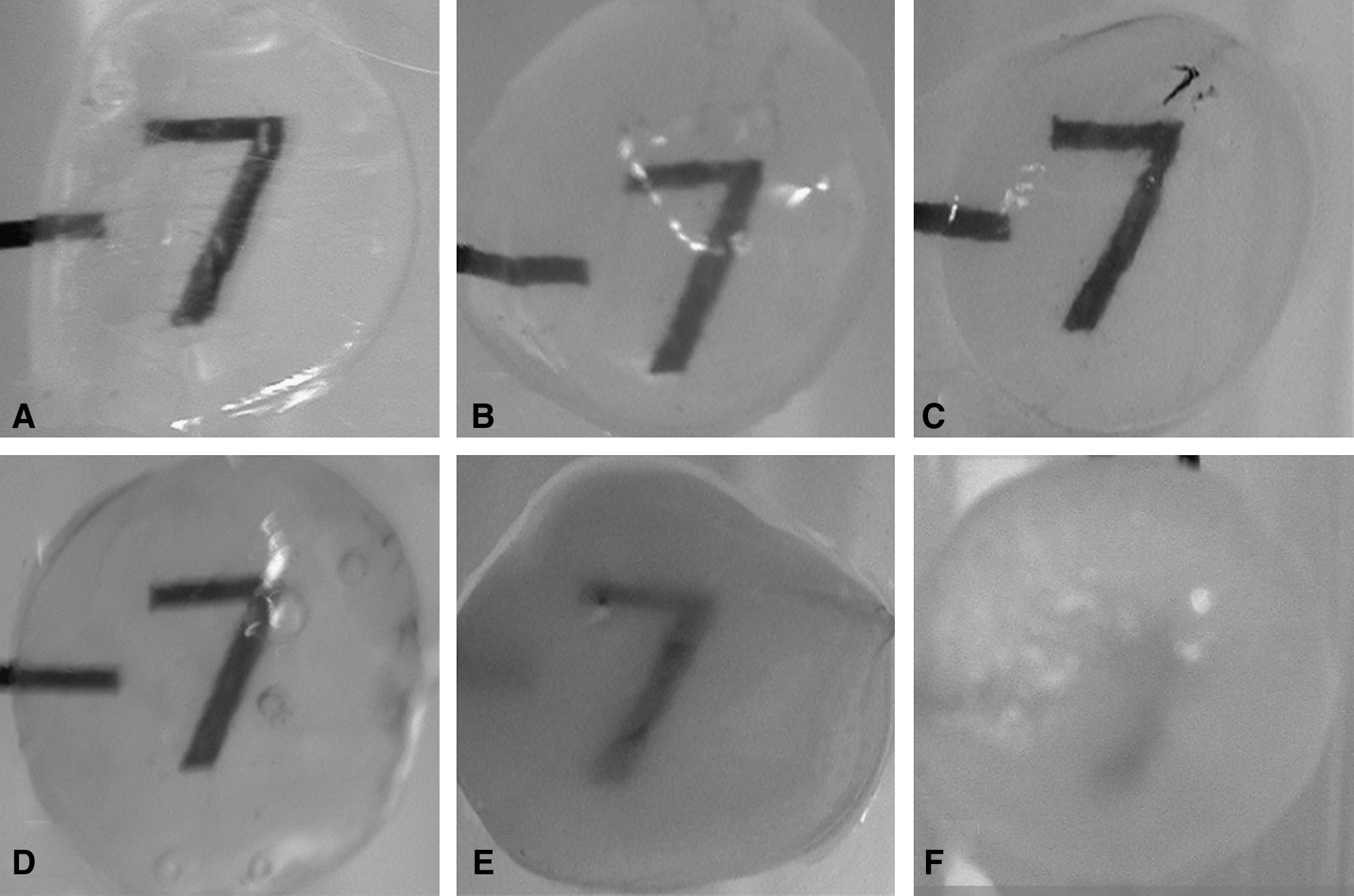
Photographs of porcine corneas without processing (
Microscopic analysis
H&E staining showed that few cells were present in the three freeze–thaw, hypertonic NaCl, hyperosmolar glycerol, and trypsin/Dispase/SDS groups, while many nuclei were present throughout the stroma of the control and freezing groups (Fig. 2). Cells were absent in the corneas of the DNase/RNase group, but the collagen structure was severely distorted, which was compatible with the opaque appearance on gross examination.
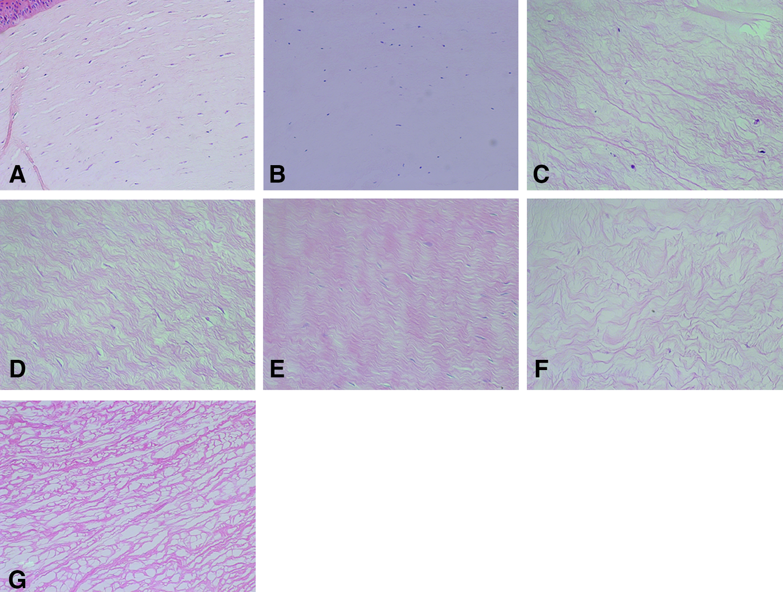
Hematoxylin–eosin staining of porcine corneas without processing (
The TUNEL assay revealed many positively staining nuclei in the porcine corneal stroma of the hyperosmolar glycerol and trypsin/Dispase/SDS groups, some positively staining nuclei in the three freeze–thaw group, and fewer nuclei in the hypertonic NaCl group (Fig. 3). The TUNEL assay showed no positively staining nuclei in the control and freezing groups, while H&E staining showed many cells. This indicated that there was no apoptosis in the cells of either group. In contrast, no nuclei were stained in the DNase/RNase group on either H&E or TUNEL staining, which suggested that neither cells nor nuclear debris were present in the corneas treated with DNase and RNase.

Terminal deoxynucleotidyl transferase-mediated nick end labeling (TUNEL) assay of porcine corneas without processing (
Transmission electron microscopy of the fresh corneas showed normal-appearing keratocytes between well-organized collagen lamellae. By contrast, marked cellular injury with apoptotic debris was noted in keratocytes from the three freeze–thaw and hyperosmolar glycerol groups (Fig. 4). Notably, there was no recognizable cell or cellular debris in the corneas of the hypertonic NaCl group, while the collagen bundles were well preserved and well aligned. Conversely, corneas from the trypsin/Dispase/SDS and DNase/RNase groups exhibited cytoplasmic disruption so severe that there were only empty cavities with no visible cellular structure. In addition, both groups exhibited severe destruction of collagen lamellae with collagen fibers lost or significantly fragmented.

Transmission electron microscopic photographs of the porcine corneas without processing (
Organ culture
The keratocytes from corneas in the three freeze–thaw group grew at a very slow rate compared to control corneas (Fig. 5). However, the corneas in the hypertonic NaCl, hyperosmolar glycerol, trypsin/Dispase/SDS, and DNase/RNase groups failed to yield growing cells for 21 days of culture.

Morphology of porcine corneal stromal cells cultured from normal cornea (
In vivo efficacy of decellularized corneal stroma in a xenograft model
Clinical course of corneal xenografts
All porcine corneas in the control, freezing, three freeze–thaw, hypertonic NaCl, and hyperosmolar glycerol groups were well integrated with the recipient rabbit corneas, and were completely reepithelialized by the host epithelium 1 week after transplantation. The porcine corneal stroma in the control and freezing groups were transparent until 2 weeks after they were grafted in the rabbits. However, opacification started to develop thereafter, and all grafted corneas became completely opaque by 4 weeks after implantation. The grafts from the three freeze–thaw group were clear for 1 month, but were rejected at 2 months after surgery. All porcine corneas in the hypertonic NaCl group remained optically clear with no sign of rejection or inflammation for 6 months after transplantation. Meanwhile, the rabbit eyes grafted with porcine corneas in the hyperosmolar glycerol group became inflamed and totally opacified with new vessel ingrowth at 3 weeks after surgery. The porcine corneal lamellae from the trypsin/Dispase/SDS and DNase/RNase groups started to melt immediately after being grafted into the rabbits, and graft clarity was not achieved. The survival times and corneal photographs of the grafts in each group are shown in Table 1 and Figure 6, respectively.
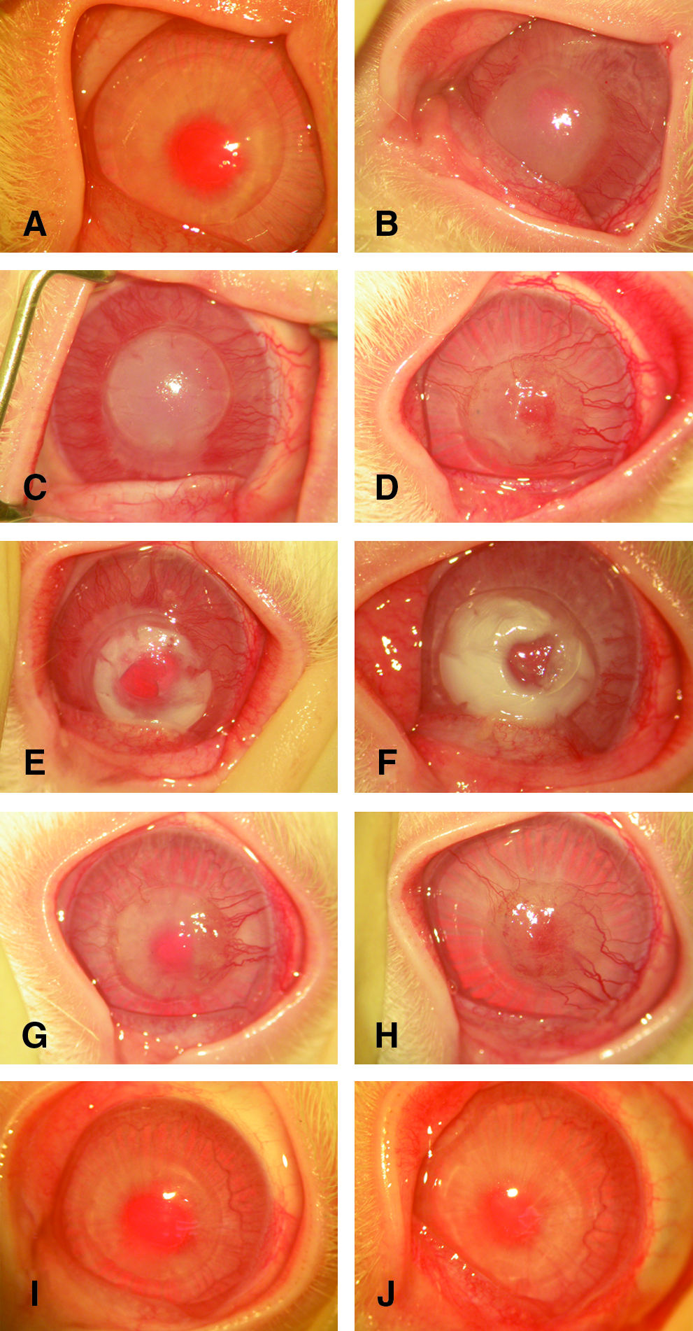
Corneal photographs of pig corneal lamellar grafts after transplantation into rabbits. Fresh porcine cornea remained clear until 2 weeks after surgery (
EDTA, ethylenediaminetetraacetic acid; SDS, sodium dodecyl sulfate.
Histological appearance of corneal xenografts
H&E staining, similar to clinical examination, showed severe infiltration of inflammatory cells after rejection. New vessel ingrowth occurred at the donor–recipient junction and extended into the graft occurred in all groups, except for the hypertonic NaCl group at 1 month after surgery (Fig. 7). Conversely, the porcine cornea in the NaCl group remained free of inflammatory cells and new vessels for more than 6 months after surgery.

Hematoxylin–eosin staining of corneas 1 month after pig-to-rabbit lamellar corneal transplantation. Massive infiltration of inflammatory cells was present at the graft–host junction in the fresh porcine cornea grafted into rabbit (
On immunohistochemistry, no CD3+ cells were found in porcine corneal grafts in the hypertonic NaCl group, while many CD3+ lymphocytes were found in the control group (Fig. 8). Moreover, on Hoechst 33342 staining, the cells were found to repopulate the decellularized porcine graft from the NaCl group 1 month after being transplanted in rabbits. This was indicative of graft repopulation by host keratocytes (Fig. 8).

CD3 and Hoechst 33342 immunohistochemistry. Many CD3+ cells were found in the rejected graft of fresh porcine cornea at 1 month postoperatively (
In vivo cross-sectional imaging study and corneal thickness measurements
Cross-sectioning of the rabbit corneas implanted with porcine grafts showed that the grafts from the hypertonic NaCl group were completely integrated into the recipient bed and successfully reconstituted the rabbit corneas (Fig. 9).

In vivo cross-sectional images of the rabbit cornea grafted with porcine cornea from the hypertonic NaCl group at postoperative months 1 (
Sequential corneal thickness measurements showed that the thickness of the grafted porcine corneal lamellae increased along with the increase in total corneal thickness (Fig. 10A). In addition, the grafted rabbit corneas grew normally compared to the contralateral nongrafted eyes (Fig. 10B).

The whole corneal thickness of the grafted eye in comparison to the graft thickness (
Discussion
The optimal corneal substitute for lamellar transplantation or tectonic corneal patching must (1) have mechanical and optical characteristics similar to human cornea, (2) be nonimmunogenic, (3) be nontoxic to surrounding ocular tissues, and (4) have functionally active ECM. The corneal ECM is important for two reasons. First, the ECM acts as a biological scaffold that provides the appropriate microenvironment for corneal repair and regeneration during wound healing. 16 Second, the ECM, composed mainly of collagen and representing >70% of the dry weight of the cornea, is essential for corneal transparency because of the regular arrangement of uniformly thin collagen fibrils into lamellae.17,18 Therefore, much effort has been expanded to develop efficient ECM for tissue-engineered corneal equivalents, although none has been clinically usable.2,3 Thus, we believed that pig corneal stroma, which has physical and refractive properties similar to those seen in human corneas, 6 would be a good ECM substitute for human corneas with defects or opacities. However, as we discovered in our previous and current studies, the porcine cornea was rejected within 4 weeks postoperatively, even if only anterior lamella was being transplanted.11,12 Given that the cells were highly antigenic, whereas the collagen was weakly antigenic,19,20 we determined to diminish the immunogenicity of the porcine cornea by cell destruction using physical (freezing, repetitive freezing–thawing), chemical (hypertonicity, hyperosmolarity), or enzymatic treatments. As a result, we found that the combination of 1.5 mol NaCl and 0.05% trypsin/0.02% EDTA achieved the best outcome in terms of reducing immunogenicity and preserving collagen structure. Moreover, the porcine cornea decellularized by the aforementioned method was well integrated into the host cornea and remained transparent without inducing inflammation or rejection for 6 months after being partially transplanted into the rabbit. Freezing at −20°C for 1 week was not sufficient to remove cells from the porcine cornea, which was rejected at the same time as the fresh pig cornea. Also, some fraction of cells remained after three episodes of freezing–thawing, and thus these cells were able to induce rejection 2 months after transplantation. Graft treatment with hyperosmolar glycerol or trypsin/SDS/Dispase induced severe cell apoptosis and produced much apoptotic cell debris, which triggered an inflammatory response in the rabbit eyes 21 and led to early rejection. Enzymatic treatments with trypsin/SDS/Dispase and DNase/RNase disrupted the collagen structure so severely that almost all collagen fibers were lost or fragmented in the porcine corneas. Treated porcine corneas lost their transparency and melted away immediately after transplantation.
A notable finding of this study was that the decellularized porcine corneal stroma in the NaCl group was recellularized with host corneal stromal cells after transplantation. The thickness of the graft itself as well as the whole corneal thickness of the grafted eye increased in accord with the growth of the contralateral nongrafted eye. This suggests that corneal stromal cells repopulating the porcine corneal stroma are functionally active, producing ECM components. Recellularization of a decellularized graft is important because keratocytes play an active role in the maintenance and metabolism of the corneal stroma.16,22,23 In addition, this decellularized porcine corneal stroma could be used as a matrix for culturing human corneal cells in vitro and making a tissue-engineered cornea.
Hypertonic saline treatment may exert its effect on reducing antigenicity as well as keratocyte killing. A recent study showed that full-thickness corneal grafts engineered from porcine collagen induced innate immunological responses with severe retrocorneal membrane formation in mice. 24 In contrast, we did not see any inflammatory or immune responses in the present study. Hypertonic saline might decrease the antigenicity of the collagen of the porcine cornea in our study, or it is possible that hypertonic saline exerts antiinflammatory/immunomodulatory properties as reported in previous studies. Hypertonic saline diminishes the expression of immune activation-associated genes in leukocytes 25 and suppresses neutrophil activation by inhibiting adhesion molecules on the ECM26,27 and attenuating neutrophil adhesion/migration.26–28 Moreover, many in vivo studies have shown that pretreatment with hypertonic saline exerts protective effects on organs such as lung, 29 liver, 30 pancreas, 31 and brain32,33 when they are in a hyperinflammatory state, by inducing the production of the antiinflammatory cytokine interleukin-10, by inhibiting tumor necrosis factor-α production, and by suppressing leukocyte migration/activation. 28 Therefore, a study is currently underway to determine if hypertonic saline may have exerted such an antiinflammatory/immunomodulatory effect on corneas in our study, in addition to destroying cells by causing osmotic shock.
Many acellular biologic materials have been produced and used to replace various tissues in humans, such as skin, heart valve, and peritoneum. However, the appropriate decellularization procedure depends on the type of organ or species of tissue donor. We decellularized rabbit corneas by the same methods used in the present study and found that all methods, including hypertonic saline treatment, induced significant damage in the collagen structure. Further, the rabbit corneal graft failed to integrate into the host cornea and melted after allotransplantation in rabbit eyes (data not shown). Given that even mild disruption of the corneal collagen structure can lead to graft opacification and lysis, we believe that customized treatment protocols are necessary to effectively remove cells without producing pro-inflammatory apoptotic debris and to minimally interrupt collagen for corneal lamellar grafts of various thicknesses in various species' donor tissue.
There are some reports showing that cryopreservation or freezing–thawing procedure decreased allo- or xenoantigenicity in the grafts of trachea, vessels, and cornea. Likewise, in the present study, some porcine corneas after three-time freezing–thawing survived for more than 3 months in rabbits. Therefore, we are also performing experiments using porcine corneas decellularized with a combination of hypertonic saline and three freezing–thawing to see the survival in rabbit eyes and have noted successful engraftment in rabbit eyes for more than 6 months (data not shown). Also, based on our ex vivo and in vivo experiments, a study is being performed using a decellularized porcine cornea for anterior lamellar transplantation in primates.
Footnotes
Acknowledgment
This study was supported by a grant from the Korea Health 21 RND project, the Ministry of Health and Welfare, Republic of Korea (Project No. A04-0004-AZ1205-06A3-00100B).
Disclosure Statement
No competing financial interests exist.