
Abstract
Whole-organ culture of a sensory organ in a rotating wall vessel bioreactor provides a powerful in vitro model for physiological and pathophysiological investigation as previously demonstrated for the postnatal inner ear. The model is of specific relevance as a tool for regeneration research. In the immature inner ear explant, the density was only 1.29 g/cm3. The high density of 1.68 g/cm3 of the functionally mature organ resulted in enhanced settling velocity and deviation from its ideal circular orbital path causing enhanced shear stress. The morphometric and physical properties, as well as the dynamic motion patterns of explants, were analyzed and numerically evaluated by an orbital path index. Application of a novel buoyancy bead technique resulted in a 6.5- to 14.8-fold reduction of the settling velocity. The deviation of the explant from its ideal circular orbital path was adjusted as indicated by an optimum value for the orbital path index (−1.0). Shear stress exerted on the inner ear explant was consequently reduced 6.4- to 15.0-fold. The culture conditions for postnatal stages were optimized, and the preconditions for transferring this in vitro model toward mature high-density stages established. This buoyancy technique may also be useful in tissue engineering of other high-density structures.
Introduction
Sensory organs and, in particular, the inner ear remain technically demanding for culturing cells, tissues, and whole organs. As recently reported by our research group, introduction of simulated microgravity culture conditions has advanced the evolution of culture techniques for the inner ear. Successful culture of the 1-week-old postnatal mouse inner ear as an entire organ for up to 7 days was demonstrated. 6 The physical properties of the RWV bioreactor environment support preservation of the integrity of the three-dimensional structure of inner ear tissue, including the cochlear duct.
Investigations in whole-organ culture such as the evaluation of ototoxicity are comparable with conventional inner ear tissue culture7,8 or in vivo investigations. 9 Nevertheless, the 1-week-old postnatal inner ear of the mouse is immature, and several obstacles must be overcome before whole-organ culture of the mature inner ear can be realized.
At postnatal day 7 (p7) the inner ear has undergone 2 weeks of differentiation after terminal mitosis, 10 which occurs at embryonic day 14. At p7 the inner ear has grown to its near final size and shows differentiation of all cell types comparable to mature developmental stages. 11 Functional maturation reaching adult hearing thresholds requires another 2 weeks of development until p21. 12 Whole-organ culture of the functionally mature 3-week-old inner ear is therefore desirable. Due to the ossification process of the inner ear capsule starting at the end of the first postnatal week, there is a significant age-dependent increase in mass and physical density of whole inner ear organ explants. This dramatically alters the hydrodynamic conditions in the RWV. Deviations of the inner ear explants from the ideal circular orbital path are increased and lead to an enhanced shear stress, as well as possible disruption of the organ caused by collisions with the vessel wall.
In this study, age-dependent morphometric dimensions and physical properties of whole inner ear organ explants were measured for the postnatal stages p7, p14, and p21. The dynamics of age- and mass-dependent motion were analyzed and further numerically characterized by introduction of an orbital path index (OPI). By applying a novel buoyancy bead technique, simulated microgravity culture conditions were optimized. The provided analysis aids understanding of the mechanical aspects of the motion of inner ear explants in the RWV. This will facilitate development of the optimal operating control conditions employing this bioreactor system. The technique describes the basic physical preconditions that must be established to transfer the previously described method of cultivating 1-week-old whole inner ear organs 6 toward functionally mature stages of higher density. This buoyancy bead technique may also be useful in engineering large and high-density organoids as the size of growing specimens plays a critical role regarding surface flows and related shear stress. 13 Accordingly, porous microcapsule scaffolds have been developed for bone tissue engineering, 14 and the use of microcarriers lighter than water for osteoblast cultures has been demonstrated to be advantageous in avoiding wall collisions. 15
Materials and Methods
Animals
Mice with a C57BL/6 genetic background (Charles River, Sulzfeld, Germany) were used and obtained from breeding colonies of an in-house animal facility. Animal use for organ explantation was notified to and approved by the Committee for Animal Experiments of the Regional Council (Regierungspräsidium) of Tübingen, Germany.
Gross and micro-dissection of the inner ear organ
Mice pups at p7, p14, and p21 were killed by ether inhalation and decapitated. The complete inner ear bony labyrinth capsules were dissected from the skull base. Micro-dissection was carried out as previously described. 6 In this study, an additional preparation step was carried out to mount single buoyancy polystyrene beads on the bony labyrinths. A thin bony lamella, covering about one-third of the subarcuate fossa aditus, was microdissected and removed. This preparation step allowed for insertion of polystyrene beads of ≤2.4 mm diameter into the subarcuate fossa by partially squeezing the bead (Fig. 1A–E).
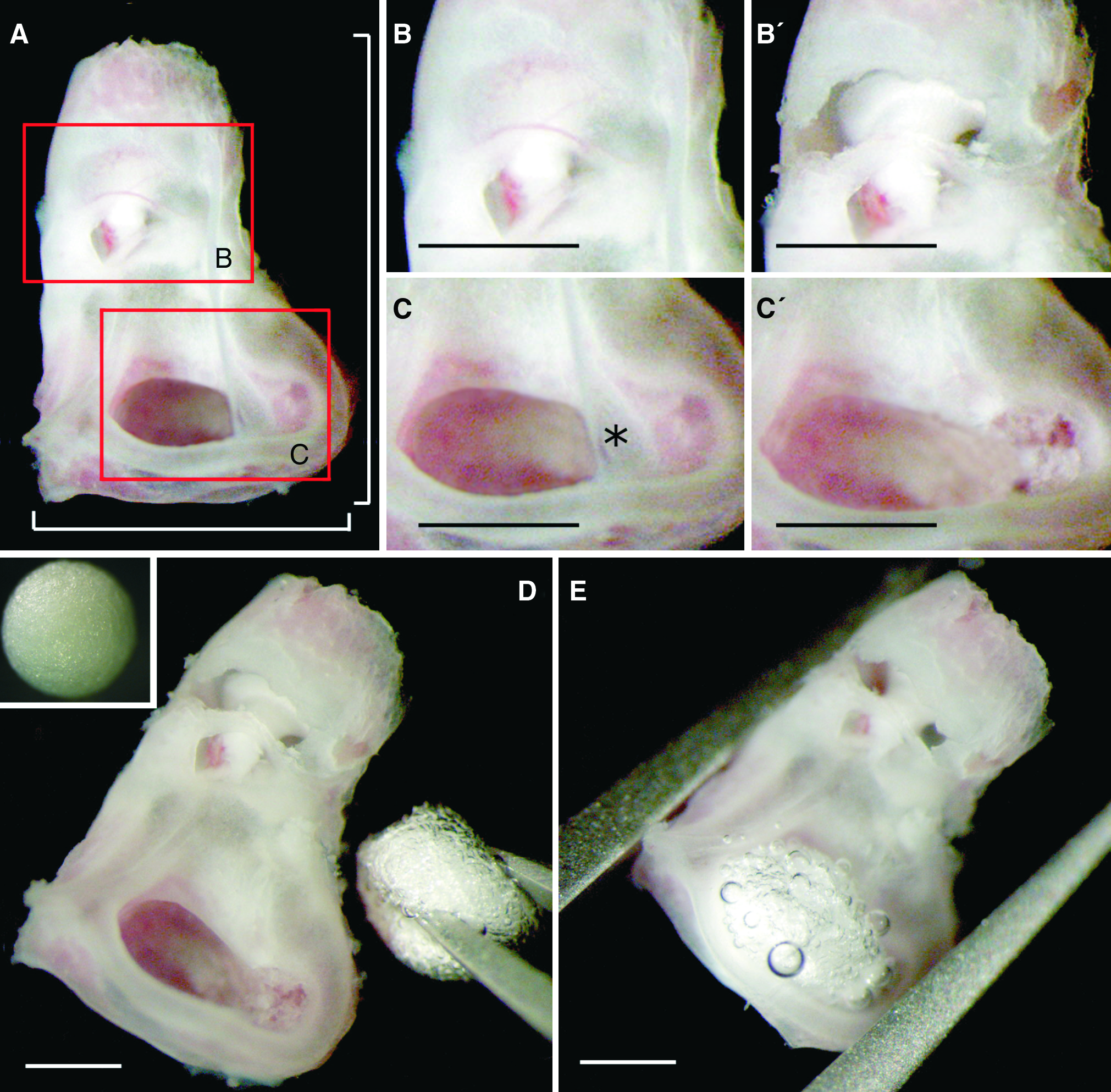
Modified dissection technique for postnatal day 14 (p14) and p21 inner ear explants and insertion of buoyancy beads into the subarcuate fossa. Gross and micro-dissection of the bony labyrinths was carried out as previously described for p7 explants.
6
(
Sampling and insertion of polystyrene beads into the subarcuate fossa
Polystyrene bead (BASF, Ludwigshafen, Germany) diameter was measured using a surgical microscope (OPMI 1-FC; Carl Zeiss, Jena, Germany), a digital camera (GP-KS162 HDE; Panasonic, Osaka, Japan), and the AxioVision 4.6 software (Carl Zeiss). Beads were manually sorted by size. A deviation of 1.5% of the diameter was tolerated. For sterilization beads were incubated in 70% ethanol for 12 h with no alterations in size. They were then spread in a dish for drying under sterile conditions. Standardized insertion into the subarcuate fossa was done using a fine Dumont forceps.
Arrangement of organ culture system
Motion dynamics were investigated in a four-station Rotary Cell Culture System (RCCS™; Synthecon, Houston, TX) at room temperature (20°C). The applied bioreactor vessel type was a horizontally rotated high-aspect-ratio vessel, with a volume of 55 mL and a radius of culture space of 5.0 cm. The transparent front wall allowed for observation of inner ear organ explants during culture. To measure inner ear organ trajectories, concentric circles ranging from radius 0.5 to 4.5 cm were labeled onto the transparent front wall of the vessel at 5-mm intervals. The culture medium was Neurobasal™ medium (Invitrogen, Carlsbad, CA) supplemented with 1× B27 supplement 16 (Invitrogen), 5 mM glutamine, 10 mM HEPES (Invitrogen), and 100 U penicillin (Sigma, St. Louis, MO) as previously described. 6
Measurements of the mass of inner ear explants
Mass (miee) measurements of inner ear explants were carried out using a high-precision balance (BP 211D; Sartorius, Göttingen, Germany).
Morphometric measurements of inner ear explants
Morphometric measurements were made using a surgical microscope (Carl Zeiss), a digital camera (Panasonic), and the Axiovision 4.6 processing software (Carl Zeiss). Microscopic measurements were calibrated using an object slide labeled with a 2-mm precision scale (Dr. Johannes Heidenhain GmbH, Traunreut Oberbayern, Germany).
Terminal settling velocity of native and buoyancy bead-inserted inner ear explants
After an initial acceleration phase (15 cm) in a medium-filled glass cylinder (Hirschmann GmbH & Co. KG, Eberstadt, Germany; dimensions: volume 1000 mL, height 47 cm, and diameter 6.5 cm), the time required for passing a 30-cm settling distance was measured three times and a mean value recorded.
Calculated volume of the inner ear explants
Calculated volume (Viee) of the inner ear explants was reconstructed using bright field microscopy data of inner ear sections from a C57BL/6 mature, juvenile mouse (Mountain, DC, personal communication) obtained from the website “EarLab @ Boston University” (
Calculated age-dependent physical density of inner ear explants
The age-dependent mean physical density (ρiee) of the inner ear explants was calculated using the following equation:
For calculations, the age-dependent mean value of measured inner ear masses, miee, was used. Viee was obtained as described in the section Calculated volume of the inner ear explants.
Calculation of maximum shear stress on the inner ear explant
The maximum shear stress (τmax) exerted on a particle is a function of the terminal settling velocity Vs.
3
In this study, maximum shear stress exerted on the explant was estimated from3,14,17,18
where
Vs = age-dependent terminal settling velocity (cm/s) (mean values from the section Terminal settling velocity of native and buoyancy bead-inserted inner ear explants).
r = equivalent radius of the inner ear explant (cm).
μ = dynamic viscosity of the culture medium (≈0.01 dyn·s/cm2 s at 20°C).
An error estimation for the calculation of τmax was done by inserting the SD of the experimentally obtained value Vs into Equation 2.
The equivalent radius (r) for the inner ear explant was estimated by equalizing the inner ear volume Viee (from the section Calculated volume of the inner ear explants) with that of an ideal sphere; r was then calculated by the spherical equation
Calculation of ideal volume and radius of polystyrene buoyancy beads
Before carrying out settling and rotation culture experiments, the age-dependent ideal volume and radius of buoyancy beads were calculated to obtain a combined explant–bead density of 1.0 g/cm3. If the overall density of the inner ear explant–bead complex is equal to 1.0 g/cm3 (=1.0 mg/mm3), the following equation is true:
where
ρiee+ib = overall physical density of the inner ear explant–bead complex
mib = mass of ideal bead (mg)
miee = mass of inner ear explant (mg) (mean values from the section Measurements of the mass of inner ear explants)
Vib = volume of ideal bead (mm3)
Viee = volume of inner ear explant (mm3) (from the section Calculated volume of the inner ear explants)
The polystyrene beads (BASF) had a physical density, ρib, of 0.015 g/cm3. Inserting the known physical density for the beads and replacing mib by the term ρib · Vib, Equation 4 was solved for the ideal bead volume:
The error σVib for Vib was defined as the SD of Vib and estimated as
The radius of the ideal bead rib was then calculated using the spherical equation (according to Equation 3). The error for rib was estimated as
Analysis of the motion dynamics of inner ear explants in the RWV
Age- and density-dependent motion patterns of inner ear explants were characterized by measuring two representative parameters: (1) the distance between the center of the explant's orbital path and the spin axis of the RWV (dis) and (2) the maximum of the almost periodically changing diameter of the orbital path (dia) (Fig. 2). For explant–bead complexes, bead sizes with resulting optimized terminal settling velocities for each individual postnatal stage were used.
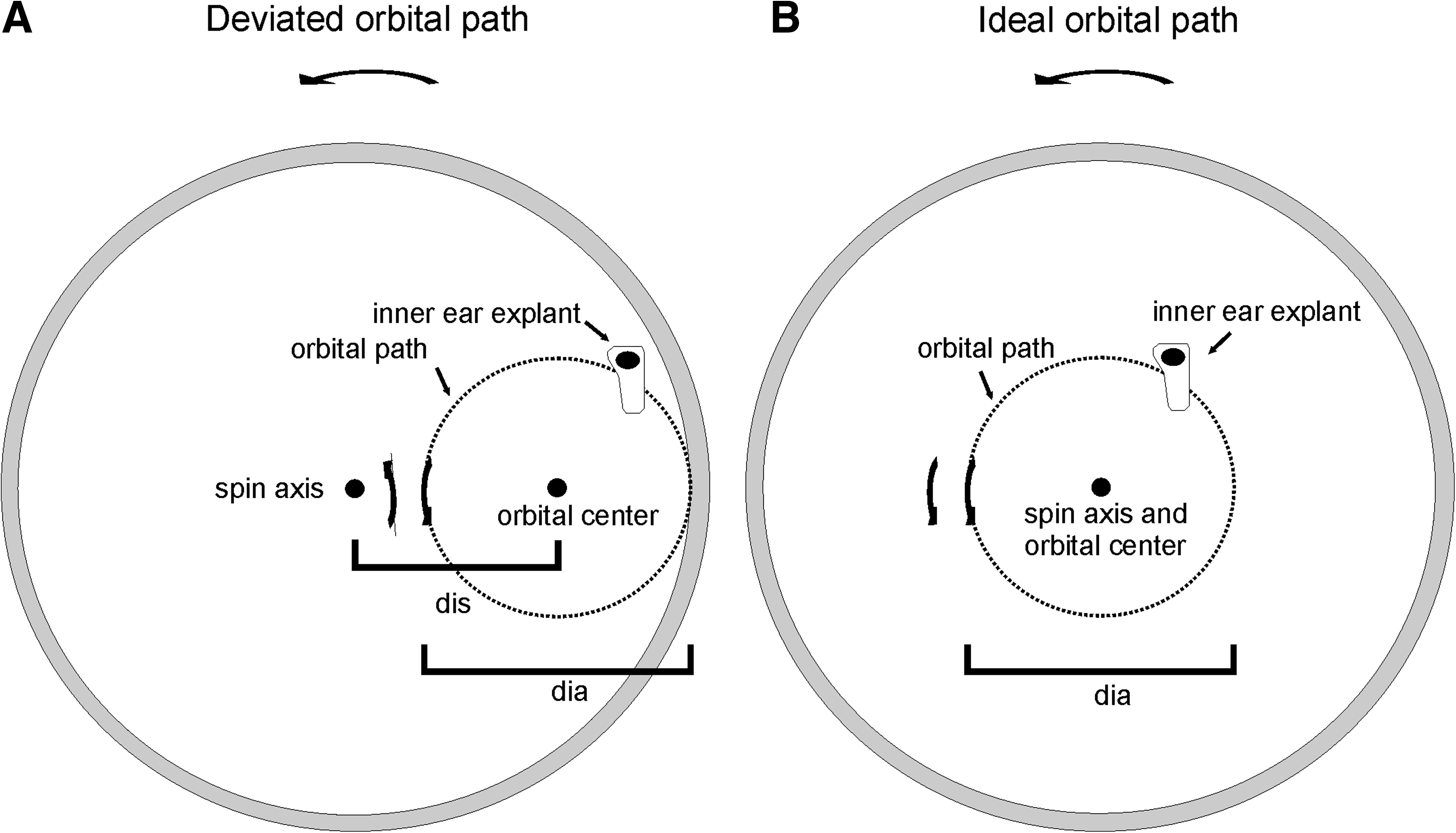
Schematic illustration of two exemplary inner ear explant motion patterns in the rotating wall vessel (simplified). (
Measurements were done by a stepwise increase of the revolution speed from 5 to 50 rpm at intervals of 5 rpm. Single measurements were repeated three times.
Orbital path index
Optimum low shear stress motion was attained if the inner ear explant traveled on a circular orbital path around a center exactly matching the spin axis of the vessel.2,19 As a numerical indicator for the deviation of the explant's motion from this ideal path, an OPI was defined as
where
dis = distance of orbital center from the spin axis of the vessel
dia = maximum diameter of orbital path
The index is negative if the spin axis of the vessel is inside the orbital path. For explants with positive index values, downward motion of the particle occurs before passing over the spin axis. It is opposed to the upwardly vectored motion of the circular fluid flow, resulting in enhanced shear (Fig. 2). By enhancing the revolution speed, the index switches from positive to negative values and asymptotically approaches the optimum value OPI = −1.0 with further speed. For OPI = −1.0 (optimum value), the center of the orbital path exactly matches the spin axis of the rotating vessel, indicating the inner ear explant traveling on a circular orbital path around the spin axis (ideal path) (Figs. 2 and 6).
Quantification and statistical analysis
Quantification of empirically obtained measurements are mean ± SD for the inner ear organ. Differences between experimental groups were assessed by the paired Student's two-tailed t-test (*p < 0.05 and **p < 0.01 values were considered significant).
Results
Age-dependent mass of inner ear explants
The mass of inner ear explants increased from stage p7 up to p21, as a result of proceeding ossification (Fig. 3A). The mass for postnatal stage p7 was 7.57 ± 0.23 mg (mean ± SD) (n = 6). For postnatal stage p14, the mass increased to 8.57 ± 0.32 mg (n = 9). Mass of p21 inner ear explants increased further to 9.85 ± 0.24 mg (n = 6). The differences between the three postnatal stages were highly significant (p < 0.01).
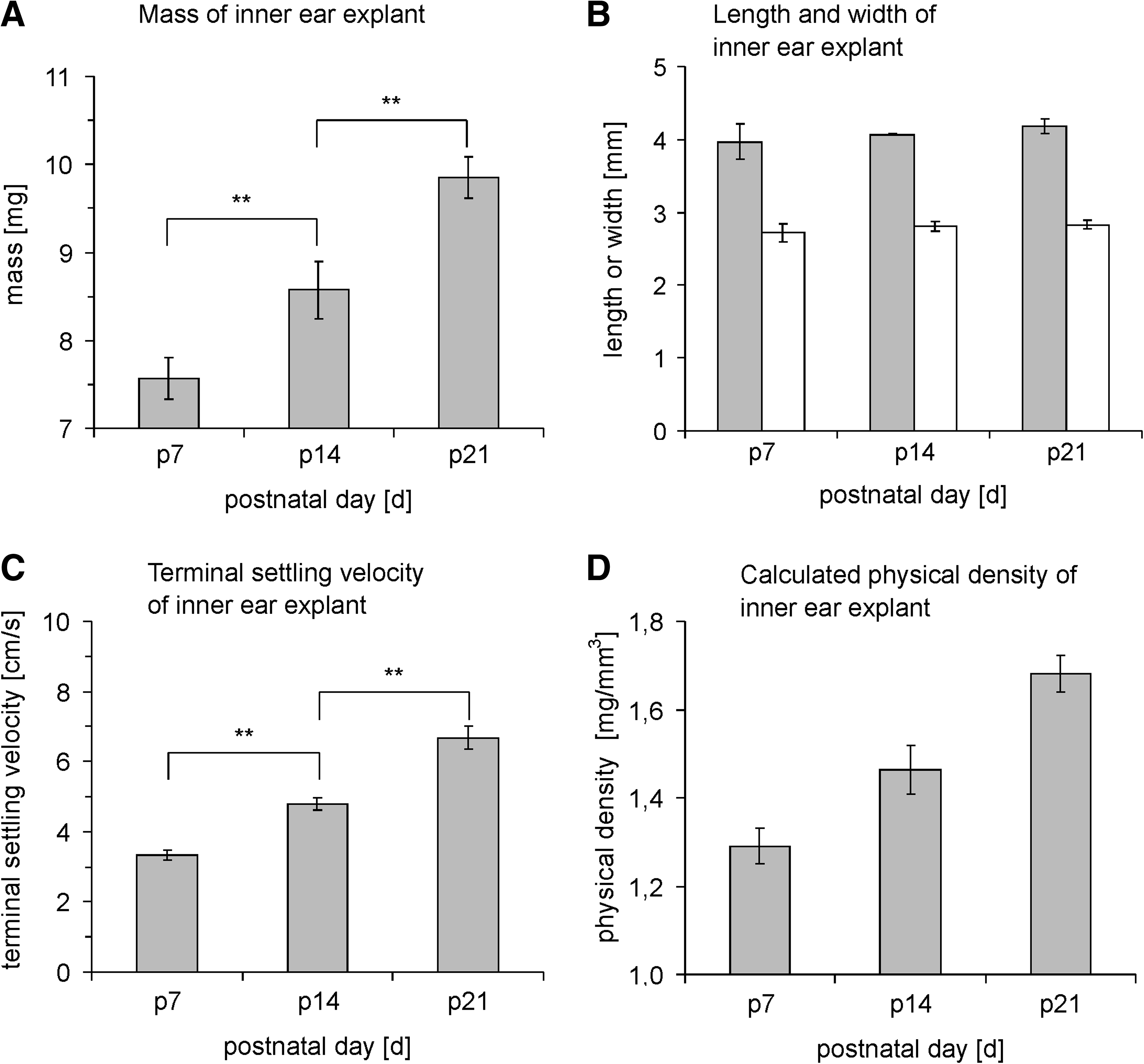
Morphometric dimensions and physical properties of inner ear explants for the postnatal stages p7, p14, and p21 including empirically measured parameters mass (
Age-dependent length and width of inner ear explants
The age-dependent length and width of inner ear explants were measured for the three postnatal stages also (Figs. 1A and 3B). The length was 3.97 ± 0.24 mm (n = 9) for stage p7, 4.07 ± 0.01 mm (n = 6) for stage p14, and 4.18 ± 0.10 mm (n = 6) for stage p21. The width was 2.72 ± 0.13 mm for stage p7, 2.81 ± 0.07 mm for stage p14, and 2.83 ± 0.06 mm for stage p21. There were no significant differences for measured length and width for p7, p14, and p21 postnatal stages, confirming that the final size was already reached at p7.
Age-dependent terminal settling velocity (Vs) of inner ear explants
The physical density of the bony labyrinth increased because of the increase in mass at constant size. This resulted in a rise of the terminal settling velocity for older postnatal stages (Fig. 3C). The terminal settling velocity was 3.34 ±0.13 cm/s for stage p7 (n = 6), 4.80 ± 0.18 cm/s for stage p14 (n = 6), and 6.68 ± 0.33 cm/s for stage p21 (n = 6) inner ear explants. Differences between the postnatal stages were highly significant (p < 0.01).
Viee of inner ear explants
Viee of the inner ear explants was 5.86 ± 0.01 mm3 (mean ± SD) and reconstructed as described in the Materials and Methods section.
Calculated age-dependent physical density of the inner ear explants
Due to increase in mass but constant size values for length and width (i.e., constant volume) of the inner ear explant, the physical density increased continuously for maturing postnatal stages. The calculated density was 1.29 ± 0.04 g/cm3 for postnatal stage p7, 1.46 ± 0.06 g/cm3 for p14, and 1.68 ± 0.04 g/cm3 for p21 (Fig. 3D).
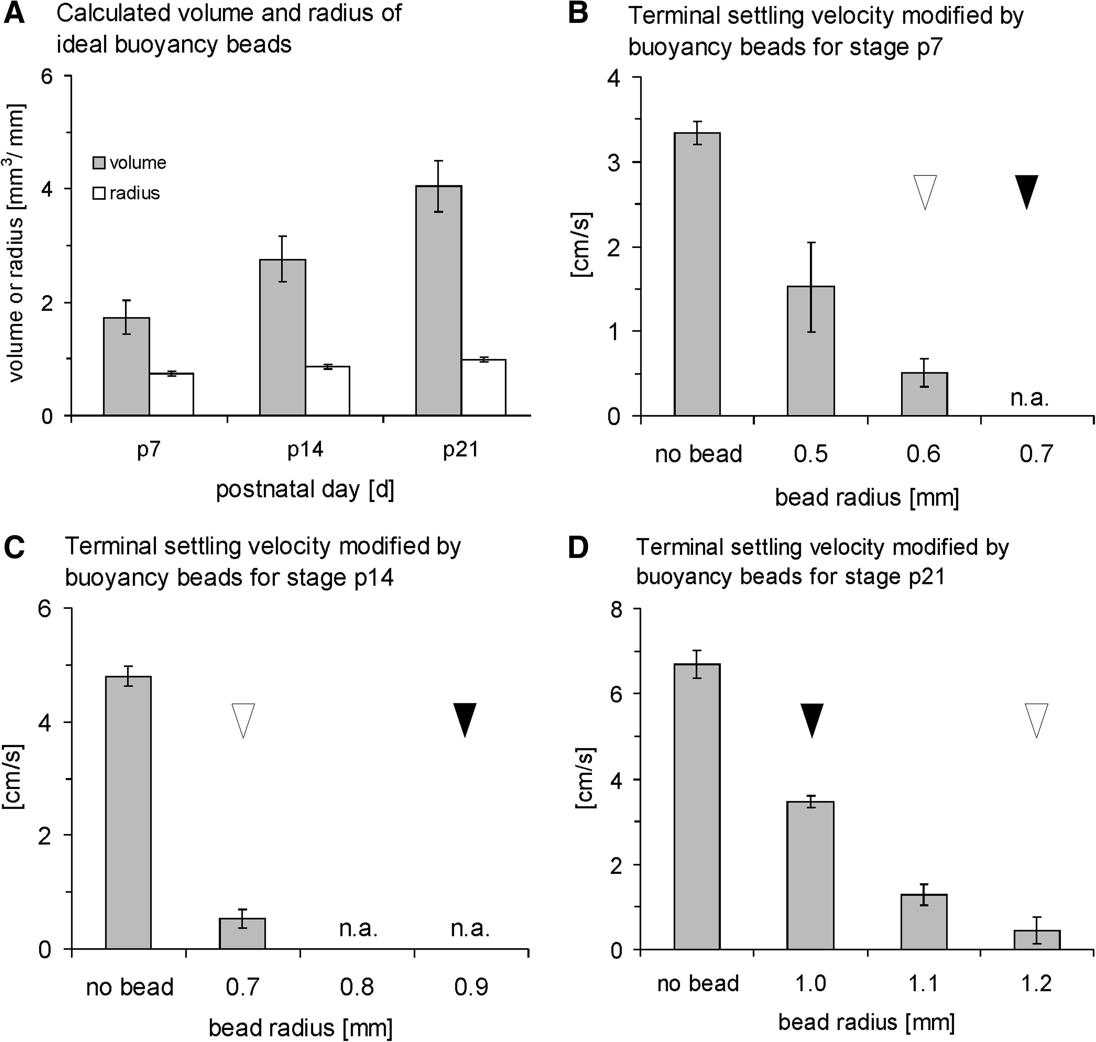
Calculated ideal volume (Vib) and radius (rib) of polystyrene buoyancy beads
The ideal volume of polystyrene buoyancy beads (Vib) was calculated to adjust the overall physical density of the inner ear explant–bead complex to the physical density of the culture medium (1.0 mg/mm3). The calculated ideal bead volume, Vib, was 1.73 ± 0.30 mm3 (mean ± SD) for postnatal stage p7, 2.76 ± 0.40 mm3 for p14, and 4.05 ± 0.45 mm3 for p21. The calculated corresponding radius rib was 0.74 ±0.04 mm for postnatal stage p7, 0.87 ± 0.04 mm for p14, and 0.99 ± 0.04 mm for p21 (Fig. 4A). Differences between all groups were highly significant (p < 0.01).
(
Age-dependent terminal settling velocity modified by insertion of buoyancy beads
The calculated ideal sizes of buoyancy beads were tested experimentally. The stage-dependent terminal settling velocity of inner ear explant–bead complexes was measured for buoyancy beads of three radii (1) equal to (2) 0.1 mm smaller or larger and (3) 0.2 mm smaller or larger than the calculated ideal bead radius depending on its buoyancy effect (Fig. 4B–D). For this experiment the calculated ideal bead radius was rounded up to the first decimal place.
Minimum terminal settling velocity for stage p7 inner ear explants was achieved using beads of radius r = 0.6 mm (Vs = 0.51 ± 0.17 cm/s (mean ± SD), n = 5). Insertion of beads of the calculated ideal size r = 0.7 mm led to floating of the inner ear explants. Applying beads of radius r = 0.5 mm resulted in a mean terminal settling velocity of Vs = 1.52 ±0.53 cm/s (Fig. 4B). For p14 inner ear explants, insertion of the calculated ideal bead sizes (r = 0.9 mm) and beads of radius r = 0.8 mm led to floating of the explant–bead complex. Minimum settling velocity of Vs = 0.54 ± 0.16 cm/s was obtained by applying 0.7-mm beads (Fig. 4C). The mean terminal settling velocity for stage p21 was 3.46 ± 0.15 cm/s for the calculated bead radius 1.0 mm and 1.28 ± 0.26 cm/s for bead radius 1.1 mm. The minimum terminal settling velocity for this stage was 0.45 ± 0.31 cm/s, obtained by applying 1.2-mm beads (Fig. 4D). Calculated and experimentally obtained bead sizes were close by 0.1 mm (p7) or 0.2 mm (p14 and p21). All differences in terminal settling velocity between bead size groups were highly significant (p < 0.01) (Fig. 4B–D).
Calculated maximum shear stress exerted on inner ear explants
Maximum shear stress (τmax) on inner ear explants was estimated for native explants versus explant–bead complexes with bead radii optimized for minimum settling velocity as described in the section Age-dependent terminal settling velocity modified by insertion of buoyancy beads. Shear stress in the RWV increased proportionally to the terminal settling velocity of native explants, and resulted in 0.45 ±0.02 (mean ± SD), 0.64 ± 0.02, and 0.90 ± 0.04 dyn/cm2 for the postnatal stages p7, p14, and p21, respectively. Shear was dramatically reduced by buoyancy beads, resulting in 0.07 ±0.02 (p7), 0.07 ± 0.02 (p14), and 0.06 ± 0.04 (p21) dyn/cm2. Differences of shear stress between native inner ear explants of different ages as well as differences between inner ear explants and inner ear explant–bead complexes for each age group were highly significant (p < 0.01). There were no significant differences in shear stress between age groups of inner ear explant–bead complexes (Fig. 5).
Calculated shear stress on the surface of inner ear explants. Age-dependent shear stress is reduced 6.4- to 15.0-fold for explant–bead complexes compared with native inner ear explants. Differences of shear stress between native inner ear explants of different ages as well as differences between inner ear explants and inner ear explant–bead complexes for each age group were highly significant (p < 0.01). There were no significant differences in shear stress between age groups of inner ear explant–bead complexes. SDs are represented by error bars.
Analysis and optimization of motion of inner ear explants in the RWV
Motion of the inner ear explants was observed as orbiting around a center within the rotating vessel with an almost periodically changing diameter. Centers of the orbital paths were in the right half of the vessel in case of counter-clockwise rotation. Depending on the physical density and revolution speed, explants exceeded the downward motion of the circular fluid flow during the downward phase of the vessel rotation, and periodically collided with the vessel wall in the lower-right quadrant (Fig. 2). The angle of impingement with the vessel wall for particles of high physical density was large, and resulted in enhanced mechanical stress with possible disruption. By insertion of buoyancy beads, explant–bead complexes rotated on approximately circular orbits along with the laminar flow of the culture medium and contacted the vessel wall only tangentially. By increasing the revolution speed of the culture vessel, the center of the orbital path of the explant approached, but did not reach, the spin axis of the RWV. There remained a distance of about 0.25 cm for p7, 0.50 cm for p14, and 1.0 cm for p21 native explants even at the maximum revolution speed of 50 rpm (Fig. 6A–C). For explant–bead complexes, the distance between the centers of the orbital paths and the spin axis was minimized even for slow revolution speeds (e.g., ≤0.25 cm at revolution speed 25 rpm for all stages). For higher revolution speeds, the center of the orbital path matched the spin axis, and the explant–bead complexes rotated on a nearly circular orbital path around the spin axis (Fig. 6A–C). The maximum diameters of the orbital paths increased at higher rotational speeds (Fig. 6D–F). Explants without beads did not follow an orbital path for slow vessel rotational speeds, but adhered to the vessel wall (dis = 5.0 cm). Explants with inserted polystyrene beads orbited even at the slowest vessel rotation speed of 5.0 rpm (Fig. 6A–C).
Distances between orbital centers and the spin axis (dis) (
Orbital path index
As a numerical indicator for deviation of the motion of the explants from the ideal path, an OPI was defined (see the section Orbital path index in Materials and Methods). By increasing the revolution speed, the index switched from positive to negative values before finally approaching the optimum value OPI = −1.0 (Fig. 6G–I). For native explants, OPI became negative only for higher revolution speeds, that is, 15 rpm for p7, 20 rpm for p14, and 30 rpm for p21. OPI values close to the optimal value −1.0 (defined as the interval of OPI more than −1.3 and less than −1.0) were reached only for very high revolution speeds, that is, 40 rpm for p7 and 45 rpm for p14. For native p21 explants, OPI values between −1.3 and −1.0 were not reached. The optimum OPI value −1.0 was not reached for all three stages, even at the maximum revolution speed of 50 rpm. For explant–bead complexes, OPI was already negative at a rotational speed of 5 rpm for all three stages. For explant–bead complexes, OPI values close to the optimum value (interval of OPI more than −1.3 and less than −1.0) were reached at 20 rpm for p7, 20 rpm for p14, and 10 rpm for p21. The optimum value OPI = −1.0 was reached at 35 rpm for p7, 30 rpm for p14, and 35 rpm for p21 (Fig. 6G–I).
Discussion
Simulated microgravity culture conditions in RWV bioreactors provide a quiescent, low shear stress, low turbulence environment for the whole mouse inner ear organ culture.1,6 However, high-order morphologic structures are still exposed to shear stress in proportion to the speed of relative fluid flow across their surfaces. 17 Several biologic phenomena, such as the assembly of complex functional mammalian tissues, are assumed to be responsive to this surface flow phenomenon.1,2,13,20,21 The physical culture conditions of this organ culture technique were analyzed and modified by a novel buoyancy bead technique aiming at an age-dependent optimization of low shear, low turbulence conditions for the postnatal stages p7, p14, and p21.
Age-dependent morphometric and physical properties of inner ear explants
The mass of the entire inner ear explants increased during postnatal development from stage p7 to p21 because of the ossification process of the otic capsule. The morphometric dimensions of length and width of inner ear explants did not change significantly for these stages. A constant volume of the inner ear explant was therefore assumed for all three stages. As expected, the physical density (ρiee) and hence the terminal settling velocity (Vs) of the inner ear explants increased at older stages. The terminal settling velocities matched the range of those for similarly sized cartilage tissue particles. 22
Age-dependent terminal settling velocity and shear stress exerted on inner ear explants
Reduction of the terminal settling velocity of the inner ear explants by buoyancy beads appears suitable to optimize for low mechanical stress. Gravity, centrifugal, and Coriolis forces are reduced, leading to a decrease of wall impacts and shear stress.2,3,17 The measured shear stress levels in this study were of the order of magnitude reported for other tissue particles cultured in RWVs.14,18,19,22,23
Three bead radii close to and equal to the calculated ideal bead radius for each of the postnatal stages were employed. The small aberrations of the experimentally obtained optimum bead radii from the calculated ideal bead radii probably resulted from squeezing during insertion into the subarcuate fossa. Other factors may include the approximations that had to be made for volume, density, and ideal bead size calculations of the inner ear explants. An age-dependent 6.4- to 15.0-fold reduction of maximum shear stress to values of 0.07 (p7), 0.07 (p14), and 0.06 (p21) dyn/cm2 was achieved by the buoyancy bead technique. Reduction of shear stress has been demonstrated to be beneficial for cell growth and three-dimensional tissue assembly. 1
Analysis and optimization of motion of inner ear explants in the RWV
The trajectories of inner ear explants were observed as orbital paths around a center with an almost periodically changing diameter, as viewed from the nonrotating external reference frame. The motion of particles with a density higher than the culture medium has been consistently described as the orbiting regime. 22 Particles of a physical density higher than that of the culture medium deviate from the ideal circular orbital path around the spin axis when observed from the nonrotating external reference frame, resulting in enhanced surface flows. 2 Spiralling caused by Coriolis forces leads to periodic collisions with the vessel wall, causing mechanical stress and possible disruption of the organs.13–15,17,24,25 The rate of outward spiralling increases with increasing particle radius, vessel rotation rate, and increasing terminal settling velocity.2,17 For inner ear explants without beads, no orbiting occurred for slow vessel rotational speeds, and the explants adhered to the vessel wall or oscillated at a stationary point in the vessel according to the settling regime as previously defined. 22
Depending on physical density and revolution speed, the native explants exceeded the downward motion of the circular fluid flow during the downward phase of the vessel rotation, and periodically collided with the vessel wall at a large impingement angle, whereas insertion of polystyrene beads led to a strong reduction of the impingement angle with only tangential contacts with the vessel wall. Matching the physical density of the rotating particle and the culture medium as close as possible is one of the important preconditions for optimized suspension culture in the RWV.3,15,25 Several motion analyses of particles with varying physical densities using hollow versus solid microcarriers 17 and scaffolds fabricated of high-density/low-density microspheres14,18 revealed that shear stress is reduced for particles most closely approaching the density of the culture medium.
We defined an OPI to further numerically describe the motion characteristics of the inner ear explants. For native explants, OPI values close to the optimum of −1.0 were reached only for early (p7) and intermediate (p14) postnatal stages by applying high revolution speeds. For explant–bead complexes, OPI reached the optimum value (−1.0) at much lower revolution speeds, indicating an approximation toward a perfectly circular orbital path around the spin axis of the vessel. Running the RWV bioreactor at low revolution speeds is preferable because of the resulting prolonged suspension residence time of the particle until wall collisions15,17 and a reduced physical momentum during wall contacts potentially damaging the explant.
Conclusions
This analysis allowed an understanding of the mechanical aspects of inner ear explant motion in the RWV. Motion analysis revealed increasing deviations of inner ear explants from the ideal orbital path due to the ongoing ossification process of the otic capsule, leading to enhanced shear stress, as well as possible disruption caused by impacts with the vessel wall. Physical density and terminal settling velocity were successfully reduced by a novel bouyancy bead technique. Consequently, organ explants were adjusted to travel on almost circular orbital paths. This modified motion resulted in reduced shear stress and possible disruption during wall contacts. By the technique presented here, it will be possible to transfer the previously described method of cultivating 1-week-old whole inner ear organs in simulated microgravity conditions 6 to older, functionally mature postnatal stages with increased mass and physical density.
As size and density of growing specimens in rotating bioreactors play a critical role regarding shear stress and mechanical disruption, 13 the presented buoyancy bead technique may also be useful for engineering large and high-density organoids of other tissues.
Footnotes
Acknowledgments
We thank Stefan Münkner and Anthony W. Gummer for comments on the manuscript. This work was supported by grants from the “fortüne Program,” Faculty of Medicine, University of Tübingen, Germany (no. 1720-0-0) and the “Ministerium für Ernährung und Ländlichen Raum,” Baden-Württemberg, Germany (Project no. 0310 E).
Disclosure Statement
No competing financial interests exist.