
Abstract
This study describes the one-step isolation and expansion of marrow stromal cells (MSCs) directly onto the implantable nanofibrous scaffolds. Coverslips were first coated with either aligned or random configurations of poly L,D lactic acid, poly lactic-glycolic acid, and poly-ɛ-caprolactone and then seeded with fresh bone marrow aspirate. Colony-forming units were quantified and the differentiation capacities of the recovered cells were explored. Further optimization was provided by exploring the impact of hyperoxic (21% O2) and physiologically approximate (2% O2) on cell recovery. Aligned nanofibers in 2% O2 were identified as being superior for isolation of MSCs. Isolated cells formed colonies following the direction of nanofibers, indicating potential for guided tissue regeneration. The isolated MSCs demonstrated retention of multipotency. These findings offer a rapid, cost-effective method of producing a stem-cell–seeded scaffold for regeneration of multiple tissue types.
Introduction
Biomimetic nanofibre-based scaffolds bear a close resemblance to native extracellular matrix (ECM) structure as ECM typically comprises micro and nanoscale fibers. By engineering synthetic biodegradable matrices that mimic the ECM structure, electrospun nanofibers have great potential in tissue engineering. 7 To date many different cell lines have been cultured on electrospun nanofibre surfaces, including chondrocytes, 8 fibroblasts,9,10 hepatocytes, 11 osteoblasts, 12 and stem cells of various origins including mesenchymal,13,14 neural, 15 and embryonic. 16 It is considered that nanofibers provide greater opportunity for focal adhesions by cells, by creating a nanoscale fibrous environment providing a greater area for substrate–cell contact. In addition to attachment, both the proliferative and differentiation properties of MSC appear unaltered by nanofiber substrates.17,18
The apparent success of other in vivo studies19–21 indicates the potential of electrospun nanofibers. We sought to explore if nanofibers could be used to recover MSC directly from BMA without an initial recovery step on generic tissue culture plastic (TCP). This has the potential to vastly improve the efficiency and speed with which primary MSCs can be seeded, expanded, and differentiated on an implantable biodegradable scaffold. Our results demonstrate that nanofibers can be used to isolate multipotent MSC from BMA reproducibly and that this is optimal when aligned nanofibers are used in combination with physiologically relevant oxygen tension (2% O2).
Materials and Methods
Electrospinning of nanofibers
A variety of biodegradable materials were electrospun to coverslips in both random and aligned orientations. All electrospun materials were Food and Drug Administration–approved polymers, commonly used in tissue engineering applications. The electrospinning parameters, including solvents used for dissolution of polymers, are outlined in Table 1.
PCL, poly-ɛ-caprolactone; PLDLA, poly L,D lactic acid; PLGA, poly L,D lactic-glycolic acid; PVA, poly vinyl alcohol.
BMA extraction and preparation
All BMA samples used in the following experimentation were sourced commercially and were obtained from healthy volunteers (Lonza, Slough, United Kingdom). Five independent volunteer samples were used for experimental testing on the nanofibrous substrates.
BMA seeding onto nanofibrous materials
Nanofibre scaffolds were sterilized in 70% industrial methylated spirits for 1 h, air dried, and stored at room temperature until use. Nanofiber-coated coverslips were then placed in nonadherent 60 mm2 Petri dishes (Sterilin, Aberbargoed, United Kingdom) and seeded with 105 bone marrow mononuclear cells/cm2 in 6 mL Dulbecco's modified Eagle's medium (DMEM) supplemented with L-glutamine and 5% fetal bovine serum (FBS) and exposed to either atmospheric (21% O2) or physiological normoxia (2% O2) in 7% CO2 at a constant 37°C (RS Biotech, Irvine, United Kingdom). After 7 days, 3 mL medium was removed and replaced with 3 mL fresh medium. MSC isolation was quantified after 14 days of culture by fixation in 95% methanol/phosphate-buffered saline (PBS) for 15 min followed by histological staining and visual inspection. For differentiation studies, MSCs were transferred into the appropriate media without passaging (see below).
Differentiation studies
Differentiation into adipocytes was induced by supplementing DMEM with 2% FBS, 1% L-glutamine, 1% nonessential amino acids, 0.5 μM dexamethasone (Sigma, Poole, United Kingdom), 10 μg/mL insulin (Sigma), 60 μM indomethacin (Sigma), and 0.5 mM isobutylmethylxanthine (Sigma).
Differentiation into chondrocytes was induced with DMEM/F12 supplemented with, 1% L-glutamine, 1% nonessential amino acids, 1% sodium pyruvate (Sigma), 40 μg/mL L-proline (Sigma), 1% insulin transferin sodium selenite (Sigma), 50 μg/mL ascorbic acid-2-phosphate (Sigma), 0.1 μM dexamethasone, and 10 ng/mL transforming growth factor-β3 (Peprotech, London, United Kingdom).
Differentiation into osteoblasts was induced by supplementing DMEM with 2% FBS, 1% L-glutamine, 1% nonessential amino acids, 0.1 μM dexamethasone, β-glycerol-phosphate (Sigma), and 50 μM ascorbic acid-2-phophate (Sigma).
Medium was changed every 3 days for a period of 20 days, from the initial application of differentiation media. Samples were examined using histological stains appropriate for each lineage at 5, 10, and 20 days of differentiation. 6
Histological evaluation
Nanofiber-coated coverslips were fixed using 10% neutral buffered formalin (Sigma), rinsed twice in PBS and then stored at 4°C in PBS until use. The CFU were quantified by Giemsa staining.
Giemsa stain
Fixed nanofiber-coated coverslips were stained with 100% Giemsa solution (Sigma) for 1 h and then washed till clear in tap water; CFU were identified by the violet/purple nuclear stain. CFU were quantified by visual inspection.
Adipogenesis
Fixed nanofiber-coated coverslips previously maintained in adipogenic media were prepared by rinsing in 60% isopropyl alcohol. Staining was performed with a 0.18% Oil Red ‘O’ stain (Sigma) in 60% isopropyl alcohol for 5 min. The stain was poured off and the sample rinsed in tap water until no residual stain remains. The presence of lipid appears red and is typical of adipogenic differentiation.
Chondrogenesis
Fixed nanofiber-coated coverslips previously maintained in chondrogenic media were stained in 0.1% Alcian Blue (pH 1.5) overnight at room temperature. They were then rinsed in distilled water to remove excess stain. Sulfated glycosaminoglycans appear blue and are a typical feature of chondrogenesis.
Osteogenesis
Fixed nanofiber-coated coverslips previously maintained in osteogenic media were stained in 1% Alizarin Red solution for 5 min. The samples were then rinsed in distilled water. Calcium deposition appears red and is a characteristic of a mineralizing osteoblast.
Light microscopy
All light microscopy was performed using a bright field Nikon Eclipse TS-100 light microscope equipped with a Canon EOS 400D digital SLR camera. Representative images were taken for various samples at × 10, × 20, and × 40 magnifications in different areas of the samples.
Field emission scanning electron microscopy
Samples were coated with gold using an Emscope 200 (Emscope, Ashford, United Kingdom) sputter coater for 2 min. The samples were then analyzed using a Hitachi F4500, (Hitachi, Maidenhead, United Kingdom) field emission scanning electron microscope (FESEM). A minimum of three different areas on two separate subsamples of electrospun nanofiber were imaged for each polymer type and orientation. Fiber diameters were then calculated using generic Image J software. 22
Image analysis
FESEM images of nanofibers were accessed using Image J; scale calibration of the images was performed to determine nanometers per pixel on each image. A line drawing tool was then used to measure cross sections of individual fibers. Fibers present on each image were measured in at least two separate areas to account for variation of fiber diameter along their length. This was repeated for two separate nanofiber subsamples and a minimum of three different areas of each subsample. Once the data for the measurements were collected for each polymer/orientation, the average nanofiber diameter and standard deviations were calculated.
Results
Analysis of nanofibers
Electrospun nanofibers were analyzed using a FESEM. At least 60 measurements were taken for each polymer in any orientation. The morphologies and alignment of the resulting fibers are demonstrated in Figure 1. As anticipated we successfully electrospun nanofibers of varying polymer composition into aligned and random configurations. The diameter and alignment of the fibers determined using Image J software are demonstrated in Table 2. The majority of nanofibers assessed were below 500 nm diameter. About 2% poly L,D lactic-glycolic acid (PLGA) nanofibers were >1 mm in diameter compared to 0.5% PLGA nanofibers <200 nm, indicating the effect of polymer concentration on electrospinning of polymers.

Field emission scanning electron microscope imaging illustrating the typical morphologies of electrospun fibers in both random and aligned conformations; 10% poly-ɛ-caprolactone (
R and A indicate random and aligned orientation, respectively. Ø indicates diameter.
CFU on nanofibrous surfaces
The isolation of MSCs was evaluated by nuclear staining of cell colonies. The number of CFU recovered at 2% O2 was greater than at 21% O2 in all instances (Fig. 2A). Previous studies have demonstrated that transcriptional markers for cell attachment and survival are upregulated in 2% O2 providing potential mechanistic insight. 23 Giemsa staining revealed that the number of CFU varied substantially between nanofiber substrates. Specifically; PLGA-aligned > poly-ɛ-caprolactone (PCL) > poly L,D lactic acid > PLGA-random> poly vinyl alcohol ≥control. MSCs recovered in 2% O2 had maintained a characteristic MSC morphology (Fig. 2B). Further, CFU recovered on aligned fibers displayed a similar alignment (Fig. 2C). Optimal colony recovery was observed on the aligned PLGA nanofibers. In addition the alignment of nanofibers demonstrably guided the growth of the CFU indicating that nanoscale topographical cues may be useful for the generation of aligned tissues. 24
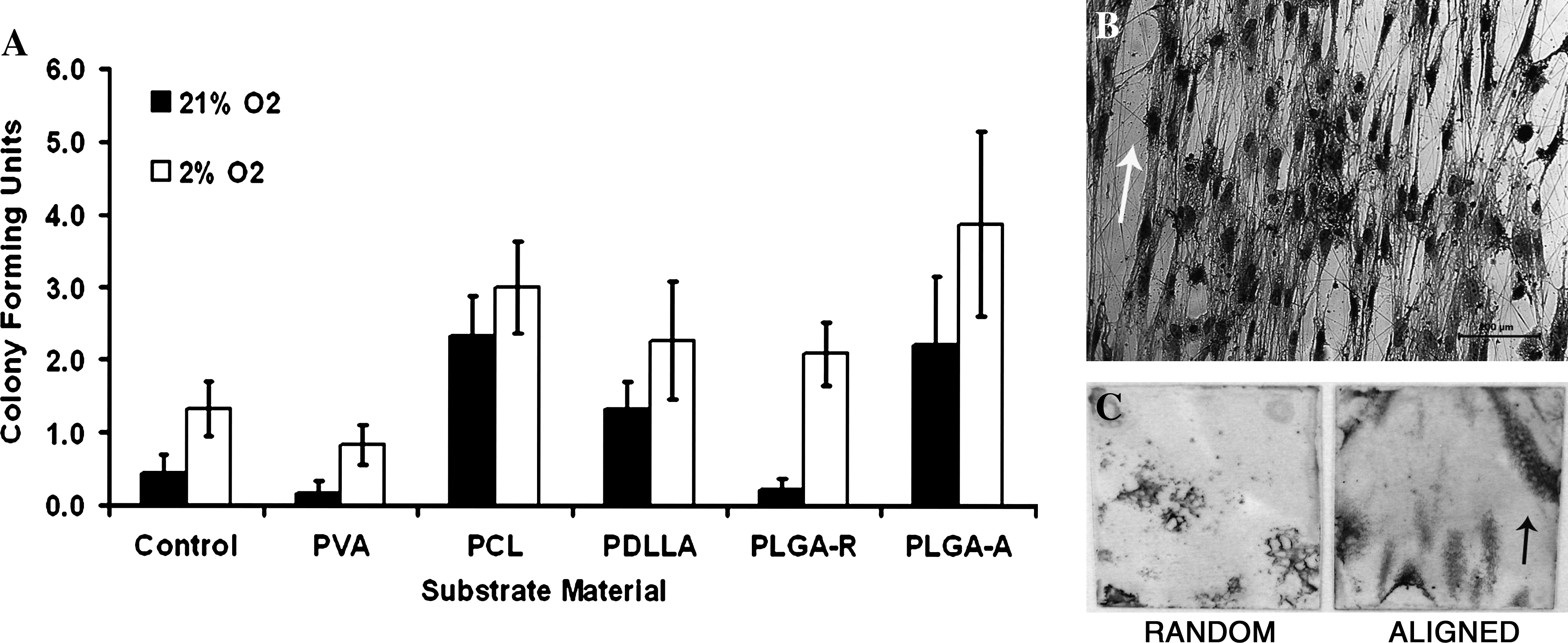
Marrow stromal cells recovered in optimal conditions display retention of multipotency. Quantification of colony-forming units on different nanofiber scaffolds at different oxygen tensions. Control indicates uncoated glass coverslip. (n = 4) (
Differentiation studies
To confirm that the isolated cells were multipotent, MSC nanofiber-coated coverslips with CFU attached were transferred into differentiation media. MSCs were differentiated on generic TCP as controls. Confirmation of differentiation was provided by histological staining: adipogenic (Oil Red O), chondrogenic (Alcian Blue), and osteogenic (Alizarin Red) (Fig. 3).
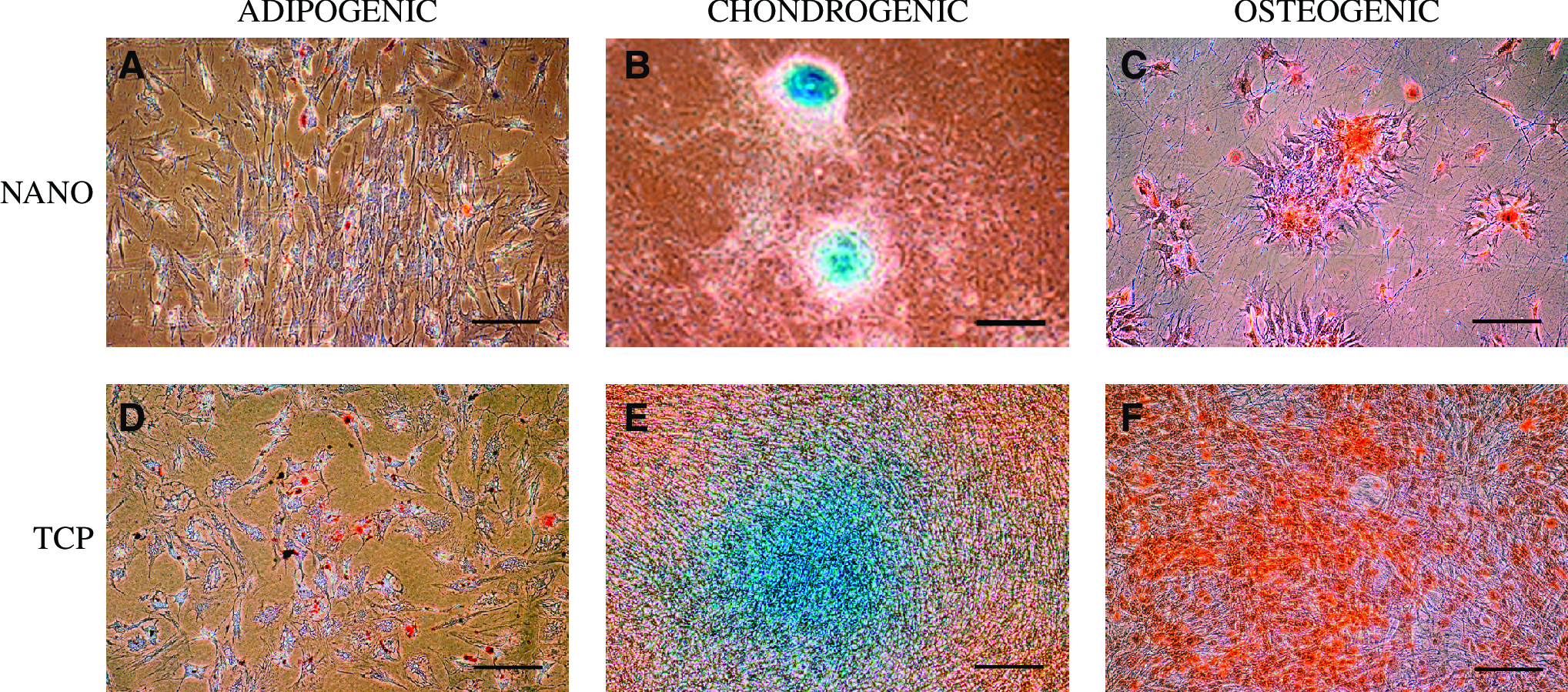
Positive staining of adipogenic, chondrogenic, and osteogenic differentiated cells on nanofibers (
Discussion
The number of descriptions of potential uses and applications of stem-cell therapy has continued to grow in recent years. A simplification and potential increase of the recovery rate of MSCs from BMA, the most convenient and abundant source of adult stem cells, by manipulation of the isolation substrate could accelerate therapeutic usage for widespread clinical applications. 25 It is known that cells respond to topographical cues at a nanoscale24,26–28; we aimed to investigate the effect of nanoscale materials on the isolation of MSCs directly from BMA. The nanofibers used in this study ranged from 100 to 500 nm diameters, as these were reported to promote adhesion. 29 This appears to agree with the findings presented here and support the notion that nanotopography can facilitate cell adhesion and exert control of cell guidance. A combination of nanofiber diameter <500 nm and alignment of those fibers appears most favorable for the isolation of stem cells from BMA, although isolation on random nanofibers is possible. Recently, it was shown by Chen et al. that the diameter of nanofibers can affect the adhesion of NIH3T3 fibroblastic cells on different fiber diameters of micro- to nanoscales. It was indicated that the greater specific surface area provided by nanofibers improved cell attachment, when compared to microfibers. 30 It is possible that the larger surface area provides the opportunity for more focal contacts for cells, increasing the potential for attachment. Chan et al. investigated the early adhesive behavior of human MSCs (hMSCs) on nanofiber substrates in comparison to cast films, uncoated coverslips, and TCP. It was found that the use of nanofibers allowed a greater number of stem cells to attach within a 30-min period, than other surfaces. Collagen-coated surfaces appeared particularly favorable, attributed to integrins within the collagen associated with cell–cell and cell–ECM adhesion. 31 Recently Ma et al. 29 indicated that bone marrow hematopoietic stem cells could be isolated using nanofibrous scaffolds. PLGA nanofibers, combined with collagen, were found to be particularly effective, proving to be significantly better than TCP control surfaces over at 60 min cell-surface contact period.
Our differentiation results demonstrate that the adherent cells on the nanofiber substrates can be differentiated into various skeletal and adipogenic lineages within a period of 20 days. This indicates that it is possible to isolate multipotent cells directly from BMA to nanofibrous substrates, while maintaining their differentiation capacity. The morphology of isolated and differentiated MSCs on nanofibers was compared to that of a control surface, in this case standard TCP. Comparable cell morphologies and responses to staining were observed in each case, indicating that isolation of stem cells on aligned nanofibers does not result in abnormal tendencies for MSC differentiation. Previously, Xin et al. used PLGA nanofibers to attachment of bone marrow–derived first passage hMSCs. In agreement with our findings, presented here, it was possible to successfully differentiate these cells to chondrogenic and osteogenic lineages. 32
It was possible to prepare electrospun nanofibers of similar diameters, approximately 100 to 500 nm in diameter (not including 2% PLGA), from different materials, by manipulation of various parameters. Investigation of the effect of increasing polymer concentration was demonstrated using 0.5% and 2% PLGA, showing a clear increase in fiber diameter with increasing polymer concentration. Previous work by Tan et al. has also shown that increasing the polymer concentration will result in larger diameter fibers. 33
Generally, it appears that alignment of nanofibers results in a decrease in nanofiber diameter. Bashur et al. observed that mandrel aligned PLGA nanofibers could either decrease or increase diameter subject to alignment, depending on the polymer concentration. 34 It was thought that contact of fibers with the rotating mandrel resulted in a “drawing effect” whereby fibers were extruded/pulled, making them thinner. This was observed using 11% PLGA, and not 5%. A hypothesis for this phenomenon was not provided. The instances of increasing fiber diameter with alignment (PCL and 2% PLGA) occur in combination with a >3 kV decrease in electrospinning voltage. It was thought that decreasing the voltage resulted in less bifurcations of the electrospinning jet. Therefore, typically, the resulting fibers (even subject to a drawing effect) were larger than fibers spun at a higher voltage. 35 Aligned PLGA nanofiber substrate exhibiting highest CFU in our study might be partially ascribed to the lower fiber diameter (Table 2). Subsequent experimentation was performed to determine if fiber alignment was a dominant factor in enhancing isolation of adherent cells. CFU were more frequently observed on aligned PCL, PLA, and PLGA when compared to their randomly oriented counterparts (n = 1; data not presented). It was considered that orientation of fibers and their diameter are important factors for cell adhesion.
The variability of quantity of stem cells within a BMA cell population can cause some variability in results, which limit the ability to identify statistical differences between samples. However, the trend identified in this study appears that aligned fibers with diameter <500 nm appear to be most successful at isolating adherent cells from BMA. In addition, the findings indicate that nanofibrous surfaces are favorable for the attachment of stem cells, compared to TCP, in agreement with previous studies.29,31 This, in combination with isolation at physiologically relevant oxygen levels (2% O2), resulted in the identification of a direct, efficient method for isolation of stem cells from BMA. A recent review by Placzek et al. indicated the diversity of factors involved in the interactions of stem cells with cells and surfaces 36 ; therefore, further optimization of stem-cell isolation could be achieved by chemically modifying nanofibers to potentially increase cell–fiber adhesion. Characterization of stem cells over prolonged culture on nanofibers after isolation would be desirable to investigate the potential of implantation of hMSC-seeded nanofibers. The method described here provides a favorable alternative to conventional techniques for MSC isolation. It may also prove favorable for the expansion of MSCs and certainly the development of oriented cell growth and potentially tissue growth.
The use of a physiological oxygen concentration (2% O2) environment for recovery of MSC from BMA directly onto nanofibrous substrates enhanced CFU recovery in all instances. Previous reports have described that reduced O2 tensions promote enhanced CFU formation and proliferative potential of MSC although to our knowledge no report has yet to combine physiological normoxia and nanofibre substrate technology.37–39 Though increased CFU formation was observed in 2% O2, no obvious mechanism exists to explain this result. We have recently observed through microarray-based transcriptional profiling that MSC recovered in 2% O2 upregulated genes associated with cell adhesion pathways (Unpublished data, Forsyth, N.R.). This would provide potential mechanistic insight for the improved CFU numbers. Importantly, though we observed enhanced CFU recovery in 2% O2 with all substrates tested, the key enhancements coincided with aligned versus random nanofibers. This argues that though 2% O2 was important, the alignment of the nanofiber was the critical factor in this study.
In conclusion, this report provides the first description of recovery of hMSCs directly onto nanofibers. The combination of aligned nanofibers and 2% O2 was superior for the recovery of multipotent MSC. This one-step technique, for the rapid production of stem-cell seeded, tissue engineering constructs, will be readily applicable to regenerative medicine cell-based therapeutics.
Footnotes
Acknowledgment
This work was supported by the European Commission (European Union Project “Expertissues”) Project NMP3-CT-2004-500283).
Disclosure Statement
No competing financial interests exist.